Vascular Density Predicts Fistula Risk in Salvage Laryngectomy: Study

USA: The risk of pharyngocutaneous fistula following salvage laryngectomy, a surgery performed after unsuccessful primary treatment of laryngeal cancer, remains a significant concern in clinical practice. A recent study focusing on pharyngeal mucosal margin vessel counts has shed new light on predicting this complication, offering potential advancements in postoperative care and patient outcomes.
The researchers revealed that higher blood vessel density in pharyngeal mucosal margins predicts increased fistula formation risk post-salvage laryngectomy.
The study, published in Otolaryngology-Head and Neck Surgery, established a threshold of 33.9 vessels/×10 field (62% specificity, 75% sensitivity) in identifying patients at heightened risk. Those exceeding this threshold were at a 5-fold higher likelihood of developing fistulas within 30 days post-op.
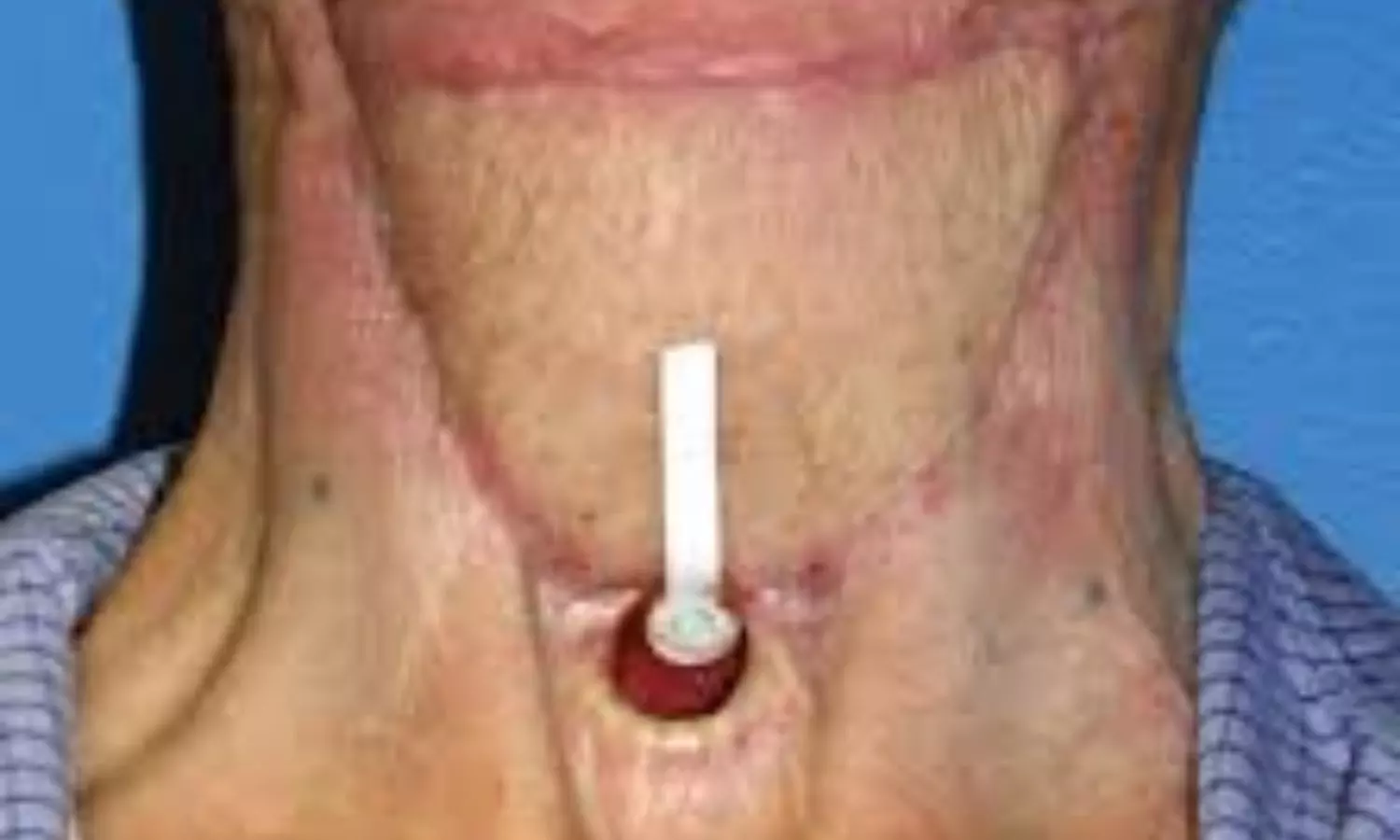
A pharyngocutaneous fistula is characterized by an abnormal connection between the pharynx and the skin of the neck and can lead to prolonged hospital stays, increased healthcare costs, and potential delays in adjuvant therapy. Andrew D.P. Prince, the University of Michigan Health System, Ann Arbor, Michigan, USA, and colleagues aimed to evaluate vessel counts in the pharyngeal mucosal margins of patients who underwent salvage laryngectomy to establish whether mucosal vascularity might predict fistula risk.
For this purpose, the researchers conducted a retrospective cohort study at a tertiary medical center. They identified patients who underwent salvage total laryngectomy at their institution between 1999 and 2015. For each patient, pharyngeal mucosal margins from laryngectomy specimens were histologically evaluated, and vessel counts were performed on 5 ×10 images.
The main outcome measure evaluated was the occurrence of fistula within 30 days following surgery, with mean vessel counts analyzed as the primary explanatory factor.
The following were the key findings of the study:
- Seventy patients were included, and 40% developed a postoperative fistula.
- There was a large difference in the mean vessel count in patients who did develop a fistula (48.6 vessels/×10 field) compared to those who did not (34.7 vessels/×10 field).
- A receiver operative characteristic curve found that a cutoff value of 33.9 vessels/×10 field provided a sensitivity of 75% and specificity of 62% to predict the likelihood of fistula occurrence (area under the curve = 0.71).
- In a binary logistic regression, patients with vessel counts greater than 33.9 had a 5-fold increased risk of developing fistula.
- Histologically, vessels in the pharyngeal mucosa of patients who developed fistulas were more disorganized.
The findings showed that patients with higher mean mucosal margin vessel counts are at increased risk of fistula after salvage laryngectomy.
“The precise mechanism remains unclear, but vascular disorganization is theorized to play a role in impaired wound healing. Counting vessels could aid in stratifying the risk of fistula development and inform postoperative care strategies,” the researchers wrote.
Reference:
Prince ADP, Heft Neal ME, Buchakjian MR, Chinn SB, Stucken CL, Casper KA, Malloy KM, Prince MEP, Rosko AJ, McHugh JB, Spector ME. Pharyngeal Mucosal Margin Vessel Counts Predict Pharyngocutaneous Fistula in Salvage Laryngectomy. Otolaryngol Head Neck Surg. 2024 Jun 17. doi: 10.1002/ohn.865. Epub ahead of print. PMID: 38881401.


