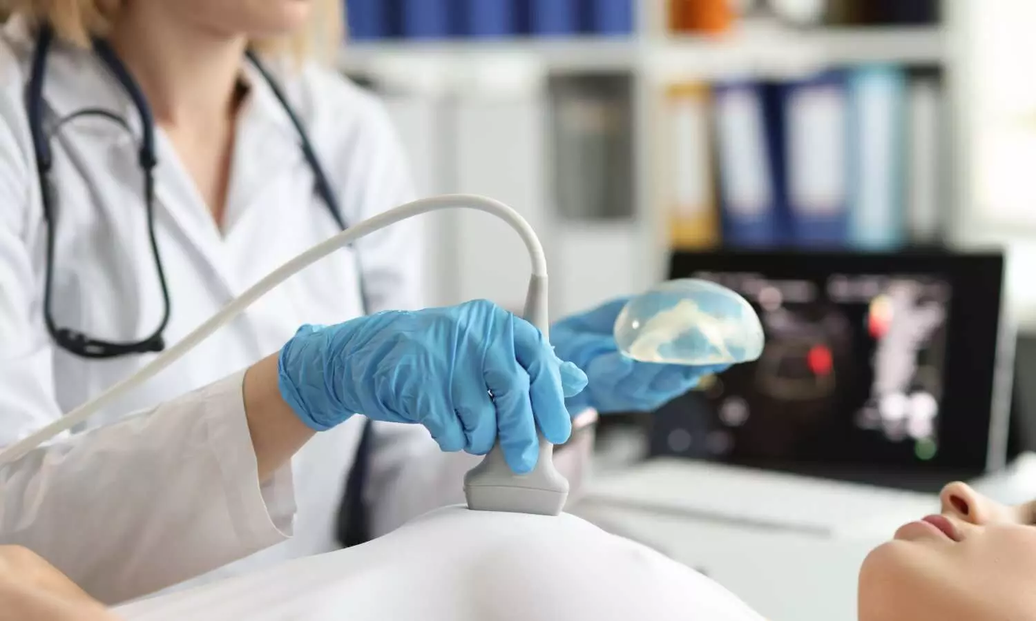
Ultrasound and mammography scores over AI for Early Breast Cancer Detection in Dense Breasts, Study suggests

South Korea: Using a combination of mammography and supplemental ultrasound (US) provides superior outcomes compared to relying solely on artificial intelligence (AI)-enhanced mammography assessment for screening women with dense breasts, according to a recent study.
The study, published in the Radiology Journal, found that although AI enhanced the specificity of mammography interpretation, the combination of mammography and supplemental ultrasound identified more early breast cancers that were node-negative and not detectable by AI-enhanced mammography alone.
AI aids radiologists in interpreting screening mammography but cannot completely overcome its inherent low sensitivity in women with dense breasts,” the researchers wrote.
The effectiveness of AI compared to supplemental ultrasound for screening women with dense breasts remains uncertain. Previous studies indicate that AI systems can assist radiologists in interpreting images. However, some research suggests that AI, like human readers with mammography interpretation, may face challenges in detecting malignant lesions in dense breast tissue.
Against the above background, Jung Min Chang, Seoul National University Hospital Healthcare System Gangnam Center, Seoul, Republic of Korea, and colleagues aimed to compare the performance of mammography alone, mammography with AI, and mammography plus supplemental US for screening women with dense breasts, and to examine the characteristics of the detected cancers.
The study enrolled 5,707 asymptomatic women with an average age of 52.4 years, all aged 40 and above. Between January 2017 and December 2018, these women underwent mammography along with supplemental whole-breast handheld ultrasound at a primary healthcare center.
During the study, five breast radiologists performed sequential readings for mammography alone, and mammography assisted by an AI system. Additionally, a dedicated breast radiologist reviewed the mammography marks either independently or with AI assistance to verify lesion identification.
The reference standard consisted of histologic examination and 1-year follow-up data. The study compared the cancer detection rate (CDR) per 1000 screening examinations, as well as sensitivity, specificity, and abnormal interpretation rate (AIR), across mammography alone, mammography with AI, and mammography plus the US.
The researchers reported the following findings:
- Among 5707 asymptomatic women (mean age, 52.4 years ± 7.9), 33 had cancer (median lesion size, 0.7 cm).
- Mammography with AI had higher specificity (95.3%) and lower AIR (5.0%) than mammography alone (94.3% and 6.0%, respectively).
- Mammography plus US had a higher CDR (5.6 versus 3.5 per 1000 examinations) and sensitivity (97.0% versus 60.6%) but lower specificity (77.6% versus 95.3%) and higher AIR (22.9% vs 5.0%) than mammography with AI.
- Supplemental US alone helped detect 12 cancers, mostly stage 0 and I (92%, 11 of 12).
“Supplemental screening ultrasound revealed additional early breast cancers that were node-negative and not detected by mammography and artificial intelligence in women with dense breasts, albeit with an increase in false-positive findings,” the researchers concluded.
Reference:
https://doi.org/10.1148/radiol.233391


