
Rare case unravels uncommon Hematological Complication of typhoid Fever: A report
India: In a medical anomaly that has puzzled experts, a recent
case report has unveiled an unusual hematological complication associated with
typhoid fever, shedding new light on the complex manifestations of this
infectious disease. This discovery underscores the importance of vigilant
monitoring and comprehensive assessment in managing typhoid fever cases,
especially in regions where the disease remains endemic.
Typhoid fever, caused by the bacterium Salmonella enterica
serotype Typhi, is a systemic illness characterized by fever, abdominal pain,
and gastrointestinal symptoms. While complications such as intestinal
perforation and encephalopathy are well-documented, hematological abnormalities
beyond leukopenia and thrombocytopenia are relatively rare.
The case report published in the Journal of the Association of Physicians of
India describes the case of a young female
who presented with complaints of severe left upper quadrant pain after being
diagnosed with typhoid fever.
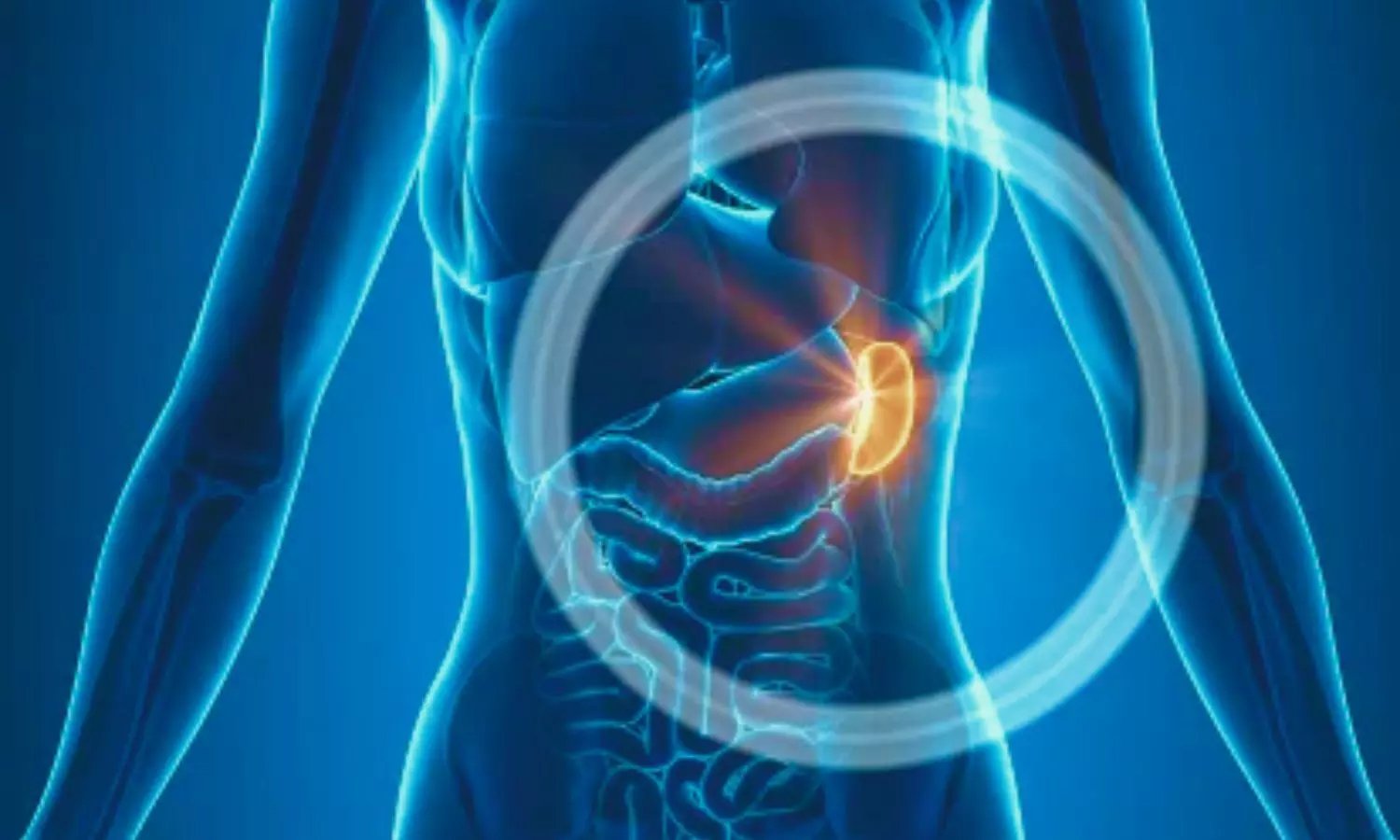
Computed tomography (CT) showed
multiple wedge-shaped splenic infarcts. She was treated with antibiotics and
was also started on antiplatelets. She completely recovered with this
management, and antiplatelets were tapered off on subsequent visits.
The case is of a 25-year-old female
patient with no known comorbidities who was referred to a hospital with complaints
of high-grade fever for 9 days, which was associated with loose stools. She was
diagnosed with typhoid fever from the referring hospital and was started on ceftriaxone.
She then started complaining of chest pain, left-sided abdominal pain, and
breathlessness. She had a family history of rheumatoid arthritis in her mother.
On examination,
she was oriented and conscious, febrile, and had a temperature of 104°F. Her
abdominal examination showed tenderness over the left hypochondrium, and her
respiratory system showed signs of left-sided pleural effusion.
She was admitted,
and baseline blood investigations were conducted. Her blood culture confirmed
the diagnosis of Salmonella Typhi, which was sensitive to the
antibiotic. Her serology for other tropical illnesses, such as dengue, malaria,
leptospirosis, and scrub typhus, was negative.
On further
evaluation, an abdomen ultrasound showed hepatomegaly, mild splenomegaly with an evolving abscess of size 8 × 7 cm. It was followed by contrast-enhanced
computed tomography (CBCT) of the chest and abdomen, which revealed multiple
wedge-shaped splenic infarcts and mild pleural effusion. The echocardiogram was
unremarkable, and the thrombophilia panel was within normal range.
She
was treated with antibiotics, nonsteroidal anti-inflammatory drugs, and fluid
resuscitation and was put on oral antibiotics on discharge. A rheumatologist and hematologist took an opinion to exclude hypercoagulable states,
blood-borne malignancy, autoimmune diseases, and collagen vascular diseases.
She
was initiated on antiplatelets and continued for 6 months. Her follow-up
ultrasound of the abdomen revealed a normal splenic echotexture and size.
“Splenic infarction is an unusual complication of typhoid
fever, and there are only a few literature mentioned for the same,” the
researchers wrote. “Common presentation is left-sided abdomen pain and
tenderness. CT scan is the imaging modality of choice for suspected patients.”
“It is also crucial to exclude other common causes of
splenic infarction. We have found a benefit with antiplatelet
therapy and supportive treatment in our patient,” they concluded.
Reference:
John S, V A. An Unusual Hematological Complication of
Typhoid Fever Case Report. J Assoc Physicians India 2024;72(5):101-102.

