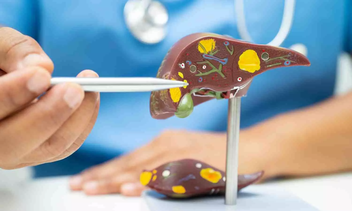
Photon-counting detector CT is an Accurate Alternative to Quantify Liver Fat, finds study

A new study published in the journal of Radiology found that counting photons when evaluating liver fat in individuals with fatty liver disease, computed tomography (CT) may be used instead of magnetic resonance imaging (MRI). Photon-counting detector (PCD) CT provides a consistent CT value and might overcome the restriction of traditional energy-integrating detector CT in precisely measuring liver fat because of protocol-induced CT value variations. To improve the accuracy of fat measurement across different PCD CT procedures in relation to MRI proton density fat fraction (PDFF), Huimin Lin and colleagues carried out this investigation in order to create and validate a universal CT to MRI fat conversion formula.
The viability of fat measurement in phantoms with different nominal fat fractions was assessed in this prospective investigation. Between September 2023 and March 2024, 157 persons with suspected metabolic dysfunction–associated steatotic liver disease (MASLD) and 500 asymptomatic subjects were recruited. 6 groups with varying CT protocols (90, 120, or 140 kVp tube voltage and standard or reduced radiation dosage) were randomly allocated to the participants.
The training cohort consisted of 51% (53 of 104) of the subjects in the 120-kVp standard-dose asymptomatic group, whereas the validation cohort consisted of the remaining asymptomatic patients. To estimate the CT-derived fat fraction (CTFF), a method for quantifying fat from CT to MRI was developed using the training cohort. The asymptomatic validation cohort, subcohorts stratified by radiation dosage, tube voltage, and body mass index, as well as the MASLD cohort, were used to assess CTFF agreement with PDFF and its error. Further analysis was done on the factors affecting CTFF error.
Excellent agreement between CTFF and the nominal fat fraction was observed in the phantoms (intraclass correlation coefficient: 0.98; mean bias: 0.2%). There were a total of 122 MASLD participants and 412 asymptomatic participants in all. The following formula was developed to convert CT to MRI fat: MRI PDFF (%) = −0.58· CT (HU) + 43.1. CTFF and PDFF showed excellent agreement in all comparisons (mean bias values < 1%). Tube voltage, radiation dosage, body mass index, and PDFF had no effect on CTFF error. The MASLD cohort similarly showed agreement between CTFF and PDFF (mean bias, -0.2%). Overall, across several protocols, the standardized CT value from PCD CT demonstrated a strong and impressive agreement with MRI PDFF, and it might be a reliable substitute for measuring liver fat.
Source:
Lin, H., Xu, X., Deng, R., Xu, Z., Cai, X., Dong, H., Yan, F., & Weintraub, E. (2024). Photon-counting Detector CT for Liver Fat Quantification: Validation across Protocols in Metabolic Dysfunction–associated Steatotic Liver Disease. In K. Fowler (Ed.), Radiology (Vol. 312, Issue 3). Radiological Society of North America (RSNA). https://doi.org/10.1148/radiol.240038


