Optical imaging devices useful for diagnosis of periodontal tissue and hard tissue in orthodontic field: Study

South Korea: A recent study published in the Journal of Clinical Medicine has shed light on the potential application of non-invasive optical imaging methods in orthodontic diagnosis.
Jae Ho Baek from F.E.S. Research Lab in Ulsan, Republic of Korea, found that non-invasive optical diagnostic devices, including optical Doppler tomography (ODT) and optical coherence tomography (OCT), can be used in clinical practice during orthodontic treatment. They also introduced a new diagnostic paradigm differentiating microstructural changes in tissues in orthodontic diagnosis.
The investigator notes the importance of the early diagnosis of microscopic changes in soft and hard tissues, including periodontal tissue during orthodontic treatment to prevent iatrogenic side effects like periodontal diseases and root resorption. Cervical periodontal tissue is suggested to be the most critical area that reacts first to orthodontic forces or mal-habits, and it is also the place of bacteria deposition in the early stage of periodontal diseases.
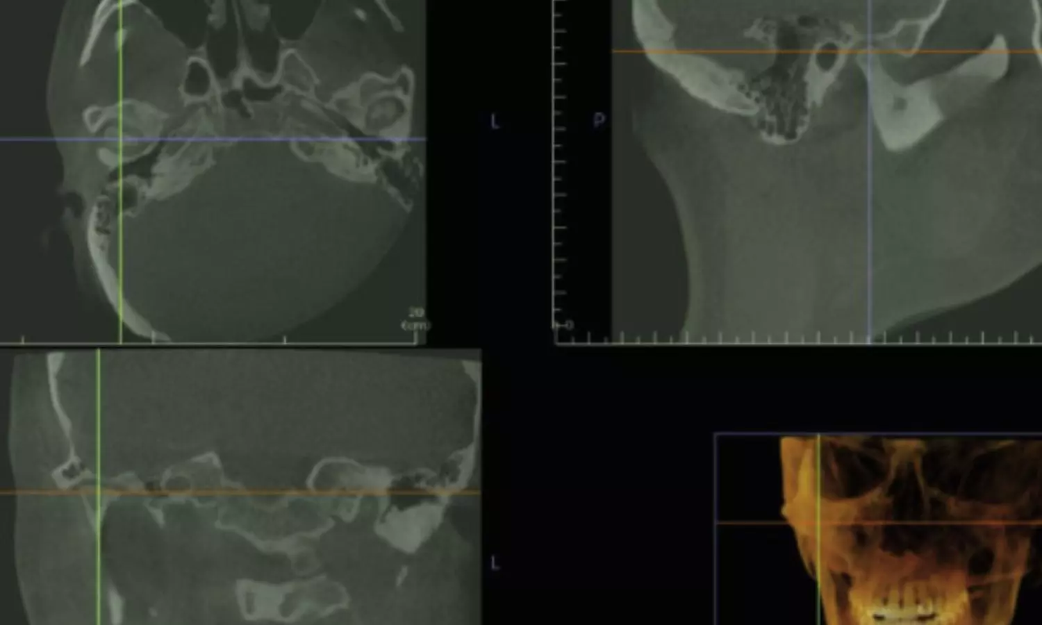
The early diagnosis of hard tissue changes, such as demineralization is also essential in maintaining a patient’s health during orthodontic treatment. Many diagnostic devices, including radiographic equipment and intra-oral scanners, help diagnose these problems but have certain limitations in precision and invasiveness.
Against the above background, the study was conducted to verify the possible utilities of non-invasive diagnostic devices in the orthodontic field that can compensate for these limitations.
For this purpose, non-invasive optical diagnostic devices were used in vivo with human and animal examinations for soft and hard tissues, including optical Doppler and coherence tomography. These devices can provide real-time three-dimensional images at the histological scale.
In conclusion, optical diagnostic imaging devices have sufficient objective potential for diagnosing soft tissue, including periodontal tissue and hard tissue, in orthodontics. In addition to solving some technical problems, the study results open a new horizon in orthodontic diagnosis through ongoing research to understand the correlation between changes in periodontal tissue and tooth movements during orthodontic treatment or pathologic progress.
The investigator found ODT and OCT can provide two-dimensional or three-dimensional images of hard tissue and periodontal changes during orthodontic treatment at the histological level non-invasively in both animal experiments and humans in vivo.
Specifically, it was found that these devices can be used during orthodontic treatment in several areas, such as estimating microscopic changes in periodontal tissue, which are essential to evaluate biomechanical effects, enamel demineralization, and the early diagnosis of periodontal diseases.
“It is a task that must be solved in the future to collect more data by applying these devices to orthodontic patients with removable or fixed appliances and then verifying the correlation between the results and existing information to establish standards for objective data analysis,” the study stated.
Reference:
Baek, J. H. (2023). Potential Application of Non-Invasive Optical Imaging Methods in Orthodontic Diagnosis. Journal of Clinical Medicine, 13(4), 966. https://doi.org/10.3390/jcm13040966


