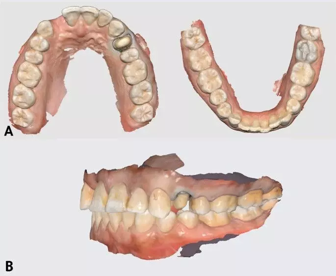
Dental Breakthrough: Lab-Grown Teeth May Offer a Natural Alternative to Fillings and Missing Teeth

Scientists have recreated the early environment of tooth development
in the lab, using a specially designed material that enables dental cells to
communicate, offering new insights into how natural teeth could one day be
grown outside the human body.

In a recent scientific development, researchers at King’s College London have made significant progress toward enabling adults to regrow their own teeth. Published in the ACS Macro Letters journal, this research introduces a promising alternative to traditional dental treatments by offering a potentially natural method for tooth repair and regeneration.

For over a decade, scientists at King’s College London’s
Faculty of Dentistry, Oral & Craniofacial Sciences have been exploring the
possibilities of lab-grown teeth. Their latest collaborative study with
Imperial College London has led to the development of a novel biomaterial that
replicates the natural conditions of early tooth development. This innovation
allows cells to communicate in a way that initiates the formation of tooth
structures.

Xuechen Zhang, from the Faculty of Dentistry, Oral &
Craniofacial Sciences, King’s College London, and author of the study said: “Fillings
aren’t the best solution for repairing teeth. Over time, they will weaken tooth
structure, have a limited lifespan, and can lead to further decay or
sensitivity. Implants require invasive surgery and good combination of implants
and alveolar bone. Both solutions are artificial and don’t fully restore
natural tooth function, potentially leading to long-term complications.”

Discussing possible approaches to using this breakthrough in
clinical settings, Xuechen added: “We have different ideas to put the teeth
inside the mouth. We could transplant the young tooth cells at the location of
the missing tooth and let them grow inside mouth. Alternatively, we could
create the whole tooth in the lab before placing it in the patient’s mouth. For
both options, we need to start the very early tooth development process in the
lab.”

Corresponding author of the study, Dr Ana Angelova Volponi
from King’s College London, emphasised the broader implications of this
research: “As the field progresses, the integration of such innovative
techniques holds the potential to revolutionise dental care, offering
sustainable and effective solutions for tooth repair and regeneration.”

Reference: Zhang X, Contessi Negrini N, Correia R,
Sharpe PT, Celiz AD, Angelova Volponi A. Generating Tooth Organoids Using
Defined Bioorthogonally Cross-Linked Hydrogels. ACS Macro Lett. 2024 Dec
17;13(12):1620-1626. doi: 10.1021/acsmacrolett.4c00520. Epub 2024 Nov 12. PMID:
39532305; PMCID: PMC11656705.

Powered by WPeMatico

Can heart failure with preserved ejection fraction be prevented?

An international team of researchers at the University of Manchester, Baylor College of Medicine and collaborating institutions has discovered a natural mechanism that protects the heart from heart failure with preserved ejection fraction (HFpEF), a serious condition in need of effective treatment. The team reports in Circulation, a journal of the American Heart Association, that when the cardioprotective mechanism fails, it promotes the development of HFpEF. Importantly, restoring the mechanism prevents the progression of the condition. The findings provide a promising therapeutic target to prevent and treat this life-threatening disease.

“HFpEF is a complex, multifactorial disease associated with metabolic stress. One of the factors involved is the toxic accumulation of lipids in heart cells,” said co-author Dr. Tamer Mohamed, associate professor of surgery and director of the Laboratory for Cardiac Regeneration at Baylor. “This study was the result of a productive multidisciplinary collaboration. We worked with slices of human hearts affected by HFpEF, a method developed in my lab at Baylor, integrating the findings with the results from studies in mouse and rat models of the condition to better understand what drives this lipid overload.”

The team began by assessing gene changes in hearts from people diagnosed with metabolic stress along with various cardiovascular complications and found alterations in the expression of 488 genes. “We found particularly relevant the simultaneous reduction of the expression of genes XBP1 and EDEM2 in human HFpEF hearts,” Mohamed said.

The link between EDEM2 and other conditions has been studied, but its role in the heart remains unexplored.

“We discovered that the XBP1 protein regulates the Edem2 gene in animal models and that EDEM2 was downregulated in hearts from individuals with metabolic disorders and in mouse models,” Mohamed said. “Further evidence supported the involvement of EDEM2 and XBP1 in cardiovascular diseases and lipid regulation.”

To investigate whether XBP1 and EDEM2 were directly involved in lipid toxicity and HFpEF, the researchers removed the Xbp1 or the Edem2 gene in mice. Consequently, these mice became more vulnerable to metabolic stress-induced heart lipid toxicity and cardiac dysfunction. “It was exciting to see that restoring XBP1 or EDEM2 alleviated lipid overload in the heart and reversed HFpEF,” Mohamed said.

This comprehensive study uncovered that XBP1 and EDEM2 work together to maintain a healthy lipid balance in heart cells. The findings provide molecular and functional evidence that deficiency of XBP1-EDEM2 leads to lipid toxicity in the heart, accelerating the onset and development of heart failure in metabolic disorders.

“Our findings support further study of the XBP1s and EDEM2 pathway as a promising therapeutic target for mitigating cardiac lipotoxicity and progression of HFpEF,” Mohamed said.

Reference:

Oveena Fonseka, Rida Raja, XBP1s-EDEM2 Prevents the Onset and Development of HFpEF by Ameliorating Cardiac Lipotoxicity, Circulation, https://doi.org/10.1161/CIRCULATIONAHA.124.072194.

Powered by WPeMatico

Digital Replication technique may accurately restore original crown morphology, suggests study

Researchers have found in a new study that Digital replication technique successfully restored the original crown morphology with high accuracy. However, the dimensional accuracy of all-ceramic crowns varied with the type of CAD/CAM porcelain material used, with lithium disilicate glass-ceramic demonstrating the most superior results.

The study aimed to evaluate the three-dimensional accuracy of computer-aided design and computer-aided manufacturing (CAD/CAM) in fabricating all-ceramic crowns using various cuttable materials, assessed through reverse engineering. The original resin tooth morphology of the left maxillary mesial incisor and left maxillary first molar from a standard resin tooth model, along with the two corresponding prepared teeth, were scanned and imported into exocad software. The digitally reproduced crown morphology was utilized to fabricate crowns from cut porcelain-reinforced resin ceramic (Uh group, Upcera Hyramic), lithium disilicate glass-ceramic (e.max group, IPS e.max Cad), and zirconia ceramic (Ue group, Upcera Explore). All specimens were subsequently rescanned, and the root mean square (RMS) values were calculated after overlaying with the original crown CAD data using the 3D analysis software Geomagic Studio 2013 to compare the dimensional accuracy. Results: Among the mesial incisor and first molar specimens, the e.max group demonstrated the highest dimensional accuracy of the all-ceramic crowns, followed by the Ue group, while the Uh group exhibited the lowest accuracy. The differences in dimensional accuracy among the three groups were statistically significant (P < 0.05). The digital replication technique effectively restored the original crown morphology with a high degree of accuracy. For the same CAM pattern, the dimensional accuracy of all-ceramic crowns varied depending on the CAD/CAM porcelain material, with lithium disilicate glass-ceramic showing the superior results.

Reference:

Wang, H., Qu, W., Wang, T. et al. Accuracy analysis of all-ceramic crowns with different materials in CAD/CAM digital replication mode. BMC Oral Health 25, 491 (2025). https://doi.org/10.1186/s12903-025-05892-9

Keywords:

Digital, Replication, technique, accurately, restore, original, crown morphology, suggests, study, Wang, H., Qu, W., Wang, T, Computer-aided design and fabrication, All-ceramic crowns, Three-dimensional deviation, Accuracy

Powered by WPeMatico

Early Gestational Diabetes Raises Postpartum Diabetes Risk, finds study

Researchers have discovered in a new study that women diagnosed with gestational diabetes mellitus (GDM) earlier than 24 weeks of pregnancy, termed early GDM, are almost twice as prone to developing diabetes mellitus (DM) post-delivery as those diagnosed with later pregnancy. The findings demonstrate that early GDM not only has a greater risk of postpartum diabetes but also is linked to more cases of impaired glucose tolerance following delivery. The study was published in the Journal of Diabetes and Its Complications by Sarah J. and fellow researchers.

GDM is usually screened for at or after 24 weeks’ gestation, but increased early diagnosis has been more frequent as prenatal care remains more proactive. But whether early detection was linked to poorer outcomes postpartum has been subject to ongoing research until now.

The research was a systematic review and meta-analysis of the evidence. Researchers reviewed PubMed, EMBASE, WHO International Clinical Trials Registry Platform (ICTRP), ClinicalTrials.gov, and the Cochrane Central Register of Controlled Trials from inception until March 2023. Researchers included observational studies and randomized controlled trials that contrasted the postpartum rate of DM among women diagnosed with early GDM (earlier than 24 weeks) compared to women with standard GDM (diagnosed at or later than 24 weeks).

The primary outcome was the diagnosis rate of postpartum diabetes. Secondary outcomes included postpartum visit attendance, completion and interpretation of oral glucose tolerance tests (OGTT), and glycated hemoglobin (HbA1c) levels. A total of 11 studies involving 1,573 women with early GDM and 3,340 with standard GDM were included in the final analysis.

Key Findings

Early GDM group (n = 1,573):

  • Postpartum diabetes: 12.8%

  • Impaired glucose tolerance: 24.7%

  • Mean age: 30.2–37.0 years

  • Mean BMI: 24.4–35.6 kg/m²

Standard GDM group (n = 3,340):

  • Postpartum diabetes: 6.1%

  • Impaired glucose tolerance: 13.0%

  • Mean age: 28.9–32.8 years

  • Mean BMI: 23.6–36.0 kg/m²

  • Risk of postpartum diabetes: Almost twice (RR = 1.89) in early GDM

  • Risk of impaired glucose tolerance: Almost twice (RR = 1.84) in early GDM

  • Follow-up duration: Primarily 5–12 weeks after delivery, one study up to 6 years

This study affirmed that women with GDM diagnosed earlier than 24 weeks of pregnancy have a considerably increased risk of developing diabetes after delivery compared with those diagnosed after this time point. With approximately 13% of early GDM patients advancing to postpartum diabetes compared with slightly over 6% in usual GDM, the results underscore the necessity for increased vigilance, early intervention, and prolonged postpartum surveillance in this at-risk population.

Reference:

Weingarten, S. J., Levy, A. T., Al-Kouatly, H. B., & McLaren, R. A., Jr. (2025). Postpartum diabetes mellitus among patients with early gestational diabetes: A systematic review and meta-analysis. Journal of Diabetes and Its Complications, 109038, 109038. https://doi.org/10.1016/j.jdiacomp.2025.109038

Powered by WPeMatico

Yoga Effective Alternative to Strength Training for Knee Osteoarthritis: JAMA

Researchers have found in a randomized clinical trial that patients with knee osteoarthritis (OA) experienced comparable benefits from yoga as from conventional strength training. The findings suggest that yoga is noninferior to strength training and may be considered a viable treatment option for managing knee OA. A recent trial from Southern Tasmania, Australia, showed that yoga was noninferior to conventional strengthening exercise for decreasing knee pain at 12 weeks in adults over 40 with knee OA. The study was conducted by Bedru J. and colleagues published in JAMA Network Open.

Knee osteoarthritis is a prevalent degenerative joint disease leading to pain, stiffness, and loss of mobility, particularly in elderly populations. Exercise is an integral part of OA treatment, yet there has been limited evidence for the relative effects of different exercise types, including yoga and strengthening, on patients with knee pain and functional recovery.

This parallel-arm, active-controlled, single-center randomized clinical trial enrolled 117 patients, 58 to yoga and 59 to strengthening. All patients had a baseline knee pain score of 40 or greater on a 100-mm visual analog scale (VAS), which is moderate to severe pain. The average age was 62.5 years (SD 8.3), and 72.6% were women.

The intervention lasted for 24 weeks, during which the participants went through 12 weeks of supervised yoga or strengthening exercise (two in-person sessions and one home session weekly), followed by another 12 weeks of unsupervised home sessions. The main outcome was the difference in knee pain during 12 weeks measured with the VAS. A prespecified non inferiority margin of 10 mm was used to assess whether yoga was not inferior to strengthening by a clinically significant amount. Secondary outcomes were WOMAC pain, function, and stiffness scores, patient global assessment, physical performance tests, depression (PHQ-9), neuropathic pain, and quality of life (AQoL-8D).

Results

  • The trial discovered that the 12-week difference in VAS knee pain between groups was similar.

  • The difference between pain decrease using yoga compared with strengthening was −1.1 mm (95% CI, −7.8 to 5.7 mm) and stayed within the prespecified margin of noninferiority, which demonstrated that yoga was no worse than strengthening exercises at lessening pain.

Significantly, 7 of 27 secondary outcomes that were measured at 12 and 24 weeks were in favor of yoga, statistically significant. The yoga group had superior improvement in:

  • WOMAC pain: −44.5 mm (95% CI, −70.7 to −18.3)

  • WOMAC function: −139 mm (95% CI, −228.3 to −49.7)

  • WOMAC stiffness: −17.6 mm (95% CI, −30.9 to −4.3)

  • Patient global assessment: −7.6 mm (95% CI, −15.1 to −0.2)

  • 40-m fast walk test: +1.8 seconds (95% CI, 0.4 to 3.2)

  • Also, depression (PHQ-9) at 12 weeks was better in the yoga group with a between-group difference of −1.1 (95% CI, −1.9 to −0.2), and quality of life (AQoL-8D) at 24 weeks was better in the yoga group (difference of 0.04; 95% CI, 0.00 to 0.07). Mild adverse events were reported equally by both groups, and no serious adverse events were linked to the interventions.

This randomized clinical trial found that yoga was noninferior to strengthening exercises for decreasing knee pain in people with knee osteoarthritis. In addition, yoga also had modest but significant benefits in joint function, patient-reported outcomes, and mental well-being through 24 weeks. These results substantiate the role of yoga as an effective, holistic exercise approach in OA management programs.

Reference:

Abafita BJ, Singh A, Aitken D, et al. Yoga or Strengthening Exercise for Knee Osteoarthritis: A Randomized Clinical Trial. JAMA Netw Open. 2025;8(4):e253698. doi:10.1001/jamanetworkopen.2025.3698

Powered by WPeMatico

THC Exposure before Birth May Impair Infant Lung Development and Function, reveals research

Using cannabis products during pregnancy may negatively affect offspring lung development and function, according to new research. The study is published in the American Journal of Physiology-Lung Cellular and Molecular Physiology and has been chosen as anAPSselect article for April.

Cannabis products are commonly used during pregnancy, despite guidelines from the American College of Obstetricians and Gynecologists recommending abstinence of use in pregnancy given limited safety data regarding for the unborn child. The psychoactive ingredient in cannabis products, delta-9-tetrahydrocannabidol (THC) crosses the placenta and emerging data suggest prenatal THC use is associated with higher risk of stillbirth, preterm birth and low birth weight.

THC edibles are the second most common form of cannabis consumption in pregnant individuals after smoking. High-quality research on the direct effects of cannabis and THC exposure during pregnancy is lacking due to the different cannabis plant strains, range of available cannabis products, common co-use with other substances and restrictions on human cannabis research.

In this study, pregnant nonhuman primates were given a THC cookie-representing the equivalent of a “heavy human medical cannabis dose”-in addition to their regular diet. They continued to eat the cookies throughout pregnancy and after giving birth. During pregnancy, researchers measured fetal lung development with MRI.

After birth, the research team performed assessments to determine the THC-exposed infants’ lung capacity, volume and forced expiratory flow (the amount of air that is forcibly exhaled within a specific time period). They also examined fluid from the exposed newborns’ lungs and analyzed their DNA and RNA.

Some of the findings associated with prenatal THC exposure include:

  • Reduced fetal lung volume beginning mid-pregnancy in the second trimester.
  • Changes to expression in more than 700 genes in newborns’ lungs.
  • Significant decrease in key growth factors for lung development in exposed infants.

“There are several key findings that highlight the potential adverse impact of THC exposure on offspring lung function and development,” the researchers wrote. The results suggest “prenatal THC exposure may have an important influence on future respiratory health, and further studies in human populations are needed.”

Reference:

Qing Ma, Hanxiang Liu, Ming Liu, TrkB signaling promotes alveolar capillary angiogenesis following perinatal hyperoxic damage, American Journal of Physiology-Lung Cellular and Molecular Physiology, https://doi.org/10.1152/ajplung.00334.2023.

Powered by WPeMatico

Willis Covered Stent Promising in Treating Intracranial Blood Blister-Like Aneurysms: Case Series Findings

China: A recent case series published in BMC Surgery has highlighted the potential of the Willis covered stent (WCS) as a viable option for managing intracranial blood blister-like aneurysms (BBAs), a rare but challenging subset of aneurysms associated with high rates of rupture and mortality.

Sheng Guan, Department of Neurointervention, Zhengzhou University First Affiliated Hospital, Henan, Zhengzhou, China, and colleagues reviewed the medical records of 21 consecutive patients who underwent endovascular treatment with the WCS between July 2017 and July 2022. BBAs are typically located at non-branching sites of the internal carotid artery and are structurally fragile due to the absence of key arterial layers, making treatment particularly complex.

This series involved 15 female and six male patients with an average age of 51.6 years. The immediate postoperative results were encouraging—angiographic evaluations confirmed complete occlusion of all aneurysms without endoleak. While the parent artery remained patent in all cases, the procedure was not without complications. Occlusion of the ophthalmic artery was noted in five patients, accounting for 23.8% of the cases. Additionally, one patient developed delayed thrombosis of the stent three days post-surgery.

During an average angiographic follow-up period of just over nine months, all aneurysms remained completely sealed. There were no cases of aneurysm rupture, recurrence, or need for retreatment. Mild to moderate asymptomatic in-stent stenosis was detected in two individuals. Clinical follow-up, which spanned an average of 20 months, revealed excellent functional outcomes—20 patients scored a zero on the modified Rankin Scale (mRS), indicating no disability, while one scored a one.

The Willis-covered stent, designed initially for use in complex intracranial pathologies such as pseudoaneurysms and carotid-cavernous fistulas, offers a promising treatment avenue for BBAs. Its structure—a combination of a balloon-expandable stent and a polytetrafluoroethylene membrane—enables it to seal the aneurysm while maintaining vessel integrity.

Despite the encouraging outcomes, the researchers emphasized that the procedure is not without risks, particularly for technical challenges related to stent delivery and potential complications like branch artery occlusion or in-stent stenosis. Given the small sample size and limited follow-up duration, they called for larger prospective studies with longer observation periods to validate these findings and refine treatment protocols.

“The findings from the case series add to the growing body of evidence supporting the Willis covered stent as a safe, feasible, and effective option for the endovascular management of blood blister-like aneurysms,” the authors noted.

However, they cautioned that its use warrants careful attention to potential procedural challenges, including stent delivery difficulties and related complications, to ensure the best possible clinical outcomes.

Reference:

Jin, Y., Guo, X., Chen, Z. et al. Endovascular treatment of intracranial blood blister-like aneurysms with the Willis covered stent: a case series. BMC Surg 25, 140 (2025). https://doi.org/10.1186/s12893-025-02874-0

Powered by WPeMatico

Covert or latent Toxoplasma gondii infection significantly correlated with CKD, reveals research

Covert or latent Toxoplasma gondii infection is significantly correlated with chronic kidney disease, according to research published in BMC Nephrology. Patients with chronic kidney disease are susceptible to acquiring opportunistic parasites due to acquired immunodeficiency caused by uremia. Therefore, the present case–control study attempted to determine the prevalence of Toxoplasma gondii infection and the associated risk factors among patients with chronic kidney disease undergoing hemodialysis and healthy controls who were registered at the Iranian National Registry Center for Toxoplasmosis in Mazandaran Province, northern Iran. A total of 212 patients with chronic kidney disease and 200 healthy controls were enrolled in this study. Informed consent and a questionnaire were obtained from all subjects. Blood samples were collected from each participant, and the serum was screened for anti-Toxoplasma antibodies (immunoglobulin G and immunoglobulin M). Polymerase chain reaction assay was performed to detect circulating Toxoplasma gondii in the blood samples of patients and controls using the primer pair targeting the repetitive element gene. Results indicated that out of 412 participants, 67.92 percent of patients and 15.5 percent of control subjects were positive for anti-Toxoplasma immunoglobulin G, but all participants were negative for anti-Toxoplasma immunoglobulin M. Also, considering polymerase chain reaction assays with the repetitive element target, the prevalence of Toxoplasma gondii infection was 24.1 percent in case subjects, while none of the control subjects tested positive. Among the polymerase chain reaction-positive cases, 34 (66.7 percent) had Toxoplasma immunoglobulin G positivity. The results from the multiple multinomial logistic regression revealed that the seroprevalence of anti-Toxoplasma gondii immunoglobulin G antibodies in patients with chronic kidney disease was 3.12 times higher than in healthy controls (odds ratio = 3.12; 95 percent confidence interval = 0.43, 14.8; P < 0.001). Also, there was a significant association between the seroprevalence of Toxoplasma gondii infection and age, having a cat at home, and the level of glomerular filtration rate in these patients. The findings demonstrate a highly significant association between latent Toxoplasma gondii infection and chronic kidney disease, mostly in the late stages. Thus, regular screening for Toxoplasma gondii infection in these patients is strongly recommended to prevent the reactivation of latent infections. A combination of serological screening, chemoprophylaxis, and polymerase chain reaction follow-up for patients at risk of reactivation should effectively reduce the likelihood of latent infection reactivation.

Reference:

Montazeri, M., Fakhar, M., Sedighi, O. et al. Latent Toxoplasma gondii infection and associated risk factors among patients with chronic kidney disease: a registry-based study. BMC Nephrol 26, 163 (2025). https://doi.org/10.1186/s12882-025-04079-2

Keywords:

Covert, latent, Toxoplasma, gondii, infection, significantly, correlated, CKD, reveals research, BMC Nephrology, Montazeri, M., Fakhar, M., Sedighi, O, Chronic kidney disease, Glomerular filtration rate, Hemodialysis, Latent toxoplasmosis, Toxoplasma gondii

Powered by WPeMatico

HEALEY ALS Trial: Pridopidine Shows No Impact on Disease Progression in ALS

USA: A recent study evaluating pridopidine for treating amyotrophic lateral sclerosis (ALS) has found no significant effect on disease progression. Conducted as part of the HEALEY ALS Platform Trial, the 24-week investigation assessed whether pridopidine could slow functional decline in individuals with ALS, a neurodegenerative disorder characterized by progressive muscle weakness and loss of motor function.

The findings were published online in JAMA (The Journal of the American Medical Association) on February 17, 2025.

Amyotrophic lateral sclerosis is a progressive and fatal neurodegenerative disease with limited treatment options. The sigma-1 (σ1) receptor has gained attention as a potential therapeutic target due to its role in cellular protection and neurodegeneration. Jeremy M. Shefner, MD, PhD, Department of Neurology, Barrow Neurological Institute, Phoenix, AZ, and colleagues aimed to evaluate the effects of pridopidine, a selective σ1-receptor agonist, in slowing ALS progression and preserving motor function.

For this purpose, the researchers evaluated pridopidine as part of the HEALEY ALS Platform Trial, a multicenter, phase 2/3 randomized study conducted at 54 sites in the US from January 2021 to July 2022. A total of 163 participants with ALS were assigned to receive either pridopidine or a placebo, with an additional 122 placebo participants included in the analysis. Participants received either 45 mg of oral pridopidine twice daily or a matching placebo for 24 weeks.

The primary outcome measured disease progression using a model integrating functional decline and survival, while secondary outcomes assessed respiratory function, bulbar symptoms, and survival-related measures.

The key findings of the study were as follows:

  • The trial included 162 patients with a mean age of 57.5 years, of whom 35% were female.
  • A total of 136 participants (84%) completed the study.
  • There was no significant difference between pridopidine and placebo in disease progression (DRR: 0.99, 95% credible interval: 0.80-1.21, probability of DRR <1: 0.55).
  • There were no improvements in ALS functional rating scale components or survival.
  • Pridopidine showed no benefit on secondary outcomes.
  • Common adverse events included falls (28.1% in the pridopidine group vs. 29.3% in the placebo group) and muscular weakness (24.0% vs. 31.7%, respectively).

The HEALEY ALS Platform Trial findings indicate that pridopidine did not demonstrate any meaningful impact on disease progression in patients with amyotrophic lateral sclerosis. Over the 24-week study period, there was no significant difference between pridopidine and placebo in terms of disease severity, functional decline, or survival. Additionally, the treatment showed no benefit across key secondary outcomes.

“These results suggest that pridopidine may not be an effective therapeutic option for ALS, highlighting the continued need for further research to identify viable treatments that can slow disease progression and improve patient outcomes,” the authors concluded.

Reference:

Writing Committee for the HEALEY ALS Platform Trial, HEALEY ALS Platform Trial Study Group. Pridopidine in Amyotrophic Lateral Sclerosis: The HEALEY ALS Platform Trial. JAMA. 2025;333(13):1128–1137. doi:10.1001/jama.2024.26429

Powered by WPeMatico

Surgery Beneficial for Recurrent Diverticulitis Without Added Risk:JAMA

Researchers have found in a new study that among patients with recurrent diverticulitis, surgery may offer a viable option to prevent future episodes and enhance quality of life, without increasing the risk of complications. The study was published in JAMA Surgery by Alexandre S. and colleagues. This outcome is based on the 4-year follow-up data of the Laparoscopic Elective Sigmoid Resection Following Diverticulitis (LASER) randomized clinical trial.

The trial, which was carried out in six hospitals in Finland, randomized 90 patients between October 2014 and October 2018. All the patients were followed up for four years, and the outcomes were compared between October 2023 and November 2024. The primary objective was to determine whether surgical intervention has significant advantages over conservative approaches, particularly in long-term environments.

The LASER trial was an open-label randomized controlled trial of 90 adult patients (mean age ~55.6 years; 31% male and 69% female) with a clinical diagnosis of recurrent, chronic painful, or complicated diverticulitis. Patients were randomly allocated in a 1:1 ratio to either receive elective laparoscopic sigmoid resection or remain on conservative treatment. Quality of life was assessed by the Gastrointestinal Quality of Life Index (GIQLI), and recurrence and complication rates were monitored over a time frame of four years by intention-to-treat and per-protocol analyses.

Key Findings

  • 90 patients were recruited (28 men [31%], 62 women [69%]; mean age ~55.6 years).

  • 45 patients were allocated to surgery; 45 to conservative management.

  • 32% (14 of 44) in the conservative group switched over to surgery within 4 years.

  • GIQLI scores at 4 years: 115.3 (surgery) vs 109.8 (conservative); mean difference 5.54 (95% CI, −2.98 to 14.06), not statistically significant.

  • Recurrence of diverticulitis: 6 of 38 (16%) in the surgery group (10% post-surgery), vs 34 of 37 (92%) in conservative group.

  • Severe complications: 4 patients (10%) in the surgery group vs 5 patients (11%) in conservative group.

Elective sigmoid resection in patients with recurrent or complicated diverticulitis greatly lowers the risk of recurrence without a rise in severe complications. Yet, it does not provide a substantial improvement in long-term quality of life over conservative management. These results indicate that patients with severely compromised QOL can gain more from initial surgery, whereas conservative management is still a valid choice for patients with milder symptoms or intact QOL.

Reference:

Santos A, Mentula P, Pinta T, et al. Sigmoid Resection vs Conservative Treatment After Diverticulitis: Prespecified 4-Year Analysis of the LASER Randomized Clinical Trial. JAMA Surg. Published online April 09, 2025. doi:10.1001/jamasurg.2025.0572

Powered by WPeMatico