Bedwetting beyond 5? KIMS Cuddles Launches Specialized Clinic for Children

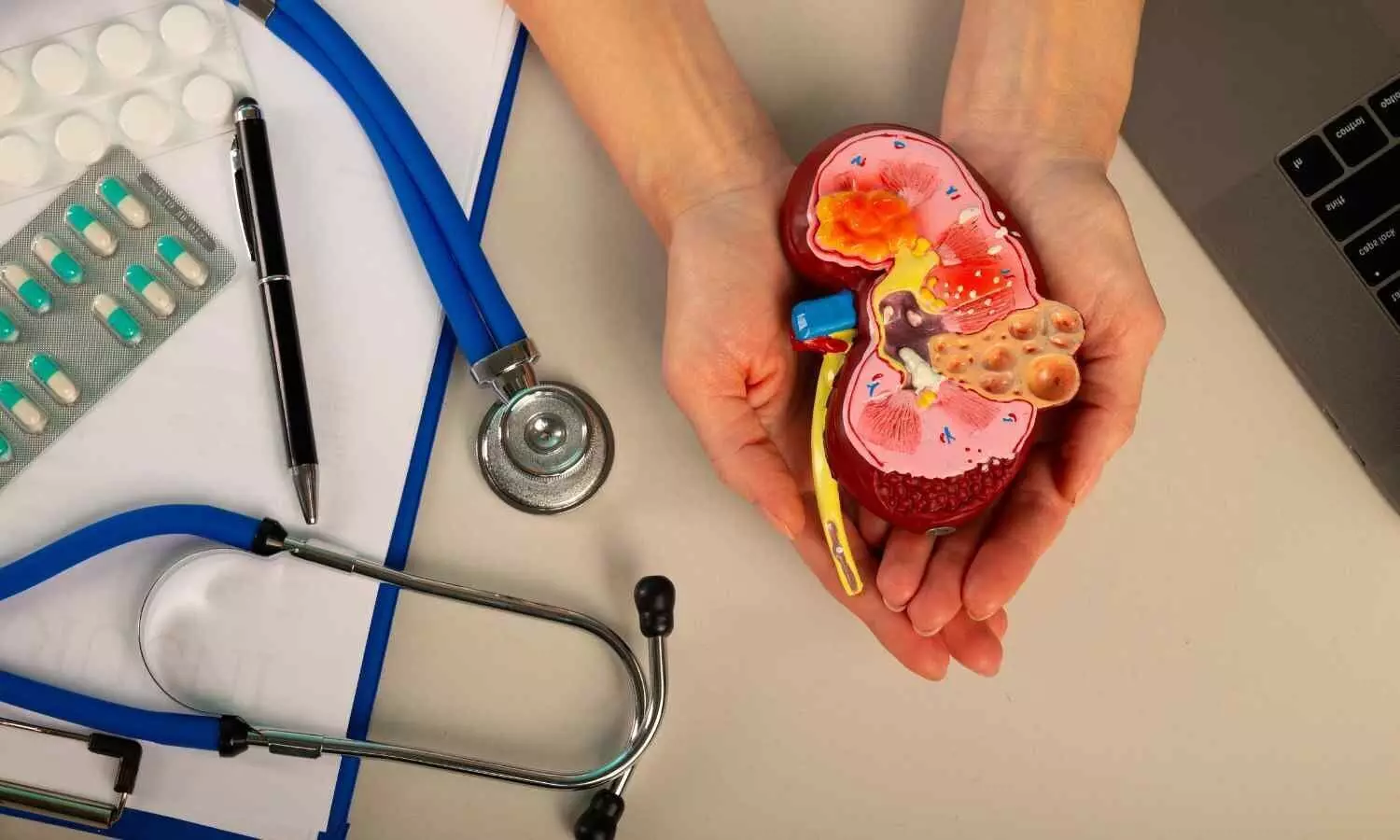
Hyderabad: Medical experts have stressed the urgent need to address pediatric nephrology and urology issues promptly, urging parents to seek medical attention at the first signs of such conditions to ensure timely treatment and prevent complications.

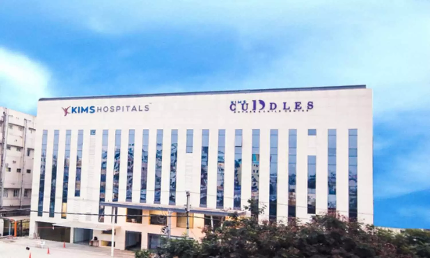
According to a UNI report, to tackle bedwetting problems in children, KIMS Cuddles has introduced a special Bedwetting Clinic, operating every Tuesday. Many children continue to wet the bed beyond the age of five but do not disclose it due to embarrassment, leading to persistent issues. The clinic aims to provide effective solutions and eliminate the social stigma surrounding the condition.

As part of the Continuing Medical Education (CME) program, a Nephro-Uro Summit 2025 was organized under the leadership of Dr Mounika Motamarri, Consultant Pediatric Nephrologist on Sunday. The event saw the participation of over 250 pediatric nephrology and urology specialists from Telangana, Andhra Pradesh, and Karnataka.

Also Read:KIMS Cuddles Doctors treat 18-Month-Old girl for Chemical Pneumonitis after mosquito repellent Ingestion

Experts highlighted that even young children can develop kidney stones, and symptoms such as persistent pain or blood in the urine should never be ignored. Some children may exhibit reddish-colored urine, requiring specific diagnostic tests.

Additionally, doctors emphasized that children with protein levels exceeding 2 mg per kg of body weight should be closely monitored. Pediatric hypertension, if untreated, can lead to kidney damage, and some cases may require surgical intervention for narrow urinary tracts, reports UNI.

Dr Mounika Motamarri, the event’s organizing Secretary, announced that the Bedwetting Clinic would be available at KIMS Secunderabad and Kondapur Hospitals every Tuesday. Several renowned doctors, including Dr Babu S. Madarkar, Dr Yog Nagendar, Dr Parag Dekate, D Aparna C., Dr VS Reddy, and Dr Nitin Chawla, attended the summit to discuss advancements in pediatric nephrology and urology care.   

Also Read:KIMSHEALTH Doctors perform posterior scoliosis correction surgery on 23-year-old Maldivian

Powered by WPeMatico

NEET 2025 Correction Window closes Today

Delhi- The National Testing Agency (NTA) is going to close the correction window for the National Eligibility and Entrance Test-Undergraduate (NEET UG) exam for the academic year 2025 today.

The correction window for NEET UG 2025 was made available from March 9 which is going to end today i.e. March 11, 2025, till 11:50 pm. During this period, registered candidates can verify their details by visiting the NTA official website. If required, they can also make corrections or modifications to their NEET UG online application form during the specified period. However, after this deadline, no further corrections will be allowed under any circumstances.

STEPS FOR NEET UG EXAM 2025 APPLICATION CORRECTION

STEP 1- Go to the NTA official website.

STEP 2- Now click on the correction window link.

STEP 3- Log in and make corrections in the required fields.

STEP 4- After this, pay the required fee and submit the form.

STEP 5- Take a printout of the acknowledgement form for further reference.

Correction Fields for the Application Form for the NEET (UG) – 2025 examination-

S.NO

ACTIONS FIELDS

FIELDS

1

Candidates shall be allowed to change any one of these two fields.

i Father Name and Qualification/Occupation.

ii Mother Name and Qualification/Occupation

2

Candidates shall be allowed to change/add all the following fields.

i Educational qualification details (Class X and Class XII).

ii State of Eligibility iii. Category.

iv Sub-category/PwD v. Signature vi. Number of attempts in NEET (UG)

3

Candidates shall be allowed to change the following based on their Permanent and Present Addresses.

i Examination City Selection ii Medium of the Examination

Meanwhile, it is to be noted that the final corrections will be applicable only after payment of any additional fees if required. In cases where changes affect the fee amount, candidates will be charged any excess fee accordingly. However, any excess payments made will not be refunded.

Powered by WPeMatico

Kerala doctor moves HC Seeking Regulations for Home Births

Kochi: A medical officer
from Malappuram, Dr Prathibha K, has approached the Kerala High Court seeking clear
legal provisions and penalties to regulate childbirth outside medical
facilities. She has urged for the establishment of proper guidelines to ensure the safety of both mothers and newborns.

The issue came to light after
a Kozhikode couple filed a complaint with the Human Rights Commission, alleging that the city corporation refused to issue a birth certificate for their baby girl due to a lack of formal proof of birth. According to the complainant, she gave birth at her rented home
in November 2024, but officials refused to issue the certificate as no formal
records of the delivery existed. Dr Prathibha has invoked Article 226 of the
Constitution, calling for judicial intervention to prevent further tragedies.

According to Onmanorama, Dr Prathibha, currently
serving as a medical officer in Tanur, Malappuram, has expressed serious
concerns about the rights of newborns, emphasizing that every child is entitled
to medical care and legal recognition. Malappuram, which records the highest
number of home births in the state, has also seen a rise in neonatal
complications, she noted in her petition. The reasons behind choosing home
births vary, but lack of medical supervision puts both mothers and infants at
grave risk. “Some mothers fear hospitals and C-sections, others adhere to
conventional beliefs, while some are reluctant to expose their bodies to
medical staff. Financial constraints also play a role. Surprisingly, many of
these women are well-educated, holding graduate or even postgraduate degrees,
yet they continue to make conservative choice,” the petition stated. 

Babies born at home face survival
challenges, seizures, and nervous system disorders, while mothers are
vulnerable to severe bleeding, cardiac arrest, infections, and complications
such as retained placenta. “Often, when home births lead to complications,
families rush the mother and baby to the hospital, claiming the birth happened
suddenly on their way. Some even keep a vehicle on standby in case of
emergencies, falsely assuring themselves of preparedness,” she says.

According to the Daily, despite the widespread
practice of home births in Malappuram, families often conceal their plans,
making it difficult for authorities to intervene.”Expectant mothers attend
routine check-ups, undergo scans, and follow medical advice, but when the time
comes, they secretly give birth at home. These deliveries often occur at night,
and when questioned, families claim the birth happened too quickly to reach a
hospital. However, many of these cases are premeditated. Some women are
influenced by past home birth experiences in their families, while others feel
emboldened after witnessing similar cases within their circles,” Dr
Prathibha says.

A Right to Information
(RTI) request filed by Kulathur Jaisingh revealed that between 2019 and
September 2024, Kerala recorded 2,931 home deliveries, with Malappuram alone
accounting for 1,244 births. In the same period, the state reported 18 neonatal
deaths, four of which occurred in Malappuram. Advocate R Gopan,
representing Dr Prathibha in court, argued that children have fundamental
rights, including access to proper medical attention at birth. He pointed out a
legal loophole that allows parents to escape accountability if complications
arise from unsafe home deliveries. While birth certificates include
“delivered at home” as an option, families exploit this provision to
obtain official documentation without medical verification.

The petition highlights
multiple past incidents of home births leading to severe complications and
fatalities in Kerala. In February 2024, a mother and baby died during delivery
in Nemom, Thiruvananthapuram. Another case in October 2024 in Thanaloor,
Malappuram, saw a woman critically injured when the baby’s head emerged before
she was rushed to a hospital. A Chalakudy, Thrissur case ended with the infant’s
death and the mother requiring intensive care, while in October 2022, a mother
and child in Chadayamangalam, Kollam, did not survive a home delivery, reports the Daily.

She further alleged the
existence of illegal birthing facilities, where women from various parts of Kerala
and beyond—including Kollam, Alappuzha, and Lakshadweep—stay for weeks to
deliver their babies in Malappuram under unsupervised conditions. “Some of
these centres have been shut down by authorities following local complaints.
Additionally, organised groups coordinate home deliveries through secret
WhatsApp chats or groups, frequently changing numbers to avoid detection. Some
agents charge as little as ₹5,000 to facilitate home births, while others offer
costlier packages lasting up to a month,” she says.

With eight years of
service, Dr Prathibha started her career as an assistant medical officer at a
Public Health Centre in Tanur. As the number of home births rises, medical
professionals like her face mounting pressure from the government to address
the issue. “Each public health centre monitors expectant mothers through a
team comprising a junior public health nurse, staff nurse, and ASHA workers,
ensuring regular health updates. However, when home births go unreported, these
records remain incomplete, leaving medical officers without answers.” She
believes that stricter legal measures are essential to address this concern. The
High Court has scheduled the next hearing for March 17, awaiting a response
from the state government on the matter.

Powered by WPeMatico

5 Senior Doctors Transferred, Appointed to 3 Medical Colleges in Jharkhand

Dhanbad: In a significant administrative reshuffle, the Jharkhand Health Department has transferred and appointed five senior doctors across three medical colleges as principals and superintendents.   

The three medical colleges include Shahid Nirmal Mahto Medical College Hospital (SNMMCH) Dhanbad, Mahatma Gandhi Memorial Medical College Hospital (MGMMCH) Jamshedpur, and Medinirai Medical College Hospital (MMCH) Palamu.  

Also read- 5 doctors transferred to Midnapore Medical College Hospital amid saline death controversy

A notification was issued by Joint Secretary Lalit Mohan Shukla in this regard, as reported by The Jharkhand Story, Prof. (Dr.) Sanjay Kumar Chourasia has been appointed as the principal of SNMMCH Dhanbad. Dr. Chourasia previously served as the superintendent of the college and now steps into the position, which was left vacant following the retirement of Dr. K.K. Lal on January 31.  

Alongside this appointment, the role of SNMMCH superintendent has been assigned to Prof. (Dr.) Dinesh Kumar Gindauria, who was heading the Surgery Department at the college. He replaces Dr. S.K. Chourasia.  

MGMMCH has also seen a leadership change in Jamshedpur with Prof. (Dr.) Ramesh Kumar Mandhan, Head of the Pathology Department, is taking over as the new superintendent. Dr. Mandhan succeeds Dr. Shikha Rani, who has been serving in the role since September 2023.

Meanwhile, MMCH Palamu has received a new superintendent as well. Prof. (Dr.) Ajay Kumar Singh, previously the Head of the Eye Department at MGMMCH, has been transferred to MMCH Palamu to take over the position from Dr. Dharmendra Kumar, who has been serving as superintendent since September 2024.

In another transfer, Prof. (Dr.) Dharmendra Kumar has been moved from MMCH Palamu to SNMMCH Dhanbad, where he will now serve as the Head of the Eye Department.

However, Dr Dharmendra Kumar’s new appointment faced backlash as he was allegedly transferred within just seven months of his appointment at MMCH Palamu. Therefore, it has been claimed that the government appointed him based on seniority.

Previously, on September 20, 2024, Dr. Dharmendra Kumar was appointed as the superintendent of MMCH Palamu following a directive from the High Court. However, he voluntarily stepped down from the position and was transferred to SNMMCH Dhanbad as the Head of the Eye Department from Palamu, reports The Jharkhand Story. 

Also read- Madras HC upholds mass transfer of ESIC doctors, overturns CAT decision

Powered by WPeMatico

Intermittent Fasting Reduces Platelet Activation and Thrombosis Risk : Study

Researchers have found in a recent study that intermittent fasting (IF) diminishes platelet activation and the risk of thrombosis significantly in patients with coronary artery disease (CAD) and in animal models. Hyperreactivity of platelets plays a critical role in thrombosis, one of the predominant causes of acute myocardial infarction (AMI) and stroke. The findings demonstrated that IF inhibits platelet activation and thrombosis by enhancing intestinal flora production of indole-3-propionic acid (IPA), which in turn modulates platelet function through pregnane X receptor (PXR)-mediated pathways. The study was recently published in the journal of Life Metabolism by Zhiyong Qi and colleagues.

The study examined the effects of IF on platelet activation and thrombotic risk in two models: patients with coronary artery disease and Apolipoprotein E knockout (ApoE−/−) mice, a general model for studying atherosclerosis. Scientists examined the way IF influences gut microbiota composition, promoting the production of IPA. The research then investigated how high IPA levels influence platelet function by binding to PXR on platelets, thus modulating intracellular signaling pathways in thrombosis. Furthermore, myocardial and cerebral ischemia-reperfusion injury models were employed to evaluate the protective effects of IF against cardiovascular events.

Key Findings

• Platelet Activation Reduction: IF significantly repressed platelet activation in models of humans and animals.

• Increased IPA Production: IF produced elevated plasma levels of IPA, which inhibited platelet activation directly.

• PXR Pathway Activation: IPA has occupied the platelet PXR receptor and reduced activity in key signaling pathways (Src/Lyn/Syk and LAT/PLCγ/PKC/Ca2+), which are key players in platelet activation.

• Thrombosis Prevention: IF lowered thrombosis risk in ApoE−/− mice, potentially indicating cardiovascular protective effects.

• Protection from Ischemia-Reperfusion Injury: IF reduced myocardial and cerebral ischemia-reperfusion injury in ApoE−/− mice, further indicating its cardiovascular protective role.

Researchers concluded that intermittent fasting decreases platelet activation and the risk of thrombosis through enhanced IPA production, which engages PXR-associated pathways in platelets. The incorporation of IF into therapeutic plans can potentially bring about better patient outcomes compared to standard pharmacologic treatment.

Reference:

Qi, Z., Zhou, L., Dai, S., Zhang, P., Zhong, H., Zhou, W., Zhao, X., Xu, H., Zhao, G., Wu, H., & Ge, J. (2025). Intermittent fasting inhibits platelet activation and thrombosis through the intestinal metabolite indole-3-propionate. Life Metabolism. https://doi.org/10.1093/lifemeta/loaf002

Powered by WPeMatico

Breakthrough cardiac regeneration research offers hope for the treatment of ischemic heart failure

Researchers in the Michael E. DeBakey Department of Surgery at Baylor College of Medicine, the QIMR Berghofer Medical Research Institute in Brisbane, Australia, and collaborating institutions report a groundbreaking discovery in cardiac regeneration that offers new hope for the treatment of ischemic heart failure. Published in npj Regenerative Medicine, the study reveals a novel approach to promoting cardiomyocyte proliferation.

“When the heart cannot replace injured cardiomyocytes with healthy ones, it becomes progressively weaker, a condition leading to heart failure. In this study, we investigated a new way to stimulate cardiomyocyte proliferation to help the heart heal,” said co-corresponding author Dr. Riham Abouleisa, assistant professor in the Division of Cardiothoracic Surgery at Baylor.

Previous studies showed that calcium plays an important role in cardiomyocyte proliferation. In the current study, Abouleisa and her colleagues explored how modulating calcium influx in cardiomyocytes would affect their proliferation.

“We found that preventing calcium influx in cardiomyocytes enhances the expression of genes involved in cell proliferation,” Abouleisa said. “We prevented calcium influx by inhibiting L-Type Calcium Channel (LTCC), a protein that regulates calcium in these cells. Our findings suggest that LTCC could be a target for developing new therapies to induce cardiomyocyte proliferation and regeneration.”

The study demonstrates that both pharmacological and genetic inhibition of LTCC can induce cardiomyocyte replication and that this occurs by modulating the activity of calcineurin, a known regulator of cardiomyocyte proliferation. This innovative approach showed promising results both in human cardiac slices grown in the lab and in live animals.

“Abouleisa’s multi-continent collaborations led to a discovery that can revolutionize the use of current medicines that regulate calcium entry to the cells, such as Nifedipine, in heart failure patients,” said Dr. Tamer Mohamed, co-author and director of Baylor College of Medicine’s Laboratory for Cardiac Regeneration.

Co-author Dr. Todd K. Rosengart, chair and professor of the Michael E. DeBakey Department of Surgery, emphasized that, “The premise of regenerating heart tissue, which once seemed like an impossible dream, is getting closer almost daily. The work of Dr. Abouleisa and the Baylor cardiac regeneration team represents a major step toward human trials that I believe are in the not-too-distant future.”

Abouleisa and her colleagues’ research highlights the importance of targeting calcium signaling pathways to unlock the regenerative potential of the heart and opens new avenues for developing cardiac regenerative therapies, potentially transforming the treatment landscape for patients suffering from heart failure.

Reference:

Lynn A. C. Devilée, Abou bakr M. Salama, Jessica M. Miller, Janice D. Reid, Qinghui Ou, Nourhan M. Baraka, Kamal Abou Farraj, Madiha Jamal, Yibing Nong, Todd K. Rosengart, Douglas Andres, Jonathan Satin, Tamer M. A. Mohamed, James E. Hudson, Riham R. E. Abouleisa. Pharmacological or genetic inhibition of LTCC promotes cardiomyocyte proliferation through inhibition of calcineurin activity. npj Regenerative Medicine, 2025; 10 (1) DOI: 10.1038/s41536-025-00389-z

Powered by WPeMatico

Daily Chlorhexidine Wipes Do Not Significantly Reduce Diabetic Foot Complications, JAMA

A news study published in JAMA has determined that daily application of 2% chlorhexidine wipes for foot hygiene in diabetic veterans did not significantly decrease the risk of new foot complications compared with standard soap-and-water wipes. Diabetic foot ulcers are a severe and prevalent complication, and about 20% of them result in infections that necessitate lower extremity amputations. This study was conducted by Alison D. and fellow researchers.

This phase 2b double-blind placebo-controlled randomized clinical trial occurred from January 2019 to January 2023 at the Baltimore Veterans Affairs (VA) Medical Center. Patients had to be diabetic veterans with a risk for diabetic foot complications but without an acute foot infection. They were ambulatory and had intact feet. A total of 175 participants (97% male, mean age 68 years) were randomly assigned in a 1:1 ratio to receive either 2% chlorhexidine wipes (n = 88) or soap-and-water wipes (n = 87). The intervention lasted for one year, during which participants were instructed to use the assigned wipes daily, followed by the application of a standardized foot lotion.

Participants were supplied with lookalike wipes for blinding. The main outcome was time to first new foot complication, occurring after randomization and defined as chronic foot ulcer, foot infection, or amputation of a lower extremity. Data analysis was performed in an intention-to-treat fashion. Follow-up occurred over one year’s median duration, during which adverse effects and intervention compliance were closely observed.

Key Findings

• In a year, 12 (14%) and 14 (16%) participants in the chlorhexidine and soap-and-water groups, respectively, developed new foot complications.

• The time to the first new foot complication was 232 days (IQR: 115-315 days) in both groups.

• No significant hazard reduction of new foot complications in the chlorhexidine group was seen compared with the soap-and-water group (hazard ratio, 0.83; 95% CI, 0.39-1.80).

• 60 adverse events were documented throughout the study, but none of them resulted from the intervention. The adherence was very high with 145 participants (83%) sustaining the intervention during the entire study period.

The study authors concluded that daily use of 2% chlorhexidine wipes did not significantly influence prevention of new diabetic foot complications among diabetic veterans in comparison with soap-and-water wipes. These findings are valuable for future research regarding how to optimize preventive interventions for diabetic foot complications.

Reference:

Lydecker AD, Kim JJ, Robinson GL, et al. Chlorhexidine vs Routine Foot Washing to Prevent Diabetic Foot Ulcers: A Randomized Clinical Trial. JAMA Netw Open. 2025;8(2):e2460087. doi:10.1001/jamanetworkopen.2024.60087

Powered by WPeMatico

Rituximab effective Steroid-Sparing Treatment of steroid-dependent or frequently relapsing nephrotic syndrome: BMC

A new study published in the journal of BMC Nephrology revealed rituximab to be an effective and safe steroid sparing medication in individuals with steroid dependent (SD)/frequent relapsing (FR) nephrotic syndrome (NS). Longer remission is attained when a follow-up maintenance dosage is administered following the initial course. 

According to the Kidney Disease: Improving Global Outcome (KDIGO) guidelines, glucocorticoid medication is advised as the first-line treatment for adult MCD since it has a positive prognosis. Although 75–90% of patients get a full response, up to 25% of steroid responders experience frequent relapses (FR), and 30–40% acquire steroid dependence.

Although corticosteroids are very effective in treating minimal change disease, many individuals develop steroid dependence or relapse often. Primary Focal Segmental Glomerulosclerosis (FSGS) has a lower response rate. An efficient substitute is needed as extended exposure to corticosteroids should be avoided and in this context, Rituximab is an agent with promise. Omri Feder and colleagues conducted a review to assess long-term safety and effectiveness of Rituximab therapy in adult patients with SD/FR NS.

This retrospective cohort research that assessed Rituximab-treated SD/FR NS patients at a tertiary institution. Rituximab was administered during induction, and the treating nephrologist made the choice about further dosages. Relapse frequency and time to first relapse were the main outcomes. Safety was evaluated.

There were 21 adults in total and out of these, 2 instances had no kidney biopsies, 5 (23.8%) had FSGS, and 14 (66.7%) had MCD. 54.6 years was the median age while 39.6 months was the median follow-up period. Rituximab significantly reduced the number of relapses when compared to prior therapy (median relapses were 0 versus 3, respectively, W = 3.70, p <.001). Before Rituximab, the time to first relapse was substantially shorter than after (median 11 vs. 536 days, respectively, W = 3.05, p =.002).

The patients who got a single course of Rituximab had a greater hazard ratio for recurrence than those who received further maintenance. Although the treatment was generally tolerated, severe COVID-19 and cholecystitis were among the significant side effects. Overall, in individuals with SD/FR NS, rituximab seems to be a safe and effective substitute for extended steroid therapy. Also, lengthier remission is attained when a follow-up maintenance dosage is administered following the first course. 

Source:

Feder, O., Amsterdam, D., Ershed, M., Grupper, A., Schwartz, D., & Kliuk-Ben Bassat, O. (2025). Long-term efficacy of Rituximab in steroid dependent and frequent relapsing adult nephrotic syndrome. BMC Nephrology, 26(1), 126. https://doi.org/10.1186/s12882-025-04035-0

Powered by WPeMatico

High-Dose Vitamin D Reduces Disease Activity in Early Multiple Sclerosis and CIS: JAMA

France: A recent randomized clinical trial, the D-Lay MS study, has demonstrated that high-dose vitamin D supplementation may significantly reduce disease activity in individuals with clinically isolated syndrome (CIS) typical of multiple sclerosis (MS). The findings highlight the potential role of vitamin D as an adjunct therapy in managing early-stage MS. 

“High-dose vitamin D not only reduced disease activity in early MS and CIS compared to placebo but also prolonged the time to disease activity, with patients on vitamin D experiencing a delay of 432 days versus 224 days in the placebo group, without any severe adverse events associated with the supplementation,” the researchers reported in JAMA.

Clinically isolated syndrome, the first neurological episode suggestive of multiple sclerosis, requires early intervention to delay disease progression. Among potential risk factors, vitamin D deficiency has been linked to increased disease activity in MS, raising interest in its therapeutic role. However, while supplementation is considered a promising strategy, existing evidence on its effectiveness remains inconclusive.

Against the above background, Eric Thouvenot, CHU Nimes, Service de Neurologie, Univ Montpellier, Nimes, France, and colleagues aimed to assess the effectiveness of high-dose cholecalciferol as a standalone treatment in lowering disease activity in patients with clinically isolated syndrome indicative of MS.

For this purpose, the researchers conducted the D-Lay MS trial, a parallel, double-blind, randomized placebo-controlled study across 36 MS centers in France. From July 2013 to December 2020, they enrolled untreated CIS patients aged 18 to 55 years, with CIS duration under 90 days, serum vitamin D levels below 100 nmol/L, and MRI findings meeting 2010 criteria for dissemination in space or oligoclonal bands. Participants were randomized to receive either 100,000 IU of oral cholecalciferol (n=163) or placebo (n=153) every two weeks for 24 months. The primary outcome was disease activity, defined by relapses or MRI changes.

Key Findings:

• Among 316 enrolled participants (median age 34 years, 70% women), 303 (95.9%) received at least one dose, and 288 (91.1%) completed the 24-month trial.

 • Disease activity was observed in 60.3% of the vitamin D group vs. 74.1% of the placebo group (HR, 0.66).

• Median time to disease activity was longer in the vitamin D group (432 vs. 224 days).

• MRI outcomes favored vitamin D over placebo:

• MRI activity: 57.1% vs. 65.3% (HR, 0.71).

• New lesions: 46.2% vs. 59.2% (HR, 0.61).

• Contrast-enhancing lesions: 18.6% vs. 34.0% (HR, 0.47).

 • There was no significant difference in secondary clinical outcomes, including relapse (17.9% vs. 21.8%).

• There were similar results in 247 patients meeting the 2017 relapsing-remitting MS criteria.

• Severe adverse events occurred in 17 patients in the vitamin D group and 13 in the placebo group, with none linked to cholecalciferol.

“The trial demonstrated that high-dose oral cholecalciferol (100,000 IU every two weeks) effectively reduced disease activity in CIS and early RRMS with a low risk of adverse events. These findings support further research on its potential as an add-on therapy in MS management,” the researchers concluded.

Reference:

Thouvenot E, Laplaud D, Lebrun-Frenay C, et al. High-Dose Vitamin D in Clinically Isolated Syndrome Typical of Multiple Sclerosis: The D-Lay MS Randomized Clinical Trial. JAMA. Published online March 10, 2025. doi:10.1001/jama.2025.1604

Powered by WPeMatico

Bilateral hearing loss prevalent in children with Down syndrome: Study

A new study published in the journal of Nature Scientific Reports showed that 4.1% of children with Down syndrome had sensorineural hearing loss (SNHL), whereas almost half (48.8%) had conductive hearing loss (CHL).

Hearing loss in DS patients can be caused by a number of circumstances. Otitis medium with effusion (OME) is the most frequent cause of CHL and a very common disease in children with DS. Due to the difficulty of diagnosing hearing impairment, parents and professionals face difficulties. To determine the prevalence of OME, permanent, and transitory hearing loss in children with DS who are receiving care at Mansoura University Children’s Hospital, Sohier Yahia and team carried out this study.

A total of 170 juvenile individuals with genetically proven DS are the subjects of this descriptive cross-sectional research. From October 2021 to October 2022, patients were gathered from the Mansoura University Children’s Hospital’s genetic outpatient clinic. A lateral X-ray of the nasopharynx with the mouth open and the neck extended was performed on all babies and children after a thorough history was taken.

After examination, any accumulated cerumen in the ears was removed. The tympanic membrane was examined otoscopically to determine whether middle ear pathology was present. Tympanometry was used to evaluate otitis media with effusion and eustachian tube dysfunction. Pure tone audiometry (PTA) and auditory brain stem response (ABR), among other appropriate hearing tests, were employed.

Of the children with DS in the study, 4.1% had sensorineural hearing loss and 48.8% had conductive hearing loss. Bilateral affection was observed in 86.5% of hearing-impaired individuals. 59.1% of individuals with CHL and 71.4% of patients with SNHL had minor hearing loss. Of the total CHL patients, 45.8% had stagnant HL, 15.7% had a regress from moderate to mild HL, and 38.6% had normalized HL.

All 7 afflicted individuals had persistent SNHL, with 4 showing a stable course and three showing a progressive one. In babies and children with DS, CHL was frequently linked to OME, adenoid hypertrophy, and upper respiratory tract infections (URTI). The majority of kids with DS have modest, bilateral hearing loss. The majority of HL is conductive. Overall, every newborn and youngster with Down syndrome should have their hearing evaluated. Even with a standard newborn hearing screening test, patients with DS should be monitored.

Source:

Yahia, S., Metawea, M., Megahed, A., ELshawaf, W., Wahba, Y., & Mahmoud, R. (2025). The prevalence of hearing impairment in infants and children with down syndrome a cross sectional study in a Tertiary Care Center. Scientific Reports, 15(1), 7570. https://doi.org/10.1038/s41598-025-90500-7

Powered by WPeMatico