Hyponatremia Signals Severity in Pediatric Urinary Tract Infections: Study

Researchers have found hyponatremia to be suggestive of a worse disease course in pediatric patients with urinary tract infection (UTI), indicating acute pyelonephritis (APN), raised levels of C-reactive protein (CRP), and renal impairment. UTI is among the most common infections encountered during childhood, with its incidence occurring in about 8% of girls and 2% of boys by the age of seven years. A recent study was conducted by Fu-Wein and colleagues published in BMC Pediatrics journal.

A retrospective review was done on pediatric patients diagnosed with UTI or APN and admitted to a medical center between October 2012 and September 2022. A total of 344 patients were classified into “hyponatremia” (serum sodium levels below normal) or “eunatremia” groups based on their serum sodium concentrations. Data collected included renal ultrasound findings, blood and urine test results, and clinical outcomes. Multivariable logistic regression models were applied to assess potential predictors of disease severity, including serum glucose and CRP levels.

Key Findings

The study involved data from 344 pediatric patients:

  • Hyponatremia Prevalence: Hyponatremia was found in 99 patients (28.8%), while normal serum sodium levels were found in 245 (71.2%) patients.

  • Association with APN: The hyponatremia group had a higher frequency of APN and renal abnormalities detected on ultrasound.

  • CRP Levels: Higher levels of CRP were observed in the hyponatremia group. A significant association was also noted in multivariable analysis (OR: 1.00; 95% CI: 1.00–1.01, p = 0.0417).

  • Serum Glucose: Hyperglycemia also has been associated with hyponatremia (OR: 1.01; 95% CI: 1.00–1.03, p = 0.0365).

  • Final Diagnosis APN: Hyponatremia was associated with markers of severe disease. No significant direct correlation was seen between the final diagnosis of APN.

Hyponatremia might provide useful biomarkers for significant diseases of hospitalized pediatric UTI. However, the present research shows that although hyponatremia has relationships with systemic inflammation and renal findings, its predictive ability is limited for APN diagnosis. These findings stress the need for further study on the mechanisms and clinical utility of hyponatremia as a predictor of disease severity.

Reference:

Liang, F.-W., Lin, Y.-J., Ho, C.-H., & Chen, Y.-S. (2024). Association between hyponatremia and disease severity in pediatric urinary tract infections. BMC Pediatrics, 24(1). https://doi.org/10.1186/s12887-024-05248-2

Powered by WPeMatico

Women with breast cancer more likely to develop periodontitis, finds study

Women with breast cancer are more likely to develop periodontitis, finds a study published in the Journal of Dental Sciences.

Epidemiologic research has linked periodontitis to several types of cancer, particularly breast cancer. Although clinical evidence indicates a higher risk of breast cancer in women with periodontitis than in those without, few studies have explored whether the risk of periodontitis is higher in women with breast cancer than in those without. In this study, we examined the incidence of periodontitis in patients with breast cancer and identified potential interventions for its prevention. This retrospective cohort study included data from the National Health Insurance Research Database of Taiwan. We identified women who received a diagnosis of breast cancer between 2010 and 2019 and included a 1:1 matched control cohort with no breast cancer. Subsequently, we analyzed the risk of periodontitis by using Cox proportional-hazards models while adjusting for sociodemographic factors, comorbidities, and treatment regimens.

Results: In 82,146 matched pairs, the breast cancer cohort was at a 51 % higher risk of periodontitis compared with the control cohort (adjusted hazard ratio = 1.51, 95 % confidence interval = 1.43–1.60). The stratified analysis revealed the same results. The risk of breast cancer was higher in younger patients than in older patients, whereas the risk of periodontitis was significantly lower in patients who underwent surgery, radiotherapy, chemotherapy, or hormone therapy compared with those who did not. Breast cancer increases the risk of periodontitis, particularly in younger patients. These patients should receive regular dental care to prevent and manage periodontitis. Anticancer treatments may mitigate the risk of periodontitis in patients with breast cancer.

Reference:

Li-Min Sun, Fuu-Jen Tsai, Cheng-Li Lin, Ya-Hsin Wu. Women with breast cancer exhibit a higher risk for periodontitis: A nationwide cohort study, Journal of Dental Sciences, 2024, ISSN 1991-7902. https://doi.org/10.1016/j.jds.2024.11.016.

(https://www.sciencedirect.com/science/article/pii/S1991790224003891)

Keywords:

Women, breast cancer, develop, periodontitis, finds, study , Journal of Dental Sciences, Breast cancer, Periodontitis, Cohort study, Li-Min Sun, Fuu-Jen Tsai, Cheng-Li Lin, Ya-Hsin Wu

Powered by WPeMatico

Probe-based confocal laser endomicroscopy aid in detecting endometriosis during surgery: Study

A recent study published in the European Journal of Obstetrics & Gynecology and Reproductive Biology highlighted the potential of probe-based Confocal Laser Endomicroscopy (pCLE) as a transformative tool in diagnosing endometriosis during both conventional and robot-assisted laparoscopic procedures. The study involved 5 women from Hospital Moriah with an average age of 35 to demonstrate the capability of this innovative technique to provide detailed, real-time imaging of cellular structures associated with the condition.

Endometriosis is a chronic gynecological condition marked by debilitating symptoms like dysmenorrhea (painful periods) and dyspareunia (painful intercourse), has traditionally required histological evaluations for definitive diagnosis. This study sought to validate utility of pCLE by complementing its intraoperative imaging with subsequent histological analyses of 214 optical biopsies collected during surgeries.

A major outcome of the research was the creation of a comparative atlas of confocal images, illustrating cellular structures like adipocytes, glandular tissue, and vascular formations associated with endometriosis. Also, surgeons used this atlas to complete a questionnaire by identifying endometriosis-specific structures in confocal images.

The responses revealed encouraging results where surgeons scored an average of over 8 out of 10 in evaluating the utility of this technique, regardless of their familiarity with pCLE. Approximately 63.1% of participants expressed confidence in pCLE which recommends its use for intraoperative diagnostics.

The study highlighted several key benefits of pCLE, including its ability to generate highly detailed, real-time images, which can significantly improve the ability of surgeons to pinpoint endometriosis lesions. The imaging provided clear views of vascular networks, a hallmark of endometriotic tissue, along with other cellular structures that are often challenging to assess with conventional methods.

This research emphasized areas for improvement where challenges like the durability of the device and its cost-effectiveness were identified as barriers to widespread adoption. The outcomes of this study suggests that refining these aspects will be critical to integrate pCLE into routine gynecological practices.

The findings indicated that pCLE could revolutionize the diagnosis and management of endometriosis, particularly in minimally invasive gynecological surgeries. As the method gains further acceptance and undergoes technological improvements, this holds promise for enhancing the diagnostic accuracy and reducing the reliance on time-consuming histological procedures. Overall, this research not only underlined the feasibility of this method but also sets the stage for its broader clinical applications.

Source:

Okita, F., Andres, M. P., Coudry, R. de A., Riccio, L. G. C., Baracat, E. C., & Abrão, M. S. (2024). Confocal Laser Endomicroscopy as a method for assessing endometriosis: A pilot study. In European Journal of Obstetrics & Gynecology and Reproductive Biology (Vol. 302, pp. 225–231). Elsevier BV. https://doi.org/10.1016/j.ejogrb.2024.09.025

Powered by WPeMatico

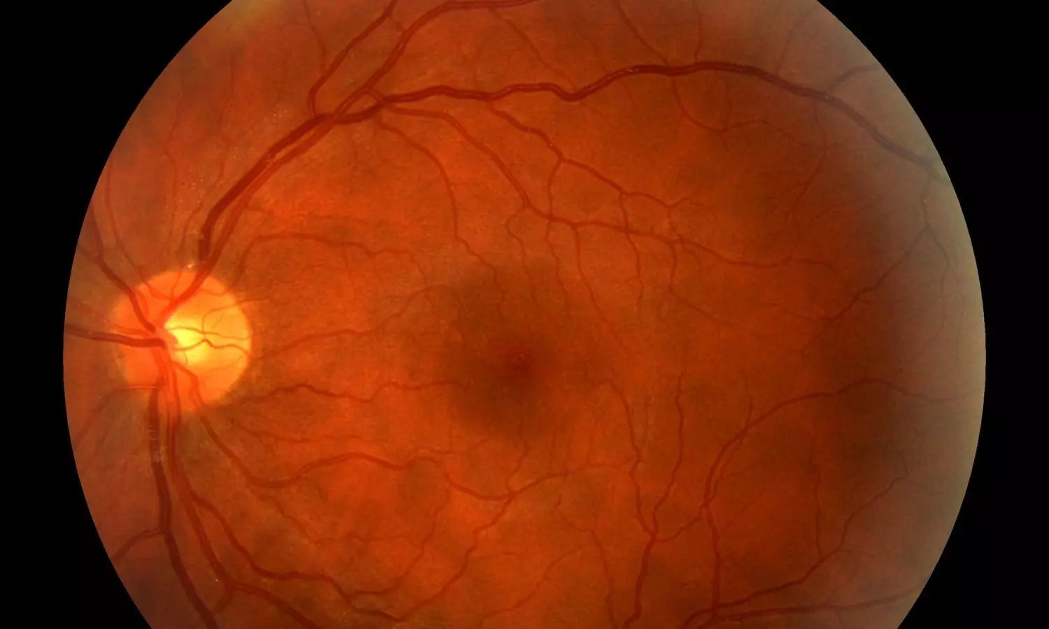
Empagliflozin has potential of lowering progression of Diabetic Retinopathy among diabetes patients: S6

A recent study published in the
journal JAMA Ophthalmology found that empagliflozin was associated with a
reduced risk of the progression of diabetic retinopathy compared to dipeptidyl
peptidase 4 inhibitor (DPP4i).

Diabetic Retinopathy (DR) is a
prevalent complication in diabetics. It is the leading cause of irreversible
vision loss among type 2 diabetics. Empagliflozin has significant protective
effects against adverse cardiorenal outcomes in previous trials. They inhibit
the retinal sodium-glucose cotransporter 2 protein and prevent retinal pericyte
loss, thus decreasing the progression of DR. As there is limited data on the
association between the role of empagliflozin and DR, researchers
conducted a study to evaluate the association between empagliflozin use and the
risk of incident nonproliferative DR (NPDR) and DR progression within the
Empagliflozin Comparative Effectiveness and Safety (EMPRISE) study.

A new-user active-comparator
cohort study was carried out based on US nationwide insurance claims data
from 2 commercial insurers and Medicare from August 2014 to September 2019.
Individuals using empagliflozin between August 2014 and September 2019 were
included in the study, with DPP4i as the comparator. Adults with T2D initiating
study drugs without prior diagnosis or treatment for proliferative DR or other
advanced retinal diseases were included. To assess incident NPDR, patients with
a history of NPDR were additionally excluded, while for the DR progression
outcome, patients were required to have a history of NPDR. The occurrence of
nonproliferative DR (NPDR) and the progression of DR were considered the
primary outcomes of measurement. Incident NPDR was defined using diagnostic
codes for mild, moderate, or severe NPDR. The DR progression outcome was described
as a composite of incident proliferative DR, vitreous hemorrhage, intravitreal
anti-vascular endothelial growth factor injection initiation, or pan-retinal
photocoagulation.

Findings:

  • About 34 239 propensity-score-matched adults
    were identified in the incident NPDR cohort and 7831 pairs in the DR
    progression cohort.
  • In the incident NPDR cohort, 35 867 patients
    (52.4%) were male, and the mean (SD) age was 65.6 (10.3) years.
  • In the DR progression cohort, 8229 patients
    (52.5%) were male, and the mean (SD) age was 67.0 (10.0) years.
  • There was no difference in the risk of incident
    NPDR between the two groups over a mean (SD) follow-up period of 8 (7.5) months
    of receiving treatment.
  • However, the empagliflozin group showed a lower
    risk of DR progression compared with those who began DPP4i therapy.
  • Multiple subgroups and sensitivity analyses also
    showed consistent results.

Thus, the study concluded that empagliflozin
is beneficial as it showed a reduced risk of DR progression compared to DPP4i
initiation. Even though empagliflozin and DPP4i were similar in reducing the
risk of incident NPDR, empagliflozin’s potential to lower the progression of
diabetic retinopathy may influence the choice of the drug for glucose-lowering
treatments in diabetic individuals.

Further reading: Tesfaye H, Paik
JM, Roh M, et al. Empagliflozin and the Risk of Retinopathy in Patients
With Type 2 Diabetes. JAMA Ophthalmol. Published online
December 05, 2024. doi:10.1001/jamaophthalmol.2024.5219.

Powered by WPeMatico

Severe maternal morbidity in first birth impacts subsequent pregnancies, reveals study

A recent Swedish study that involved over 1 million women revealed that severe maternal morbidity (SMM) during a first birth significantly lowers the probability of having a subsequent child. The findings of this study were published in the Journal of American Medical Association.

This population-based cohort study analyzed data from 1,046,974 women in Sweden who gave birth between 1999 and 2021. This study drew information from the Swedish Medical Birth Register and the National Patient Register and identified instances of SMM which was defined as severe complications occurring during delivery or within 42 days postpartum.

Of the participants, 36,790 women (3.5%) underwent SMM during their first birth. Women with SMM had a lower rate of subsequent births, with an incidence rate of 136.6 per 1000 person-years when compared to 182.4 per 1000 person-years for the women without SMM. After adjusting for factors like age and socioeconomic status, this study calculated an adjusted hazard ratio (aHR) of 0.88 by indicating that women with SMM were 12% less likely to have another child.

This study also identified specific SMM subtypes that significantly reduced the probability of subsequent births. Women who underwent severe uterine rupture (aHR, 0.48), cardiac complications (aHR, 0.49), cerebrovascular accidents (aHR, 0.60), or severe mental health conditions (aHR, 0.48) in their first birth were particularly less likely to have another child. These findings suggest that both physical and psychological maternal health challenges can profoundly affect reproductive decisions. The research included sibling comparisons to rule out familial or genetic confounding factors and to strengthen the reliability of the results. The findings suggest that the association between SMM and subsequent childbirth is unlikely to be influenced by shared genetic or environmental factors.

The study underlined the long-term impact of SMM on the reproductive health of women. The outcomes emphasize the need for tailored reproductive counseling and improved antenatal care for women with a history of SMM. Proactive management and support during subsequent pregnancies could reduce risks and provide reassurance for affected women. This large-scale study provides critical insights and highlights the importance of addressing the far-reaching effects of severe maternal complications. With adequate care and support, women who have underwent SMM can better navigate future reproductive decisions.

Source:

Tsamantioti, E., Sandström, A., Lindblad Wollmann, C., Snowden, J. M., & Razaz, N. (2024). Association of Severe Maternal Morbidity With Subsequent Birth. In JAMA. American Medical Association (AMA). https://doi.org/10.1001/jama.2024.20957

Powered by WPeMatico

Study Links Dietary Fructose to Enhanced Tumor Growth

New research from Washington University in St. Louis reveals that fructose, a sweetener prevalent in ultra-processed foods, indirectly promotes tumor growth in animal models of melanoma, breast cancer, and cervical cancer. Published in Nature, the study shows that while fructose does not directly fuel tumors, the liver metabolizes it into lipids that cancer cells need for growth.

Fructose consumption has increased considerably over the past five decades, largely due to the widespread use of high-fructose corn syrup as a sweetener in beverages and ultra-processed foods. New research from Washington University in St. Louis shows that dietary fructose promotes tumor growth in animal models of melanoma, breast cancer and cervical cancer. However, fructose does not directly fuel tumors, according to the study published Dec. 4 in the journal Nature.

Instead, WashU scientists discovered that the liver converts fructose into usable nutrients for cancer cells, a compelling finding that could open up new avenues for care and treatment of many different types of cancer.

“The idea that you can tackle cancer with diet is intriguing,” said Gary Patti, the Michael and Tana Powell Professor of Chemistry in Arts & Sciences and a professor of genetics and of medicine at the School of Medicine, all at WashU.

“When we think about tumors, we tend to focus on what dietary components they consume directly. You put something in your body, and then you imagine that the tumor takes it up,” Patti said. “But humans are complex. What you put in your body can be consumed by healthy tissue and then converted into something else that tumors use.”

Our initial expectation was that tumor cells metabolize fructose just like glucose, directly utilizing its atoms to build new cellular components such as DNA. We were surprised that fructose was barely metabolized in the tumor types we tested,” said the study’s first author, Ronald Fowle-Grider, a postdoctoral fellow in Patti’s lab. “We quickly learned that the tumor cells alone don’t tell the whole story. Equally important is the liver, which transforms fructose into nutrients that the tumors can use.”

Using metabolomics-a method of profiling small molecules as they move through cells and across different tissues in the body — the researchers concluded that one way in which high levels of fructose consumption promote tumor growth is by increasing the availability of circulating lipids in the blood. These lipids are building blocks for the cell membrane, and cancer cells need them to grow.

“We looked at numerous different cancers in various tissues throughout the body, and they all followed the same mechanism,” Patti said.

The corn syrup era

Scientists have long recognized that cancer cells have a strong affinity for glucose, a simple sugar that is the body’s preferred carbohydrate-based energy source.

In terms of its chemical structure, fructose is similar to glucose. They are both common types of sugar, with the same chemical formula, but they differ in how the body metabolizes them. Glucose is processed throughout the whole body, while fructose is almost entirely metabolized by the small intestine and liver.

Both sugars are found naturally in fruits, vegetables, dairy products and grains. They are also added as sweeteners in many processed foods. Fructose, in particular, has penetrated the American diet over the last few decades. It is favored by the food industry because it is sweeter than glucose.

Prior to the 1960s, people consumed relatively little fructose compared with today’s numbers. A century ago, an average person consumed just 5-10 pounds of fructose per year. To put it in familiar terms, that is roughly equal to the weight of a gallon of milk. In the 21st century, that number has increased to be as high as the equivalent of 15 gallons of milk.

“If you go through your pantry and look for the items that contain high-fructose corn syrup, which is the most common form of fructose, it is pretty astonishing,” said Patti, who is also a research member of Siteman Cancer Center, based at Barnes-Jewish Hospital and WashU Medicine, and the Center for Human Nutrition at WashU Medicine.

“Almost everything has it. It’s not just candy and cake, but also foods such as pasta sauce, salad dressing and ketchup,” he said. “Unless you actively seek to avoid it, it’s probably part of your diet.”

Given the rapid rise in the consumption of dietary fructose over recent decades, the WashU researchers wanted to know more about how fructose impacts the growth of tumors.

Patti and Fowle-Grider began their investigation by feeding tumor-bearing animals a diet rich in fructose, then measuring how quickly their tumors grew. The researchers found that added fructose promoted tumor growth without changing body weight, fasting glucose or fasting insulin levels.

“We were surprised to see that it had a rather dramatic impact. In some cases, the growth rate of the tumors accelerated by two-fold or even higher,” Patti said. “Eating a lot of fructose was clearly very bad for the progression of these tumors.”

But the next step in their experiments initially stumped them. When Fowle-Grider attempted to repeat a version of this test by feeding fructose to cancer cells isolated in a dish, the cells did not respond. “In most cases they grew almost as slowly as if we gave them no sugar at all,” Patti said.

So, Patti and Fowle-Grider went back to looking at changes in the small molecules in the blood of animals fed high-fructose diets. Using metabolomics, they identified elevated levels of a variety of lipid species, including lysophosphatidylcholines (LPCs). Additional dish tests showed that liver cells that were fed fructose release LPCs.

“Interestingly, the cancer cells themselves were unable to use fructose readily as a nutrient because they do not express the right biochemical machinery,” Patti said. “Liver cells do. This allows them to convert fructose into LPCs, which they can secrete to feed tumors.”

A defining characteristic of cancer is uncontrolled proliferation of malignant cells. Each time a cell divides, it must replicate its contents, including membranes. This requires a substantial amount of lipids. While lipids can be synthesized from scratch, it is much easier for cancer cells to simply take lipids up from their surrounding environment.

“Over the past few years, it’s become clear that many cancer cells prefer to take up lipids rather than make them,” Patti noted. “The complication is that most lipids are insoluble in blood and require rather complex transport mechanisms. LPCs are unique. They might provide the most effective and efficient way to support tumor growth.”

Avoiding fructose

Interestingly, over the same period of time when human fructose consumption has surged, a number of cancers have become increasingly more prevalent among people under the age of 50. This raises the question whether the trends are linked. With $25 million in support from Cancer Grand Challenges, Patti recently teamed up with Yin Cao, an associate professor of surgery at WashU Medicine, and other investigators from around the world, none of whom were involved in this study, to investigate possible connections.

“It will be exciting to better understand how dietary fructose influences cancer incidence. But one take-home message from this current study is that if you are unfortunate enough to have cancer, then you probably want to think about avoiding fructose. Sadly, that is easier said than done,” Patti said.

Aside from dietary intervention, the study authors said that this research could help us develop a way to prevent fructose from driving tumor growth therapeutically, using drugs.

“An implication of these findings is that we do not have to limit ourselves to therapeutics that only target disease cells,” Patti said. “Rather, we can think about targeting the metabolism of healthy cells to treat cancer. This has worked with mice in our study, but we would like to take advantage of our observations and try to improve the lives of patients.”

Reference:

Fowle-Grider, R., Rowles, J.L., Shen, I. et al. Dietary fructose enhances tumour growth indirectly via interorgan lipid transfer. Nature (2024). https://doi.org/10.1038/s41586-024-08258-3

Powered by WPeMatico

Microneedle Betamethasone delivery efficacious, less painful for managing mild-to-moderate alopecia areata: Study

Researchers have found that microneedle transdermal delivery of compound betamethasone is just as effective as traditional intralesional injections for mild to moderate alopecia areata (AA). A recent study was published in the Journal of the American Academy of Dermatology. This study was conducted by Rui Qiao and colleagues

Alopecia areata (AA), an autoimmune condition that causes hair loss, is a very stressing and burdensome condition for the patients. Intralesional glucocorticoid injections are one of the most commonly used treatments, but they cause severe pain. This randomized controlled trial was conducted to determine whether compound betamethasone delivered via microneedle transdermal delivery could provide a less painful yet comparable alternative for the treatment of mild-to-moderate AA.

The study included 80 AA patients with SALT scores below 50. Patients were assigned to two groups: Group A received traditional intralesional betamethasone injections, and Group B received the compound via transdermal microneedle delivery. Treatments were delivered for three months, and once a month. The main outcome that was measured was the decrease in the SALT score, whereas remission rates (SALT30/50/75/90) and pain levels by the VAS were recorded as secondary outcomes.

Key Findings

SALT Score Reduction:

  • After three months, Group A showed a mean SALT reduction of 7.000 ± 4.5017, while Group B exhibited a reduction of 8.075 ± 8.014. The difference was not statistically significant.

Remission Rates:

Remission rates for SALT30/50/75/90 were:

  • Group A: 92.50%, 90.00%, 57.50%, and 42.50%.

  • Group B: 95.00%, 87.50%, 72.50%, and 40.00%.

  • No significant differences were observed between the groups.

Pain Levels:

  • Group B showed considerable lower pain scores than in Group A (4.000 ± 1.174 versus 5.281 ± 2.098, P =0.0047).

The use of microneedle transdermal delivery for compound betamethasone presents an innovative alternative for managing mild-to-moderate alopecia areata. This treatment offers equivalent efficacy to the conventional intralesional injections, with significantly reduced pain.

Reference:

Qiao, R., Zhu, J., Fang, J., Shi, H., Zhang, Z., Nie, J., Ge, Y., Lin, T., & Jiang, Y. (2024). Microneedle transdermal delivery of compound betamethasone in alopecia areata—A randomized controlled trial. Journal of the American Academy of Dermatology. https://doi.org/10.1016/j.jaad.2024.09.059

Powered by WPeMatico

Estetrol drospirenone effective as firstline endometriosis treatment: Study

Endometriosis is a chronic inflammatory condition affecting
women of reproductive age that worsens their quality of life (QoL) because of
various chronic pelvic pain symptoms, including dysmenorrhea, intermenstrual
lower abdominal and back pain, defecation pain, and dyspareunia. Guidelines
recommend combination estrogen and progestin drugs as the first-line therapy.
Ethinyl estradiol (EE), a widely used estrogen in combined oral contraceptives
(OCs), affects the vascular endothelium and liver protein synthesis related to
hemostasis and blood pressure, increasing the risk of cardiovascular
complications. Estetrol (E4) is a native estrogen produced in fetal liver that
specifically binds to estrogen receptors (ERs) a and b. Estradiol activates
both nuclear and membrane ERa equally, whereas E4 acts as an agonist of nuclear
ERa and an ERa-dependent membrane-initiated steroid signaling antagonist.
Therefore, E4 was termed the first native estrogen with selective tissue
activity and was classified differently from selective ER modulators. The
combination of E4 (15 mg)/drospirenone (DRSP) (3 mg) achieved sufficient
ovulation suppression, with a low frequency of unscheduled bleeding and minimal
effects on lipid and glucose metabolism and hemostasis.

Estetrol/drospirenone has lower estrogenic activity than
EE-based OCs, possibly with decreased thromboembolic risk. To evaluate the
efficacy and safety of 24-week cyclic administration of estetrol (E4) (15
mg)/drospirenone (DRSP) (3 mg) in Japanese patients with endometriosis, a
24-week, multicenter, randomized, double-blind, placebo-controlled,
parallel-group study was carried across Twenty-five study centers in Japan. A
total of 162 Japanese women were diagnosed with endometriosis. Participants
were randomly allocated to the E4/DRSP group or the placebo group. In the
E4/DRSP group, participants were orally administered one tablet containing E4
(15 mg) and DRSP (3 mg) daily for 24 days, followed by one placebo tablet for 4
days for a hormone-free interval, constituting a 1-cycle regimen. One placebo
tablet was administered once daily for 28 days to participants in the placebo
group. The treatments were continued for six cycles (24 weeks) throughout the
confirmatory period. Changes in visual analogue scale (VAS) scores for the most
severe pelvic pain (lower abdominal and back pain) from baseline to six
treatment cycles at the end of the confirmatory study period were studied.

Estetrol/drospirenone showed changes in the mean VAS scores
for the most severe pelvic pain from baseline to the end of the 6-cycle
treatment. The between-group difference was significant, showing superiority to
placebo. The responder rates, R30% and R50% reductions in the VAS scores from
baseline, were higher in the E4/DRSP group than in the placebo group: 53.2% vs.
29.6% and 36.4% vs. 12.3%. Objective gynecological findings (induration of the cul-de-sac,
pelvic tenderness, and limited uterine mobility) were significantly improved by
E4/DRSP treatment, and the proportions of stable and worsened participants were
significantly lower than in the placebo group.

Estetrol/drospirenone decreased the size of endometriomas
and improved quality of life, on the basis of quality of life–related
questionnaires and global impression scores. No safety concerns were observed
with E4/DRSP treatment. Few differences were observed in the proportion of
participants with hemostasis parameters outside the reference range between the
E4/DRSP and placebo groups.

Endometriosis and its associated symptoms negatively affect
educational attainment, work productivity, career choices and success, social
life and activities, family choices, personal relationships, mental and
emotional health, and QoL (21), which may alter women’s life course
trajectories. Furthermore, there is often a prolonged delay from symptom onset
to diagnosis and treatment of endometriosis, increasing the impact on the life
course. Therefore, early diagnosis, effective intervention, and comprehensive
care are required to minimize the impact of endometriosis.

This study demonstrated that E4/DRSP is clinically effective
for EAPP treatment, with improved objective gynecological findings, QoL, and
global impressions in patients with endometriosis. Estetrol/drospirenone should
be considered a firstline endometriosis treatment.

Source: Tasuku Harada, M.D., Ph.D.,a Takao Kobayashi, M.D.,
Ph.D.,b Akihiro Hirakawa; Fertil Steril® Vol. 122, No. 5, November 2024

https://doi.org/10.1016/j.fertnstert.2024.07.011

Powered by WPeMatico

HC directs police not to arrest medical college principal, two students without credible evidence in suicide case

Lucknow: The Allahabad High Court has recently provided interim relief to the principal of Rajarshi Dashrath Medical College, along with two students from the institution, by directing the Ayodhya police to refrain from arresting them without concrete evidence related to the allegations of abetting the suicide of a contractual worker.

Apart from directing the police to gather evidence before arresting the accused, the division bench of Justice Vivek Chaudhary and Justice Om Prakash Shukla also asked the accused petitioners to fully cooperate in the investigation.

Also read- Allahabad HC Quashes Termination of Dental Surgeon, Rules “Absconded” Stigma is Adverse in Nature

As per a TOI news report, the case concerns a contractual worker who committed suicide by consuming poison on August 7, 2024. According to the deceased father, his son was physically assaulted by around 12 doctors following a dispute with the accused students. 

He claimed that the doctors reached the spot and allegedly beat him. Further, he alleged that his son was threatened to be implicated in a false case. Following his son’s suicide, the complainant approached the police but they refused to register an FIR. As a result, he gave an application in the court and after the court’s order an FIR was registered in the case on November 7, 2024.

The students argued that while they had disagreements with the deceased, it was not severe enough to lead to his suicide. Therefore, they claimed that their actions didn’t force him to commit suicide. The principal of the medical college also denied any involvement in the incident or the dispute.

Considering the facts, the bench emphasized that arrests should only be made if substantial and credible evidence is found against the accused. 

Also read- Allahabad HC slams Govt for denying Medical Care to Undertrial Prisoner during Illness

Powered by WPeMatico

Stuck in lift for an hour: Woman dies after childbirth as Meerut Hospital lift crashes

Meerut: Tragically, just minutes after giving birth to a baby girl, a 28-year-old woman died at Capital Hospital in Meerut when a malfunction in the lift’s belt caused it to crash and her neck became trapped in the lift’s gates, causing severe head and neck injuries.

Angered and devastated over her death, the family members vandalised the hospital property when doctors and staff fled the spot during the accident, as per TOI news report. 

An FIR has been registered against the hospital owner, the doctor and the hospital staff under BNS section 105 (culpable homicide not amounting to murder) for not helping to rescue the woman, according to a statement from a police officer.  

The incident occurred yesterday when the woman was being transferred to a ward on the ground floor following her delivery. As the lift descended, its belt snapped just before it reached the ground floor, causing it to plummet. The woman’s neck became severely trapped in the lift, resulting in her death on the spot. Two other hospital staff who accompanied her managed to survive and sustained minor injuries from the accident. 

Also read- Unfortunate: MBBS Students die in car accident in Alappuzha

The two hospital staff and the woman were rescued around 45 minutes or an hour after the crash and were immediately taken to another private hospital where the doctors declared her brought dead. 

The incident took a turn when the family members claimed that the hospital staff and doctors instead of helping their patient fled from the scene. They claimed that even though they pleaded for help desperately, no employee of the hospital helped them to rescue the woman. The hospital staff have been accused of negligence towards the patient. 

Her husband who is an army personnel told TOI, “We could hear her cries for help. By the time she was rescued, she was pronounced dead at another facility.”

Raged over the loss of the woman, her family members returned to the hospital and created a ruckus. The atmosphere at the hospital was chaotic as the relatives protested, shouted slogans against the hospital administration and vandalised the hospital property. Amid the ruckus, 13 patients admitted to the hospital were shifted to another hospital by their attendants. 

Subsequently, the police were informed who reached the spot and brought the situation under control. An FIR has been registered against the hospital owner, the doctor and the hospital staff under BNS section 105 (culpable homicide not amounting to murder) for not helping to rescue the woman. 

“My brother’s wife had just delivered a baby. After the lift crashed, it took almost 45 minutes to rescue her. The hospital staff fled the scene instead of calling a mechanic or providing some help. This was a horrifying and painful incident,” a relative told India Today.

Chief Medical Officer (CMO) Dr Ashok Kataria said, “The lift might have snapped due to overloading. Lift maintenance records will be scrutinised. If any negligence is found, strict action will be taken against those responsible.”

Presently, the accused hospital staff is absconding. 

Also read- 5 PG Doctors from Saifai Medical College Lose Lives after SUV collides with truck

Powered by WPeMatico