Vitamin D deficiency linked to severity of chronic rhinosinusitis with nasal polyposi, claims research

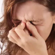
A new study published in the Ear, Nose and Throat Journal found that the severity of chronic rhinosinusitis with nasal polyposis is influenced by serum vitamin D levels. A persistent infection of the sinonasal mucosa lasting 12 weeks or more, chronic rhinosinusitis with nasal polyposis (CRSwNP) is characterized by certain symptoms and clinical indicators. This illness significantly impairs quality of life and interferes with everyday tasks.

The involvement of Vitamin D in the development of allergy disorders, such as CRSwNP, has garnered a lot of interest lately. As it influences both the innate and adaptive immune systems by altering the T-helper 1 (Th1)/Th17 response to a Th2/Treg less inflammatory profile, vitamin D has been demonstrated to have immunomodulatory effects and is crucial for the regulation of multiple immune cells, including T cells and dendritic cells, as well as for lowering inflammation through a variety of mechanisms. Thus, this study was to look into the connection between the severity of CRSwNP and serum vitamin D levels.

Serum vitamin D levels were assessed in 104 individuals with uncontrolled CRSwNP who were scheduled for functional endoscopic sinus surgery after failing all forms of medication treatment. The Lund-Mackay (LM) score, total nasal polyp scores, Sinonasal Outcome Test-22 (SNOT-22), and absolute eosinophil counts were used to compare vitamin D levels across individuals.

The average age of the 104 patients that were included was 42.09 ± 13.3 years, and 63.5% of them were men. Vitamin D levels were 57.9 ± 31.2 nmol/L on average. The mean score on the SNOT-22 was 65.49 ± 21.3. The LM score was 14.48 ± 6.64 on average. The overall score for nasal polyps was 4.3 ± 2.08. Vitamin D levels did not correlate with other factors, however, they did have a negative correlation with polyp grade (r = -.264, P =.007) and LM score (r = -.210, P =.032).

Overall, patients with CRSwNP frequently have vitamin D insufficiency or inadequacy. More evidence that low vitamin D levels are linked to more severe CRSwNP was established when the study discovered a negative correlation between low blood vitamin D levels. These early results suggest that vitamin D measurement may be useful in the clinical assessment and treatment of CRSwNP. 

Source:

Magboul, N. A., Alotaibi, M., Aldokhayel, F., Almazyad, L. M., Alkwai, K., Almutawa, N., Alotaibi, M., Alyousef, M. Y., Alsaleh, S., & Alroqi, A. (2024). Association Between Serum Vitamin D Level and Uncontrolled Chronic Rhinosinusitis With Nasal Polyposis. In Ear, Nose & Throat Journal. SAGE Publications. https://doi.org/10.1177/01455613241302892

Powered by WPeMatico

5-Point Airway Ultrasound Protocol may revolutionize Pediatric Intubation, study suggests

Confirming the correct placement of an endotracheal tube is crucial in pediatric patients due to their low tolerance to oxygen deprivation from esophageal intubation or incorrect tube positioning. Airway ultrasonography is recognized for its ability to confirm the placement of the endotracheal tube across various age groups in emergency and intensive care settings. Recent study evaluated the performance of a 5-point airway ultrasound (5-AIR USG) protocol in confirming endotracheal intubation (ETI) and endotracheal tube (ETT) positioning (ETP) in 75 pediatric patients undergoing elective surgery.

Protocol Description

The 5-AIR USG protocol involved real-time tracheal ultrasound scanning followed by bilateral pleural and diaphragmatic ultrasound scanning. The primary objective was to estimate the diagnostic sensitivity of the 5-AIR USG protocol for detecting correct (endotracheal) versus incorrect (endobronchial) ETP. Secondary objectives included estimating the accuracy of the protocol for detecting ETI (tracheal versus esophageal), comparing the time required for confirmation of ETI and ETP by the USG protocol versus quantitative waveform capnography (QWC) and 5-point auscultation, and determining the quality of visualization (QOV) score for each component of the USG protocol.

Study Findings

The study found that there were no esophageal intubations, so the accuracy of the USG protocol for ETI could not be determined. For ETP, the 5-AIR USG protocol identified 4 out of 7 endobronchial placements, resulting in a sensitivity of 100%, specificity of 57.14%, and an overall diagnostic accuracy of 96%. The mean time for confirmation of ETI by QWC was significantly longer than real-time tracheal USG (20.77 s vs 2.11 s). For ETP, the mean time for 5-point auscultation was 12.69 s versus 6.39 s for pleural USG. Adding diaphragmatic scanning increased the time to 11.45 s, and 30.68 s if a probe change was required. The 5-AIR USG protocol demonstrated high diagnostic accuracy to confirm endotracheal intubation and position in pediatric patients, though the specificity for ETP was relatively low. The protocol was faster than QWC for confirming ETI and comparable to 5-point auscultation for ETP, except when a probe change was required. The study suggests that the 5-AIR USG protocol may be a useful adjunct to clinical signs, auscultation, and QWC to confirm endotracheal intubation and position.

Key Points

1. The study evaluated the performance of a 5-point airway ultrasound (5-AIR USG) protocol in confirming endotracheal intubation (ETI) and endotracheal tube (ETT) positioning (ETP) in 75 pediatric patients undergoing elective surgery.

2. The 5-AIR USG protocol involved real-time tracheal ultrasound scanning followed by bilateral pleural and diaphragmatic ultrasound scanning. The primary objective was to estimate the diagnostic sensitivity of the 5-AIR USG protocol for detecting correct (endotracheal) versus incorrect (endobronchial) ETP. Secondary objectives included estimating the accuracy of the protocol for detecting ETI (tracheal versus esophageal), comparing the time required for confirmation of ETI and ETP by the USG protocol versus quantitative waveform capnography (QWC) and 5-point auscultation, and determining the quality of visualization (QOV) score for each component of the USG protocol.

3. The study found that there were no esophageal intubations, so the accuracy of the USG protocol for ETI could not be determined. For ETP, the 5-AIR USG protocol identified 4 out of 7 endobronchial placements, resulting in a sensitivity of 100%, specificity of 57.14%, and an overall diagnostic accuracy of 96%.

4. The mean time for confirmation of ETI by QWC was significantly longer than real-time tracheal USG (20.77 s vs 2.11 s). For ETP, the mean time for 5-point auscultation was 12.69 s versus 6.39 s for pleural USG. Adding diaphragmatic scanning increased the time to 11.45 s, and 30.68 s if a probe change was required.

5. The 5-AIR USG protocol demonstrated high diagnostic accuracy to confirm endotracheal intubation and position in pediatric patients, though the specificity for ETP was relatively low.

6. The study suggests that the 5-AIR USG protocol may be a useful adjunct to clinical signs, auscultation, and QWC to confirm endotracheal intubation and position.

Reference –

Adhiraj Baruah et al. (2024). 5-Point Airway (5-AIR) Ultrasound Protocol For Confirmation Of Endotracheal Intubation And Position In Paediatric Patients Undergoing Surgery: A Prospective Observational Study. *Indian Journal Of Anaesthesia*. https://doi.org/10.4103/ija.ija_682_24.

Powered by WPeMatico

Avoiding Prophylactic Drains After Gastrectomy Increases Risk of Postoperative Complications: ADIGE Trial

Italy: A recent study, the Abdominal Drain in Gastrectomy (ADIGE) Randomized Clinical Trial, has revealed important insights into using prophylactic drains after gastrectomy, a common surgical procedure for treating stomach cancer and other gastrointestinal conditions. The study challenges the notion that avoiding the use of abdominal drains post-surgery might reduce complications and highlights the associated risks of postoperative invasive procedures.

In their study published in JAMA Surgery, the researchers found that avoiding prophylactic drain use after gastrectomy increased the risk of postoperative invasive procedures, making it inadvisable to forgo their use.

Previous evidence indicates that prophylactic abdominal drainage after gastrectomy for cancer may help reduce postoperative morbidity and shorten hospital stays. However, this evidence comes from small studies with a significant risk of bias. More research is needed to assess whether drains effectively fulfill their primary role of identifying and managing postoperative intraperitoneal collections without necessitating reoperation or additional percutaneous drainage. In light of this, Jacopo Weindelmayer, General and Upper GI Surgery Division, Azienda Ospedaliera Universitaria Integrata, Borgo Trento, Verona, Italy, and colleagues aimed to investigate whether avoiding routine abdominal drainage would lead to an increase in postoperative invasive procedures.

For this purpose, the researchers conducted the ADIGE Trial, a multicenter, prospective randomized noninferiority study. The trial included 404 patients with gastric cancer undergoing subtotal or total gastrectomy, recruited from 11 centers in Italy between December 2019 and January 2023. Participants were randomized into two groups: prophylactic drain or no drain.

The primary outcome was reoperation or percutaneous drainage within 30 days post-surgery, analyzed through a modified intention-to-treat approach. The study aimed to test if avoiding drains increased the need for invasive procedures, with surgeons and patients blinded until gastrointestinal reconstruction.

The trial revealed the following findings:

  • Of the 404 patients randomized, 226 (57.8%) were male, with a median age of 71 years.
  • Fourteen patients were excluded due to intraoperative identification of nonresectable disease, leaving 390 patients for analysis.
  • In the modified intention-to-treat (mITT) analysis, 7.7% of patients in the drain group required reoperation or percutaneous drainage by day 30, compared to 15% of patients in the no-drain group, favoring the drain group (difference 7.2%).
  • The primary difference was due to a lower reoperation rate in the drain group (5.1%) compared to the no-drain group (12.4%).
  • Drain-related complications occurred in 4 patients.

“These findings indicate that avoiding drains may raise the risk of postoperative complications, especially invasive procedures, in this patient population. Future research identifying high-risk groups could help refine decisions regarding prophylactic drainage,” the researchers concluded.

Reference:

Weindelmayer J, Mengardo V, Ascari F, et al. Prophylactic Drain Placement and Postoperative Invasive Procedures After Gastrectomy: The Abdominal Drain After Gastrectomy (ADIGE) Randomized Clinical Trial. JAMA Surg. Published online November 27, 2024. doi:10.1001/jamasurg.2024.5227

Powered by WPeMatico

Cutaneous systemic lupus associated with increased risk for ASCVD, reveals research

Researchers have established that an association exists between cutaneous lupus erythematosus (CLE) and an increased risk for atherosclerotic cardiovascular disease (ASCVD), it is similar to that observed in patients with systemic lupus erythematosus (SLE), and is greater than among those with psoriasis. A recent study was conducted by Henry W. Chen and colleagues which was published in the journal of JAMA Dermatology.

This retrospective matched cohort study analyzed data from the IBM MarketScan Commercial Claims and Encounters Database from January 2018 through December 2020. A total of 8138 individuals with CLE were matched 10:1 with controls based on age, sex, insurance type, and enrollment duration; 24,675 had SLE and 192,577 had psoriasis.

Outcomes: ASCVD prevalence was determined by coronary artery disease, prior myocardial infarction, or cerebrovascular accident. Incident ASCVD was assessed via hospitalization events over a median follow-up of 3 years.

Statistical Analysis: Multivariable logistic regression and Cox proportional hazards models were used to adjust for age, sex, and cardiovascular risk factors.

The study yielded significant results regarding the ASCVD risk among individuals with CLE, SLE, and psoriasis:

Demographics:

  • CLE: Mean age, 49 years; 81% female

  • SLE: Mean age, 46 years; 91% female

  • Psoriasis: Mean age, 48 years; 55% female

  • Controls: Mean age, 49 years; 81% female

ASCVD Prevalence:

  • ASCVD odds were higher in the CLE than controls at 1.72 [Odds Ratio (OR): 1.72; 95% CI, 1.45–2.02; p < 0.001].

  • SLE showed the greatest odds for ASCVD, with OR = 2.41 (95% CI, 2.14–2.70; p < 0.001).

  • Psoriasis did not have a significant increase in the odds of ASCVD (OR: 1.03; 95% CI, 0.95–1.11; p = 0.48).

ASCVD Incidence Rates per 1000 person-years:

  • SLE: 24.8 (95% CI, 23.3–26.4).

  • CLE: 15.2 (95% CI, 13.1–17.7).

  • Psoriasis: 14.0 (95% CI, 13.5–14.4).

  • Controls: 10.3 (95% CI, 9.77–10.94).

Hazard Ratios for Incident ASCVD

  • SLE: 2.23 times greater risk than controls [Hazard Ratio (HR): 2.23; 95% CI, 2.05–2.43; p < 0.001].

  • CLE: 1.32 times greater risk [HR: 1.32; 95% CI, 1.13–1.55; p < 0.001].

  • Psoriasis: No significant risk increase [HR: 1.06; 95% CI, 0.99–1.13; p = 0.09].

This study found that patients with CLE had a significantly higher risk of developing ASCVD than that of SLE, though they were at a higher risk than those with psoriasis. Clinicians must consider this increased risk when managing CLE patients and take necessary measures for screening and prevention.

Reference:

Chen HW, Liu J, Yang DM, et al. Incidence and Prevalence of Atherosclerotic Cardiovascular Disease in Cutaneous Lupus Erythematosus. JAMA Dermatol. Published online December 04, 2024. doi:10.1001/jamadermatol.2024.4991

Powered by WPeMatico

Multifocal Epitheloid Hemangioma of Bone – A Rare Entity

Epithelioid hemangiomas (EHs) are rare vascular lesions which generally affect the skin and subcutaneous tissue but rarely seen in bones. It is a benign entity but intermediate grade, i.e., locally aggressive in nature. It has very confusing clinicoradiological and histopathological features which make diagnosis difficult and help us to avoid inappropriate treatment.

A 36-year-old male presented with pain and swelling over the right wrist extending toward the dorsal aspect of the hand associated with difficulty in wrist range of movements, for the past 3 months. There was no history of trauma or any twisting injury and no history of any fever. The swelling did not respond to any analgesics. Moreover, the swelling was increasing day by day, but there were no erythematous changes over the skin. Upon examination, there was tenderness over the wrist joint and carpometacarpal joints with a restricted range of movements of the wrist and multiple lobulated swelling felt over the dorsal aspect of the wrist.

After initial examination, the patient reported with X-ray and magnetic resonance imaging (MRI). A plain X-ray of the wrist showed a destructive lytic lesion over the distal radius which had ill-defined margins, lytic lesions also seen in the base of the 2nd and 3rd metacarpal base. MRI report showed an osteolytic lesion measuring 2.5 × 2.4 cm distal end of radius extending to a subarticular location with extraosseous soft-tissue component breaching the volar surface of distal radius. Multiple lytic lesions involving the trapezium, trapezoid bone, and capitate bones with associated marrow edema lytic lesions are also seen in the base of 2nd and 3rd metacarpals with enhancing soft tissue lesion measuring 2.9 × 2.1 cm abutting the carpal bones also seen. This was followed by whole-body positron emission tomography-computed tomography (CT) scan which revealed increased fluorodeoxyglucose uptake of standardized uptake value Max-5 in the distal radius with soft-tissue component and involvement of multiple carpal bones as described in MRI reports.

The authors performed a closed Jamshedji needle biopsy which was reported as EH. Then, he was managed with extended curettage + bone grafting + bone cementing and plating.

The patient was kept in close follow-up and there was no recurrence till 1-year post-operative period.

The authors concluded that – “EH of bone is a rare tumor and has a difficult diagnosis due to its aggressive clinicoradiological features. This case had aggressive radiological features, extensive soft tissue components, and multifocality which were looking like a malignant bone tumor with extensive soft-tissue involvement. Only after a closed J needle biopsy, expert histopathological study, and clinicoradiological correlation helped us to come to a confirmed diagnosis of EH. The case highlights clinical, radiological, and histopathological findings of EH of bone and also provides insights about the approach to these uncommon locally aggressive bone tumors. This case will help orthopedic surgeons and orthopedic onco-surgeons about the diagnostic approach and management of these rare bone tumors. An expert histopathological examination with the judicious help of an IHC panel is essential for a proper diagnosis which helps onco-pathologists and pathologists to understand the nature of the disease. Finally, coordination between orthopedic surgeons, radiologists, and pathologists (O.R.P) teams is a key for diagnosing rare bone tumors and its management.” 

Further reading:

Multifocal Epitheloid Hemangioma of the Bone – A Rare Entity

Bibhudutta Malla et al

Journal of Orthopaedic Case Reports 2024 November:14(11):4-9

https://doi.org/10.13107/jocr.2024.v14.i11.4894

Powered by WPeMatico

Study exposes link between genetic risk of depression and heart disease in women

Women who have a high genetic risk of depression are more likely to develop heart disease, University of Queensland researchers have found.

During a study that analysed genetic and health data from more than 300,000 people, Dr Sonia Shah and Dr Clara Jiang from UQ’s Institute for Molecular Bioscience found women who had a high genetic risk of developing depression also had a high risk of developing heart disease, even in the absence of a depression diagnosis.

Dr Shah said these results exposed a difference in the risk for women compared to men.

“In our study, the link between the genetic risk of depression and developing a cardiovascular disease was seen even among women who had never been diagnosed with depression or taken any psychiatric medications,” Dr Shah said.

“However, this link was not observed in men, despite an overall greater proportion of men developing heart disease.

“The variation between men and women could also not be explained by differences in traditional risk factors such as BMI, smoking, high blood pressure or high cholesterol.

“Our research highlights the need to understand this relationship separately in men and women.”

Dr Jiang said that despite heart disease being the leading cause of death for women globally, they have historically been underrepresented in cardiovascular research and clinical trials.

“This has led to a bias towards men in our knowledge and approach to cardiovascular health, and as a result, women are going under-diagnosed and under-treated,” Dr Jiang said.

Dr Shah said while the risk of heart disease increases for women after menopause, this study highlighted that women who have depression should be assessed for heart disease risk regardless of their menopausal stage.

“Our research found that the higher risk of developing coronary artery disease, where blood vessels narrow because of the build-up of plaque, was present regardless of whether the women were pre-menopausal or post-menopausal at recruitment,” Dr Shah said.

“Frequent heart health checks are especially important for women who have a history of depression.”

During the study, researchers developed genetic predictors of psychiatric disorders using data from large-scale genetic studies including the psychiatric genomics consortium, genetic health and biopharmaceutical company 23andMe, and UK BioBank, a large-scale biomedical database and research resource containing anonymised genetic, lifestyle and health information from half a million consenting UK participants.

Reference:

Jiayue-Clara Jiang,  Rachana Nitin, Sex-Specific Association Between Genetic Risk of Psychiatric Disorders and Cardiovascular Diseases, Circulation Genomic and Precision Medicine, https://doi.org/10.1161/CIRCGEN.124.004685.

Powered by WPeMatico

Mechanical rotation chair method superior to traditional manual BPPV diagnostics for diagnosing complex BPPV: Study

A recent groundbreaking study published
in Frontiers in Neurology revealed that the mechanical rotation chair (MRC)
method is superior to traditional manual BPPV diagnostics for diagnosing
complex Benign paroxysmal positional vertigo (BPPV).

Benign paroxysmal positional
vertigo (BPPV) is the most common vestibular disease that causes vertigo due to
the dislodgement of otoconia. BPPV is classified into different types based on
the anatomical location of otoliths and the endolymphatic flow. A
clinician diagnoses BPPV using traditional manual diagnostics. With the advent of
various diagnostic methods, the management of BPPV has improved a lot. Hence,
researchers have conducted a study to compare traditional manual BPPV
diagnostics (MD) with MRC when using videonystagmography goggles with both
modalities.

A prospective open-label randomized diagnostic crossover study involved adults with typical BPPV
symptoms at a tertiary University Hospital-based outpatient clinic in Denmark. Participants
underwent traditional manual and MRC diagnostics with videonystagmography
goggles by randomly assigning participants to the two modalities with a
duration gap of 30 minutes between the two procedures. Identifying the sensitivity,
specificity, positive predictive value (PPV), and negative predictive value
(NPV) of the traditional procedure with MRC was the primary outcome of
measurement. Agreement between the two modalities was the secondary outcome of
measurement.

Findings:

  • MRC diagnostics demonstrated a significantly
    higher sensitivity for detecting BPPV across all participants (p=0.00).
  • Compared to MRC diagnostics, traditional MD
    displayed a sensitivity of 69.5%, specificity of 90.9%, PPV of 88.0%, and NPV
    of 75.8%.
  • Both the diagnostics showed an overall
    inter-modality agreement of 80.5%.
  • Both modalities detected the unilateral
    posterior canal BPPV equally well (p=0.51).
  • The traditional MD significantly underperformed
    for non-posterior BPPV.
  • Underperformance was also seen in the subgroups
    referred by ENTs (trend) compared to MRC.
  • Traditional methods significantly underperformed
    in uncooperative patients when compared to MRC.

Thus, the study concluded that even
though traditional methods are valuable first-line diagnostic methods, MRC was equally
beneficial and more helpful in specific situations like ENT referrals,
uncooperative patients, and non-posterior BPPV. The researchers emphasized incorporating
advanced diagnostics like MRC with traditional methods to improve diagnostic
accuracy and optimize patient outcomes.

Further reading: Is Diagnostics
of Benign Paroxysmal Positional Vertigo with a Mechanical Rotation Chair
Superior to Traditional Manual Diagnostics? A Randomized Controlled Crossover
Study. doi: 10.3389/fneur.2024.1519837.

Powered by WPeMatico

Significantly Higher Salivary Opiorphin Concentrations Linked to Burning Mouth Syndrome, Study Reveals

Iran: A recent study published in Clinical & Experimental Dental Research found that salivary opiorphin levels are elevated in patients with Burning Mouth Syndrome (BMS) compared to healthy individuals. Additionally, a majority of BMS patients report experiencing anxiety and depression.

Burning Mouth Syndrome is a chronic intraoral burning sensation lasting over 2 hours daily for at least 3 months, often accompanied by oral dryness, taste changes, and dysesthesia. Its global prevalence is 1.73%, rising to 8% among clinical patients, predominantly affecting middle-aged and elderly women. Psychological factors, anxiety, depression, and neuropathy are linked to BMS, though its exact cause remains unclear. Misdiagnosis and improper treatment can worsen symptoms, highlighting the need for multidisciplinary care.

No specific paraclinical parameters exist for diagnosing Burning Mouth Syndrome, which relies solely on clinical findings. Recent studies suggest that opiorphin, a natural analgesic in saliva, may influence BMS by modulating pain perception and exhibiting antidepressant effects. Considering this, Maryam Amirchaghmaghi, Mashhad University of Medical Sciences, Mashhad, Iran, and colleagues examined the association between salivary opiorphin levels, pain severity, and psychiatric scores to explore its role in BMS pathogenesis and treatment response.

For this purpose, the researchers conducted a cross-sectional study involving 28 BMS patients and 40 healthy individuals referred to the Department of Oral and Maxillofacial Medicine, Faculty of Dentistry, Mashhad, between September 2018 and August 2019. Participants with confirmed BMS, based on clinical and psychiatric evaluations, were included. Unstimulated salivary opiorphin levels were measured using ELISA, and statistical analysis was performed with SPSS version 24.

The investigation revealed the following findings:

  • Salivary opiorphin levels were significantly higher in BMS patients (2.16 ± 0.30 ng/mL) compared to healthy subjects (1.80 ± 0.36 ng/mL).
  • Opiorphin levels in BMS patients showed no significant change after psychiatric therapy.
  • There was no significant association between opiorphin levels and variables such as age, gender, menopause, burning severity, anxiety, and depression status.

The study investigated salivary opiorphin concentrations in BMS patients and their association with anxiety and depression levels. The findings revealed that most BMS patients exhibited some degree of anxiety and depression. Initial opiorphin levels were significantly higher in the BMS group compared to controls. Although opiorphin levels showed only a slight, insignificant increase four weeks after treatment, there was a significant reduction in burning sensation intensity, anxiety, and depression.

The authors acknowledge a limitation in their study due to the small number of patients available for post-treatment assessment, which limited the ability to evaluate the effectiveness of psychiatric treatment.

“Further research is needed to confirm the link between opiorphin concentration and burning sensation severity in BMS patients. Salivary opiorphin levels may serve as a noninvasive tool for monitoring treatment outcomes and assessing BMS symptoms, depression, and anxiety during long-term follow-up,” the researchers concluded.

Reference:

Dalirsani, Z., Amirchaghmaghi, M., Semnani, M. M., Talebi, M., Hashemy, S. I., Shakeri, M. T., & Taghizadeh, A. (2024). Comparison of Salivary Opiorphin in Burning Mouth Syndrome and Healthy Subjects and Its Correlation With Psychiatric Disorders. Clinical and Experimental Dental Research, 10(6), e934. https://doi.org/10.1002/cre2.934

Powered by WPeMatico

From Womb to Growth: Study reveals Implications of Neonatal Anemia on child Health

Neonatal anemia can have lasting impacts on the growth and development of children. Anemia is highly prevalent among pregnant women globally, making it a significant public health concern if it results in newborn anemia. Recent research paper aimed to investigate the impact of neonatal anemia on children’s growth and development, as well as the prevalence of anemia during pregnancy and its public health implications. Neonatal anemia has long-term effects on children, with maternal anemia during pregnancy being a common nutritional deficiency globally. The study focused on the relationship between maternal anemia during pregnancy and neonatal hemoglobin levels. Data from various databases were systematically searched for studies on this topic, leading to the inclusion of 18 articles involving 1873 patients.

Meta-analysis Findings

The meta-analysis revealed a significant association between maternal anemia during pregnancy and lower neonatal hemoglobin levels. The findings suggested that neonates of anemic mothers had hemoglobin levels 1.38g/dL lower than those of non-anemic mothers. Subgroup analyses indicated a correlation between maternal and infant hemoglobin levels, with maternal blood samples taken after delivery showing the most significant correlation with neonatal blood levels. Additionally, mothers with iron deficiency anemia had a stronger correlation with neonatal hemoglobin levels compared to other types of anemia.

Importance of Addressing Anemia

The study highlighted the importance of addressing maternal anemia during pregnancy to reduce the risk of neonatal anemia. Future research with larger sample sizes and standardized methods is needed to confirm the relationship between maternal and neonatal anemia. The authors emphasized the need for enhanced management of anemia during pregnancy and nutritional monitoring of newborns. While the study supported an association between maternal and neonatal anemia, further investigation is required to assess the effectiveness of treating maternal anemia during pregnancy and establish guidelines for iron supplementation tailored to different cultural contexts. The authors recommended future studies with uniform sampling times and comprehensive control of confounding factors to validate the findings and improve anemia management during pregnancy.

Key Points

– The research paper aimed to investigate the impact of neonatal anemia on children’s growth and development, as well as the prevalence of anemia during pregnancy and its public health implications. – In total, 18 articles involving 1873 patients were included in the study after systematically searching various databases for relevant studies on the relationship between maternal anemia during pregnancy and neonatal hemoglobin levels.

– The meta-analysis conducted as part of the study revealed a significant association between maternal anemia during pregnancy and lower neonatal hemoglobin levels. Specifically, neonates of anemic mothers were found to have hemoglobin levels 1.38g/dL lower than those of non-anemic mothers.

– Subgroup analyses also indicated a correlation between maternal and infant hemoglobin levels, with maternal blood samples taken after delivery showing the most significant correlation with neonatal blood levels. Mothers with iron deficiency anemia exhibited a stronger correlation with neonatal hemoglobin levels compared to other types of anemia.

– The study emphasized the importance of addressing maternal anemia during pregnancy to reduce the risk of neonatal anemia. The authors underscored the need for future research with larger sample sizes and standardized methods to confirm the relationship between maternal and neonatal anemia and improve anemia management during pregnancy.

– Overall, the findings supported an association between maternal and neonatal anemia, suggesting the need for enhanced management of anemia during pregnancy and nutritional monitoring of newborns. However, further investigation is recommended to assess the efficacy of treating maternal anemia during pregnancy and establish culturally tailored guidelines for iron supplementation.

Reference –

Zhao, B., Sun, M., Wu, T. et al. The association between maternal anemia and neonatal anemia: a systematic review and meta-analysis. BMC Pregnancy Childbirth 24, 677 (2024). https://doi.org/10.1186/s12884-024-06832-1

Powered by WPeMatico

Low consumption of animal products and oil tied to reduced hot flashes in postmenopausal women: Study

A low-fat vegan diet with soybean supplementation has proven effective in decreasing both body weight and hot flashes in postmenopausal women. Another study, the Women’s Health Initiative, found that increasing whole grains, fruits, and vegetables while lowering fat intake led to reduced hot flashes, especially in individuals who lost over 10% of their body weight. Recent secondary analysis aimed to assess the association between different plant-based diet indices and changes in hot flashes experienced by postmenopausal women. The study participants were randomly assigned to either a low-fat vegan diet supplemented with soybeans (n=42) or a control group (n=42) for 12 weeks. The researchers calculated three plant-based dietary indices: the plant-based diet index (PDI), the healthful plant-based diet index (hPDI), and the unhealthful plant-based diet index (uPDI). They found that all three scores increased in the vegan group compared to no change in the control group. The effect sizes were: – PDI: +9.8 (95% CI +5.8 to +13.8, p<0.001) – hPDI: +10.9 (95% CI +6.4 to +15.3, p<0.001) – uPDI: +3.6 (95% CI +0.5 to +6.6, p=0.02)

Correlation with Changes in Body Weight

The changes in all three scores were negatively correlated with changes in body weight. Importantly, the changes in PDI and uPDI were also negatively associated with changes in severe hot flashes, and these associations remained significant after adjusting for changes in body mass index. The researchers conclude that minimizing the consumption of animal products and vegetable oil may be an effective strategy to reduce hot flashes in postmenopausal women. They also suggest that the categorization of plant foods as “healthful” or “unhealthful” may be unwarranted in this context, as both the “healthy” and “unhealthy” plant-based indices were inversely associated with weight changes and vasomotor symptoms.

Insights from Findings on Low-Fat Vegan Diet

These findings provide valuable insights into the potential benefits of a low-fat vegan diet, supplemented with soybeans, for managing postmenopausal hot flashes. The study highlights the importance of considering overall dietary patterns, rather than just individual food groups, when evaluating the impact of plant-based diets on health outcomes.

Key Points

1. This secondary analysis aimed to assess the association between different plant-based diet indices and changes in hot flashes experienced by postmenopausal women.

2. The study participants were randomly assigned to either a low-fat vegan diet supplemented with soybeans (n=42) or a control group (n=42) for 12 weeks.

3. The researchers calculated three plant-based dietary indices: the plant-based diet index (PDI), the healthful plant-based diet index (hPDI), and the unhealthful plant-based diet index (uPDI). All three scores increased in the vegan group compared to no change in the control group.

4. The changes in all three scores were negatively correlated with changes in body weight. The changes in PDI and uPDI were also negatively associated with changes in severe hot flashes, even after adjusting for changes in BMI.

5. The researchers conclude that minimizing the consumption of animal products and vegetable oil may be an effective strategy to reduce hot flashes in postmenopausal women. They also suggest that the categorization of plant foods as “healthful” or “unhealthful” may be unwarranted in this context.

6. The findings provide valuable insights into the potential benefits of a low-fat vegan diet, supplemented with soybeans, for managing postmenopausal hot flashes, and highlight the importance of considering overall dietary patterns rather than just individual food groups.

Reference –

Brennan, H., Znayenko-Miller, T., Sutton, M. et al. Diet quality, body weight, and postmenopausal hot flashes: a secondary analysis of a randomized clinical trial. BMC Women’s Health 24, 620 (2024). https://doi.org/10.1186/s12905-024-03467-4

Powered by WPeMatico