Metformin Outperforms Vildagliptin in Reducing BMI and Weight in T2D Patients: Study

Researchers have found that both vildagliptin and metformin reduce hepatic steatosis effectively and improve glycemic control in newly diagnosed type 2 diabetes mellitus (T2D) patients. A recent study was conducted by Asmaa and colleagues which was published in the journal BMC Pharmacology and Toxicology.

Hepatic steatosis (HS), one of the most common complications of T2D, is largely associated with adiposity and glycemic dysregulation. This study was done to compare the effect of vildagliptin and metformin on hepatic steatosis through the use of HSI and ultrasound grading, as well as their consequences on metabolic parameters.

This study enrolled newly diagnosed T2D patients with a total of 246 for this randomized controlled trial. Patients were randomly allocated to the following groups:

  • Group 1: 117 patients received 50 mg of vildagliptin orally twice daily.

  • Group 2: 129 patients received 500 mg of metformin orally twice daily, with doses titrated weekly by 500 mg to a maximum of 2000 mg per day.

Assessment Parameters: baseline and 6 months followed by fasting blood glucose (FBG), HbA1c, and physical measurements: weight; BMI; waist circumference (WC); hip circumference (HC); HSI and grading of hepatic steatosis via ultrasound.

Key findings

  • 246 newly diagnosed T2D patients participated: 117 in the vildagliptin group, 129 in the metformin group.

  • Both groups showed significant improvement in FBG, HbA1c, weight, BMI, WC, HC, HSI, and hepatic steatosis grading baseline to 6 months (p < 0.001).

  • Compared with Vildagliptin, metformin proved efficacious to a greater extent in both weight reduction and BMI (p = 0.001 and p = 0.009, respectively).

  • There was a strong correlation of HSI with HbA1c, BMI, WC, HC (p < 0.001), and FBG (p = 0.008).

  • Lipid profile (total cholesterol and LDL) was the best predictor of hepatic steatosis.

Both vildagliptin and metformin were effective in terms of improving glycaemic control and lowering the degree of hepatic steatosis in newly diagnosed T2D patients. Metformin had a better effect on weight and attained a lower BMI; thus, it is likely to benefit patients who have obesity-related complications even more so than vildagliptin.

Reference:

Mohamed, A. S., Ahmad, H. M., Sharawy, M. A., & Kamel, F. M. M. (2024). The effect of vildagliptin versus metformin on hepatic steatosis in type 2 diabetic patients: a randomized controlled trial. BMC Pharmacology and Toxicology, 25(1). https://doi.org/10.1186/s40360-024-00818-7

Powered by WPeMatico

Vericiguat safe drug for use in patients with coronary artery disease, finds research

Researchers have identified that vericiguat, a new drug for heart failure, is safe in patients with coronary artery disease. A recent study showed that vericiguat is not associated with major serious adverse events despite mild alterations in blood pressure and heart rate. This study was conducted by Bin Zarti and colleagues published in the American Journal of Cardiovascular Drugs.

The study reviewed the data from three randomized controlled trials involving 151 patients on the safety profile of vericiguat versus a placebo. Clinical outcomes and adverse events were stringently analyzed to provide valuable insights in its application in cardiovascular care. The meta-analysis covered a detailed search of three major databases: PubMed, ClinicalTrials.gov, and the Cochrane Library, until March 27, 2024. Only studies that compared vericiguat with a placebo in patients diagnosed with coronary artery disease were included.

Quality assessment of the enrolled studies was performed on each, and clinical information for analysis was extracted from these studies. Statistical methods to analyze the studies conducted in this review include measures like ORs and CIs; Review Manager software version 5.4 was used in that regard. Parameters scrutinized include blood pressure, heart rate, and adverse events.

Main Results

Blood Pressure and Heart Rate

  • The systolic blood pressure decreased by 1.4-10 mmHg and diastolic blood pressure by 0.4-6 mmHg; both the changes were clinically insignificant.

  • Heart rate increased by 1.8-7 bpm, which was also of clinical insignificance.

Adverse Events

  • No serious adverse events were reported significantly with the use of vericiguat in comparison with the placebo (OR = 1.97; 95% CI = 0.39-9.91; p= 0.41).

  • There was a greater overall adverse events rate between vericiguat and the placebo group (OR = 4.04; 95% CI = 2.17 – 7.52; p< 0.001).

Clinical Trials and Population

  • Analysis involved all three randomized controlled trials with an enrollment of 151 patients.

  • Therefore, minor adverse effects notwithstanding, vericiguat appears to still be safe in managing coronary artery disease.

This meta-analysis therefore shows that vericiguat is safe for the use of patients with coronary artery disease as it is free from a significant risk of serious adverse events. The minor adverse events were, however, more frequent in the vericiguat arm, and the clinical significance remains low. More clinical studies are required to confirm these results and make vericiguat a first-line therapy in cardiovascular practice.

Reference:

Bin Zarti, M., & Tamgheli, A. (2024). Safety of vericiguat in patients with coronary artery disease: A systematic review and meta-analysis. American Journal of Cardiovascular Drugs: Drugs, Devices, and Other Interventions. https://doi.org/10.1007/s40256-024-00701-0

Powered by WPeMatico

Positive Results for Mifepristone in Treating Hypercortisolism and Type 2 Diabetes: CATALYST Trial

USA: Mifepristone (Korlym) shows significant HbA1c improvement in hypercortisolism and difficult-to-control diabetes; Corcept announces positive results in the CATALYST Trial.

Corcept Therapeutics Incorporated, a commercial-stage company, has announced promising results from the treatment phase of the Phase 4 CATALYST trial. The findings highlight the efficacy of mifepristone in addressing hypercortisolism (Cushing’s syndrome) and difficult-to-control type 2 diabetes (T2D).

The randomized, double-blind, placebo-controlled CATALYST trial met its primary endpoint, demonstrating that patients with hypercortisolism and T2D experienced a statistically significant and clinically meaningful reduction in hemoglobin A1c (HbA1c) following treatment with mifepristone. Patients treated with Korlym showed a reduction of 1.47% in HbA1c levels compared to a 0.15% reduction in the placebo group, translating into a placebo-adjusted reduction of 1.32%.

“CATALYST’s first part showed that hypercortisolism is much more common than previously assumed. The results announced today show that Korlym is a safe and effective treatment option,” Ralph DeFronzo, MD, chief of the Diabetes Division and professor of medicine at UT Health San Antonio and a CATALYST study investigator, said in a press release. DeFronzo emphasized that these findings are particularly significant given that participants were already receiving advanced diabetes therapies but continued to struggle with their disease.

The CATALYST study was designed as a two-part, Phase 4 investigation. The first part screened 1,057 patients with difficult-to-control type 2 diabetes—defined as having HbA1c levels greater than 7.5% despite optimal treatment with GLP-1 agonists and other therapies. Of these patients, 23.8% were identified as having hypercortisolism and eligible for the treatment phase. A total of 136 patients were randomized in a 2:1 ratio to receive either Korlym or a placebo for 24 weeks.

Bill Guyer, PharmD, Corcept’s Chief Development Officer, highlighted the importance of these findings: “One in four patients with difficult-to-control type 2 diabetes have hypercortisolism, and Korlym was highly effective in controlling hyperglycemia in this patient population.”

Importantly, the safety profile of Korlym observed in the CATALYST study was consistent with its existing labeling, with no new adverse events or side effects identified. Complete results from the trial are expected to be presented at a medical conference in the coming year.

Corcept’s findings underscore the potential for expanded screening for hypercortisolism and improved treatment strategies to enhance outcomes for patients struggling with these challenging conditions.

About Hypercortisolism (Cushing’s Syndrome)

Hypercortisolism, also known as Cushing’s syndrome, occurs due to excessive levels or activity of the hormone cortisol in the body. The symptoms can vary widely among individuals, but common signs include high blood pressure, central obesity, elevated blood sugar, and challenging-to-control type 2 diabetes. Many patients also report severe fatigue, muscle weakness, irritability, anxiety, depression, and cognitive impairments.

Hypercortisolism has the potential to impact multiple organ systems, making early diagnosis and treatment critical. If left untreated, it can lead to life-threatening complications.

Powered by WPeMatico

Azithromycin therapy shows positive effect on lung perfusion in infants with cystic fibrosis, finds research

A new study published in the Journal of Cystic Fibrosis found that infants with cystic fibrosis who get azithromycin exhibit a decrease in the thickness of their bronchial walls and may benefit from improved lung perfusion.

The COMBAT-CF research examined the effectiveness of azithromycin as an anti-inflammatory medication in newborns with CF. From the time of diagnosis until the age of 3, children with CF identified by newborn screening were stratified by site and randomly assigned 1:1 to maintenance therapy with azithromycin or a placebo.

The main outcome measure was structural airway illness as determined by the manual Perth-Rotterdam Annotated Grid Morphometric for CF (PRAGMA-CF) grading technique using chest computed tomography images (CTs). At 3 years old, there were no discernible variations in PRAGMA-CF sub-scores across treatment groups. On the other hand, children treated with azithromycin had fewer exacerbations, fewer days needing antibiotic therapy, and less inflammation in their bronchoalveolar lavage fluid.

Reanalyzing of the COMBAT-CF CTs utilized the AI-based BA-method and an AI-based technique to assess low attenuation regions (LAR, trapped air) was the goal of the post hoc investigation. Yuxin Chen and colleagues postulated that children receiving azithromycin would have thinner bronchial walls and lower LAR than those receiving a placebo.

The CT scans of the lungs at 12 and 36 months were examined. Measures of BA-analysis for the BA-diameters are the artery (A), bronchial wall thickness (Bwt), bronchial outer wall (Bout), and bronchial inner wall (Bin). BA-ratios: Bwt/A and Bwa/Boa (bronchial wall area/bronchial outer area) for bronchial wall thickening, and Bout/A and Bin/A for bronchial widening. An automated technique was used to analyze low attenuation regions (LAR). BA-outcomes at 36 months were compared between treatment groups using a mixed-effect model.

Analysis was done on 228 CTs (59 placebo, 66 azithromycin). When compared to the placebo group, the azithromycin group expressed increased Bin/A (p = 0.001) and lower Bwa/Boa (p = 0.0034). Because of a decrease in artery diameters, which was associated with a decrease in LAR, Bout/A (p = 0.0088) was greater.

Overall, the favorable impact of azithromycin maintenance treatment is further supported by the automated BA-analysis, which revealed a decrease in bronchial wall thickness in young children with CF receiving azithromycin. Also, the azithromycin group showed a decrease in artery diameter, indicating that azithromycin has a beneficial effect on small airway disease and related lung perfusion. 

Source:

Chen, Y., Charbonnier, J.-P., Andrinopoulou, E.-R., Sly, P. D., Stick, S. M., & Tiddens, H. A. W. M. (2024). Azithromycin reduces bronchial wall thickening in infants with cystic fibrosis. In Journal of Cystic Fibrosis (Vol. 23, Issue 5, pp. 870–873). Elsevier BV. https://doi.org/10.1016/j.jcf.2024.04.001

Powered by WPeMatico

Cardiac Connection: New study reveals association of Pre-eclampsia and Fetal Heart Defects

The pathophysiology of pre-eclampsia has traditionally been thought to result from placental dysfunction; however, a heart-related origin has been proposed recently. While a link between fetal congenital heart disease and pre-eclampsia has been established, a specific biological mechanism explaining this association remains undefined. Recent research paper explores the potential cardiac origin and pathophysiological mechanisms underlying the association between fetal congenital cardiovascular disease and pre-eclampsia. While the dominant theory has been that pre-eclampsia stems from placental dysfunction, a cardiac origin has been more recently proposed.

Biophysical and Biochemical Markers

The paper highlights various biophysical and biochemical markers of cardiac dysfunction that have been investigated as potential predictors of pre-eclampsia. Biophysical markers include echocardiography and Doppler indices, which have shown that women with pre-eclampsia demonstrate evidence of cardiac dysfunction, such as increased left ventricular wall mass and deteriorating diastolic indices. Biochemical markers include proteomic, metabolomic and genetic/transcriptomic markers.

Proteomic and Metabolomic Studies

Proteomic markers like NT-proBNP, troponins, and other proteins involved in inflammation and angiogenesis have been investigated. Metabolomic studies have found elevated levels of lipids, fatty acids, acylcarnitines, and amino acids like glutamate, which are associated with increased cardiac disease risk. Transcriptomic and epigenetic analysis has revealed shared pathways involving oxidative stress, inflammation, and immune responses between cardiovascular disease and pre-eclampsia. The paper also discusses the link between fetal congenital heart disease and maternal pre-eclampsia. Fetuses with structural heart defects like ventricular septal defects, atrioventricular septal defects, and coarctation of the aorta have a higher risk of developing pre-eclampsia. Fetal biomarkers like natriuretic peptides have been explored as predictors of fetal cardiac dysfunction and its impact on the mother.

Conclusion

In summary, this research highlights the growing evidence supporting a cardiac origin for pre-eclampsia, in contrast to the traditional placental dysfunction theory. The shared pathophysiological pathways between maternal/fetal cardiac disease and pre-eclampsia, involving inflammation, oxidative stress, and angiogenic imbalance, provide a strong rationale for further investigating the cardiac basis of this pregnancy-specific condition.

Key Points

1. The paper explores the potential cardiac origin and pathophysiological mechanisms underlying the association between fetal congenital cardiovascular disease and pre-eclampsia, in contrast to the traditional placental dysfunction theory.

2. The paper highlights various biophysical markers of cardiac dysfunction, such as echocardiography and Doppler indices, which have shown that women with pre-eclampsia demonstrate evidence of cardiac dysfunction, including increased left ventricular wall mass and deteriorating diastolic indices.

3. Proteomic markers like NT-proBNP, troponins, and other proteins involved in inflammation and angiogenesis have been investigated as potential predictors of pre-eclampsia. Metabolomic studies have found elevated levels of lipids, fatty acids, acylcarnitines, and amino acids like glutamate, which are associated with increased cardiac disease risk.

4. Transcriptomic and epigenetic analysis has revealed shared pathways involving oxidative stress, inflammation, and immune responses between cardiovascular disease and pre-eclampsia.

5. Fetuses with structural heart defects like ventricular septal defects, atrioventricular septal defects, and coarctation of the aorta have a higher risk of developing pre-eclampsia, and fetal biomarkers like natriuretic peptides have been explored as predictors of fetal cardiac dysfunction and its impact on the mother.

6. The growing evidence supports a cardiac origin for pre-eclampsia, in contrast to the traditional placental dysfunction theory, and the shared pathophysiological pathways between maternal/fetal cardiac disease and pre-eclampsia provide a strong rationale for further investigating the cardiac basis of this pregnancy-specific condition.

Reference –

Kelly M Reilly et al. (2024). Feto-Maternal Indicators Of Cardiac Dysfunction As A Justification For The Cardiac Origins For Pre-Eclampsia.. *International Journal Of Gynaecology And Obstetrics: The Official Organ Of The International Federation Of Gynaecology And Obstetrics*. https://doi.org/10.1002/ijgo.15770.

Powered by WPeMatico

Laparoscopic lumbar sympathectomy feasible, safe with less postoperative complications in Critical limb-threatening ischemia: Study

A study published in BMC Surgery suggests that laparoscopic lumbar sympathectomy is feasible, safe, and has fewer postoperative complications in Critical limb-threatening ischemia.

The treatment of critical limb-threatening ischemia (CLTI) is revascularization. Lumbar sympathectomy (LS) could be attempted when this is not amenable. Using laparoscopic techniques to perform LS adds the advantages of minimally invasive surgery. Twenty-four patients, presenting with non-reconstructable CLTI and rest pain, were randomly divided into group I (14 patients) who underwent retroperitoneoscopic lumbar sympathectomy (RPLS) and group II (10 patients) who had conventional open lumber sympathectomy (COLS). Results: RPLS patients had shorter hospital stays, fewer intraoperative complications, and less postoperative pain. However, the mean operative time was significantly longer (86.4 ± 9.1 min, p-value: 0.02) in the RPLS group but decreased with each subsequent case after that. The differences in post-operative capillary refill time, ABI, TBI, and TcPO2 were not statistically significant between both groups (p-values: 0.97, 0.13, 0.32, 0.10, respectively). However, the difference in the quality-of-life score was statistically significant; the mean (± SD) SF-36 score increased from 48 ± 6.8 to 81 ± 4.4 (p-value < 0.001) in RPLS group compared to 52 ± 8.8 to 59 ± 1.2 (p-value: 0.52) in COLS group. RPLS is feasible, safe, and has the advantages of minimally invasive surgery: minimal blood loss, less intraoperative complications, shorter hospital stay, and less postoperative pain. However, the operative time in RPLS cases is longer than in the COLS; training on the procedure is recommended to improve the learning curve.

Reference:

Shaalan, W.E., Elemam, A.A., Lotfy, H. et al. Laparoscopic versus open lumbar sympathectomy in critical limb threatening ischemia patients in Egypt. BMC Surg 24, 350 (2024). https://doi.org/10.1186/s12893-024-02618-6

Keywords:

Laparoscopic, lumbar sympathectomy, feasible, safe, less postoperative, complications, Critical, limb-threatening, ischemia, study, BMC Surgery, Shaalan, W.E., Elemam, A.A., Lotfy, H, Sympathectomy, Laparoscopic surgery, Chronic limb-threatening ischemia, Laparoscopic sympathectomy, Lumbar sympathectomy, Sympathetic denervation, Laparoscopy, Peritoneoscopy, Laparoscopic assisted surgery, Critical limb ischemia, Peripheral artery disease, Peripheral arterial disease

Powered by WPeMatico

Rapamycin enhances whole gland homeostasis and alleviates irradiation-induced parotid injury: Study

Rapamycin enhances whole gland homeostasis and alleviates irradiation-induced parotid injury suggests a study published in the Oral Diseases.

Salivary gland injury is one of the most common complications of radiotherapy in head-and-neck cancers. This study investigated the mechanism by which rapamycin prevents irradiation (IR)-induced injury in the parotid glands. Miniature pigs either received (a) no treatment (NT), (b) IR in the right parotid gland for 5 consecutive days (IR), or intraperitoneal administration of rapamycin (Rap) 1 h prior to IR (IR + Rap). Tissues were collected at three distinct time points (24 h, 4 weeks, and 16 weeks) after IR. Histological analyses, western blot, and real-time reverse transcriptase-polymerase chain reaction were performed to explore the mechanisms of IR-induced injury in the parotid gland. Results: Rapamycin treatment maintained parotid salivary flow 16 weeks post-IR, preserved the number of acinar cells, and reduced parotid tissue fibrosis, as well as reduced apoptosis levels, decreased cleaved caspase-3 expression, and increased the Bcl-2/Bax ratio in the parotid glands. Autophagy marker LC3B was upregulated by rapamycin after IR, while P62 expression was downregulated. Rapamycin reduced the expression of pro-inflammatory factors and the mesenchymal tissue fibrosis following IR. Rapamycin maintains gland homeostasis after IR by decreasing apoptosis, reducing the expression of pro-inflammatory factors, and enhancing autophagy.

Reference:

Zhu, Z., Song, J., Zhang, C., Zhang, J., & Shan, Z. (2024). Rapamycin alleviates irradiation-induced parotid injury by enhancing the whole gland homeostasis. Oral Diseases, 30, 5050–5061. https://doi.org/10.1111/odi.14948

Keywords:

Zhu, Z., Song, J., Zhang, C., Zhang, J., & Shan, Z, Rapamycin, alleviates, irradiation-induced, parotid injury, enhancing, whole gland, homeostasis.

Powered by WPeMatico

FDA approves olezarsen as first treatment of familial chylomicronemia

The US FDA has approved olezarsen (Tryngolza) as the first treatment for adults with familial chylomicronemia syndrome, a rare genetic condition that can cause triglyceride levels to soar into the thousands.

Olezarsen, a first-in-class antisense oligonucleotide, is prescribed alongside a low-fat diet.

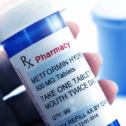
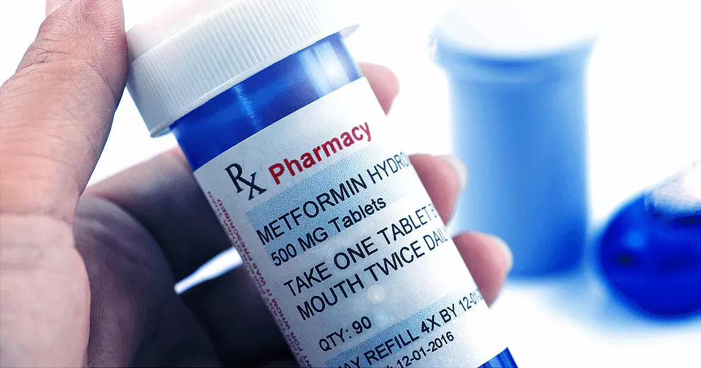
TRYNGOLZA is the first-ever FDA-approved treatment that significantly and substantially reduces triglyceride levels in adults with FCS and provides clinically meaningful reduction in AP events when used with an appropriate diet (≤20 grams of fat per day). TRYNGOLZA is self-administered via an auto-injector once monthly.

“Today’s FDA approval of TRYNGOLZA heralds the arrival of the first-ever FCS treatment in the U.S. – a transformational moment for patients and their families. For the first time, adults with FCS can now access a treatment that substantially reduces triglycerides and the risk of debilitating and potentially life-threatening acute pancreatitis,” said Brett P. Monia, Ph.D., chief executive officer, Ionis. “We are proud of our long-standing partnership with the FCS community and are grateful to the patients, families and investigators who participated in our clinical studies, enabling Ionis to make this new treatment a reality. The FDA approval of TRYNGOLZA is also a pivotal moment for Ionis, representing our evolution into a fully integrated commercial-stage biotechnology company – a goal we set out to achieve five years ago. With our rich pipeline of potentially life-changing medicines, we expect TRYNGOLZA to be the first in a steady cadence of innovative medicines we will deliver independently to people living with serious diseases.”

The FDA approval was based on positive data from the global, multicenter, randomized, placebo-controlled, double-blind Phase 3 Balance clinical trial in adult patients with genetically identified FCS and fasting triglyceride levels ≥880 mg/dL. In the Balance study, TRYNGOLZA 80 mg demonstrated a statistically significant placebo-adjusted mean reduction in triglyceride levels of 42.5% from baseline to six months (p=0.0084). Reductions from baseline to 12 months were further improved, with TRYNGOLZA achieving a placebo-adjusted 57% mean reduction in triglycerides. TRYNGOLZA also demonstrated a substantial, clinically meaningful reduction in AP events over 12 months; one patient (5%) experienced one episode of AP in the TRYNGOLZA group compared with seven patients (30%) who experienced 11 total episodes of AP in the placebo group.

TRYNGOLZA demonstrated a favorable safety profile. The most common adverse reactions (incidence >5% of TRYNGOLZA-treated patients and at a >3% higher frequency than placebo) were injection site reactions (19% and 9%, respectively), decreased platelet count (12% and 4%, respectively) and arthralgia (9% and 0%, respectively).

Results from the Phase 3 Balance study were previously published in The New England Journal of Medicine (NEJM).

“With no treatment options previously available, we were limited to relying only on extremely strict diet and lifestyle changes as the sole preventative treatment option,” said Alan Brown, M.D., FNLA, FACC, FAHA, clinical professor of medicine, Rosalind Franklin University of Medicine and Science; Balance trial investigator. “The FDA approval of TRYNGOLZA is an important moment for people living with FCS, their families and physicians who now, for the first time, have a treatment that significantly lowers triglycerides and decreases the risk of potentially life-threatening acute pancreatitis events, as an adjunct to a low-fat diet. I am excited to have a medicine I can prescribe to my patients that has been shown to change the course of their disease.”

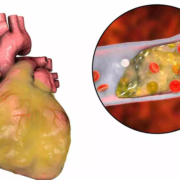
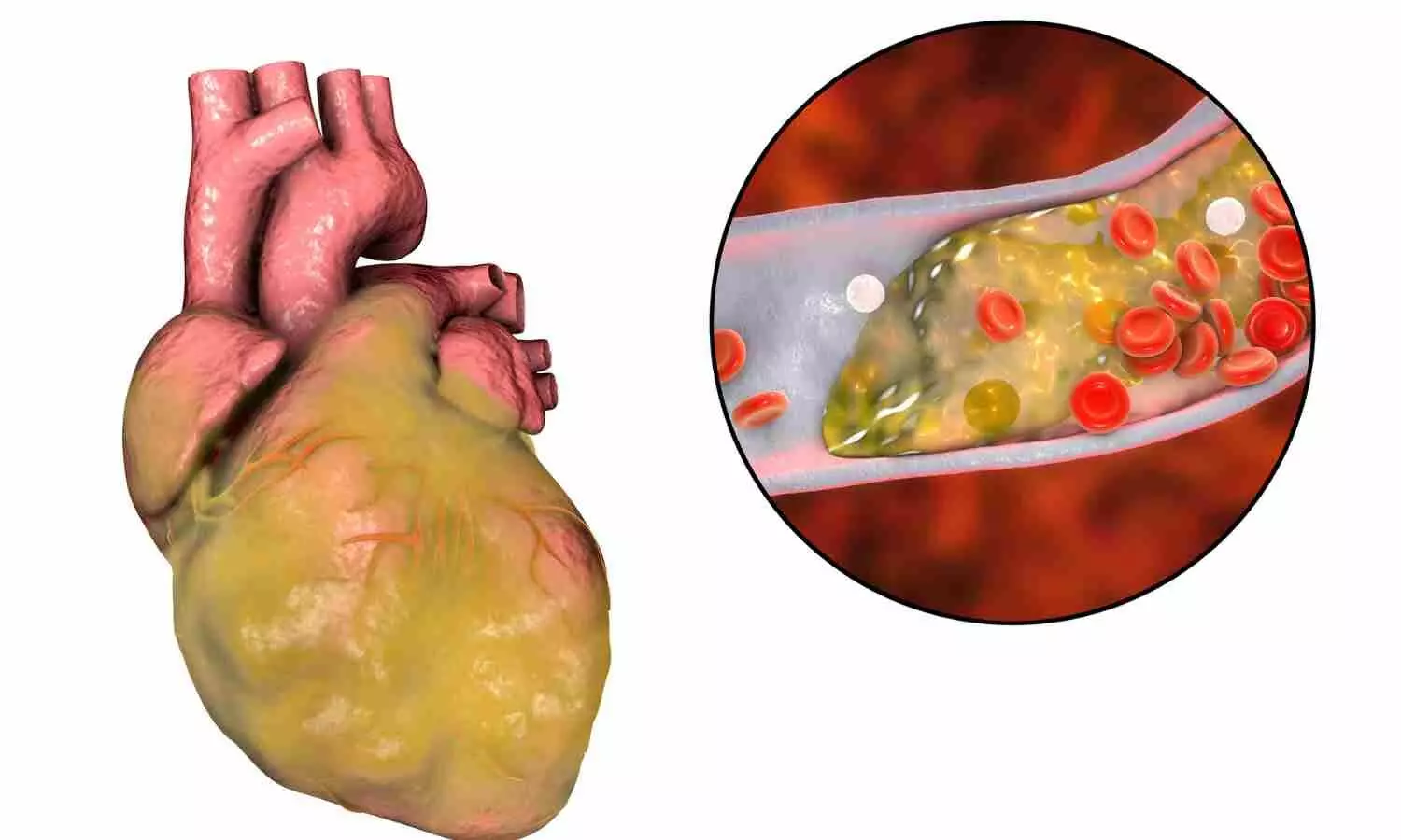
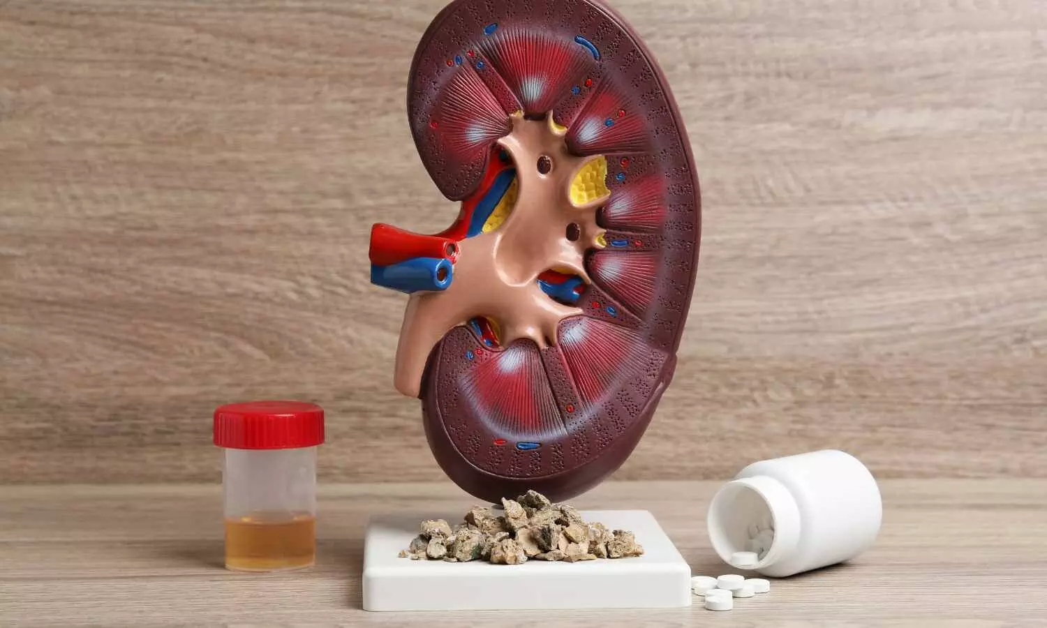
FCS is a rare, genetic, potentially life-threatening form of sHTG that prevents the body from breaking down fats and severely impairs the body’s ability to remove triglycerides from the bloodstream due to an impaired function of the enzyme lipoprotein lipase (LPL). While healthy levels for adults are below 150 mg/dL, people with FCS often have triglyceride levels of more than 880 mg/dL and often have a history of pancreatitis. Those living with FCS have a high risk of potentially fatal AP, which is a painful inflammation of the pancreas, and chronic health issues such as fatigue and severe, recurrent abdominal pain. People living with FCS can also experience psychological and financial stress, which can significantly impact their quality of life. In the U.S., FCS is estimated to impact up to approximately 3,000 people, the vast majority of whom remain undiagnosed.

“As a rare and difficult to diagnose disease, FCS has a profound impact on the lives of patients and families. Many people living with FCS have experienced severe pain their whole lives – sometimes so intense they require lengthy hospitalization stays – and struggle through life with daily fatigue, nausea, brain fog and stomach pain,” said Lindsey Sutton Bryan, co-founder and co-president, FCS Foundation. “Until now, our treatment options have been limited, relying on diet alone to try to manage triglyceride levels and keep acute pancreatitis attacks at bay. For the first time, adults with FCS have seen their hope for a treatment become a reality.”

TRYNGOLZA will be available in the U.S. before year end.

TRYNGOLZA was reviewed by the FDA under Priority Review and had previously been granted Fast Track designation for the treatment of FCS, Orphan Drug designation and Breakthrough Therapy designation. Olezarsen is undergoing review in the European Union and regulatory filings in other countries are planned. Olezarsen is currently being evaluated in three Phase 3 clinical trials – CORE, CORE2 and ESSENCE – for the treatment of sHTG. Olezarsen has not been reviewed or approved for the treatment of sHTG by regulatory authorities.

About TRYNGOLZA™ (olezarsen)

TRYNGOLZA™ (olezarsen) was approved by the U.S. Food and Drug Administration as an adjunct to diet to reduce triglycerides in adults with familial chylomicronemia syndrome (FCS). TRYNGOLZA is an RNA-targeted medicine designed to lower the body’s production of apoC-III, a protein produced in the liver that is a key regulator of triglyceride metabolism. It is the only treatment currently indicated in the U.S. for FCS, a potentially life-threatening disease

Powered by WPeMatico

First full characterization of kidney microbiome unlocks potential to prevent kidney stones, reveals study

Cleveland Clinic researchers have found definitive proof of a kidney microbiome that influences renal health and kidney stone formation, demonstrating that the urinary tract is not sterile and low levels of bacteria are normal.

The Nature Communications publication describes the rigorous multi-pronged approach a team led by Aaron Miller, PhD, and José Agudelo, MD, used to identify and characterize the small bacterial community by combining preclinical, human and dish studies.

They also identified certain bacteria within the microbiome that could promote or block kidney stone development and showed that antibiotic misuse (commonly associated with kidney stone development in a hospital setting) skewed the microbiome towards stone-promoting bacteria.

Dr. Miller hopes his team’s findings will help overturn the concept of a sterile urinary tract and lead to better prevention or treatment options for a condition that hasn’t seen major medical advances in over three decades.

“Urologic diseases like kidney stones impact 63% of the adult population and are getting worse,” he says. “The data consistently points towards bacteria. If we can’t get over the assumption of sterility, we can’t develop more effective treatments and preventative options.”

Normal bacteria levels in urine are very low, but they are rarely zero. Despite this, the urobiome – microbiome of different organs in the urinary tract including the bladder and kidneys – has been a debated topic since its discovery in human urine less than 15 years ago.

First author and urologist José Agudelo, MD, who has extensive experience in urinary stone disease, explains that previous indirect findings of a kidney microbiome gave compelling evidence but left room for doubt.

Bacterial communities need to meet three criteria to be considered a true microbiome: stability, consistency and reproducibility and being metabolically active. The team’s methods demonstrated each of these aspects for bacteria found in the urinary tract. Their research also showed that bacteria living in the urinary tract were not only there because of disease, since they found them in the urinary tract of people without evidence of urologic disease.

Other studies had shown that two species Drs. Miller and Agudelo had identified, E. coli and Lactobacillus crispatus, had been associated with the presence and absence of kidney stones, respectively. The researchers asked if the metabolic activity of their newly discovered microbial community played a role in kidney stone formation.

To see whether the kidney microbiome could influence stone formation, the researchers grew bacteria using a special chamber that mimics the movement of urine in our kidneys. They then added the “raw ingredients” of kidney stones, oxalate and calcium, to see what happened.

Several large, stone-like crystal structures formed in chambers growing E. coli. Chemical and X-ray analyses revealed these structures were indistinguishable from human kidney stones. No stones formed in the chambers growing Lactobacillus in this way. Growing the two bacteria together resulted in very small crystal structures that were structurally and chemically different from kidney stones, indicating that Lactobacillus somehow blocks E. coli’s ability to form kidney stones.

In preclinical models, the team also saw that antibiotic overuse shifted the balance of the kidney microbiome away from the healthy Lactobacillus towards the stone-forming E. coli. They believe their findings, taken together, may explain why individuals on long-term antibiotic courses are more prone to developing kidney stones.

Dr. Agudelo says his team’s findings suggest that different bacteria produce pro- and anti- kidney stone molecules, which he wants to use in new therapeutic and diagnostic techniques. He is already working to understand which bacterial metabolites influence stone formation, and how.

“If the kidney microbiome can influence kidney stones, it can likely influence other kidney diseases as well,” Dr. Miller adds. “We are already looking at microbial signatures for other kidney diseases and have even submitted a grant to investigate how certain genetic variants influence the renal microbiome and kidney disease risk in different ethnicities.

Reference:

Agudelo, J., Chen, X., Mukherjee, S.D. et al. Cefazolin shifts the kidney microbiota to promote a lithogenic environment. Nat Commun 15, 10509 (2024). https://doi.org/10.1038/s41467-024-54432-6.

Powered by WPeMatico

Tofacitinib Outperforms Azathioprine in Treating Alopecia Areata, New Study Finds

Pakistan: A recent double-blind, randomized controlled trial has provided new insights into the efficacy and safety of two alopecia areata (AA) treatments and their variants: tofacitinib and azathioprine.

The study, published in the Archives of Dermatological Research, revealed that tofacitinib efficacy was significantly higher than azathioprine in promoting hair regrowth in patients with alopecia areata and its variants. Both drugs, however, were well-tolerated, with no major differences in adverse effects between the two treatments.

Alopecia areata is an autoimmune condition characterized by sudden hair loss in small, round patches. Tofacitinib, an oral Janus kinase (JAK) inhibitor, has gained attention for its potential to treat autoimmune conditions, including AA. Azathioprine, on the other hand, is an established immunosuppressive medication commonly used to treat various autoimmune diseases. Despite their differing mechanisms, both drugs have been explored for use in treating AA, but there has been limited research comparing their relative effectiveness and safety.

To fill this knowledge gap, Majid Paracha, Department of Dermatology, Medical Teaching Institute-Lady Reading Hospital (MTI-LRH), Peshawar, Pakistan, and colleagues aimed to evaluate and compare the safety and efficacy of tofacitinib and azathioprine in patients with AA and variants.

For this purpose, the researchers conducted a double-blind randomized controlled trial (RCT) at the Department of Dermatology, Medical Teaching Institute-Lady Reading Hospital (MTI-LRH), Peshawar, Pakistan. The study included patients aged 12 years and older, diagnosed with alopecia areata (AA), alopecia totalis (AT), or alopecia universalis (AU), all of whom had at least 50% scalp hair loss for 6 months or more. Participants were randomly assigned to receive oral tofacitinib 5 mg twice daily (Group I) or oral azathioprine 2 mg/kg body weight once daily (Group II).

The primary endpoint was the Severity of Alopecia Tool (SALT) score, evaluated at baseline and again at a 6-month follow-up. Safety assessments were carried out consistently throughout the study.

The findings of the study were as follows:

  • 104 patients were randomly allocated into the tofacitinib group (n = 52) or the azathioprine group (n = 52).
  • The mean age of patients was 20.23 years in the tofacitinib group and 22.26 years in the azathioprine group.
  • The mean disease duration was 6.59 years in Group I and 7.98 years in Group II.
  • 38.5% of patients were adolescents, and 67.3% were male.
  • 50% had alopecia areata (AA), 35.5% had alopecia totalis (AT), and 14.5% had alopecia universalis (AU).
  • The mean baseline Severity of Alopecia Tool (SALT) score was 91.02 ± 10.21 in the tofacitinib group and 91.02 ± 10.63 in the azathioprine group.
  • At the 6-month follow-up, the SALT score improved to 14.1 ± 24.6 in the tofacitinib group and 63.9 ± 33.9 in the azathioprine group (difference of 11.5 points).
  • No major adverse effects were observed, and there was no significant difference in minor adverse effects between the two groups (4 adverse events in the tofacitinib group and 8 in the azathioprine group).

“The study suggests that while tofacitinib demonstrated superior effectiveness, both medications are safe options for managing AA. The findings highlight tofacitinib as a promising therapeutic alternative for patients, though further research is needed to confirm these results and evaluate long-term safety,” the researchers concluded.

Reference:

Paracha, M., Wasim, M., Noor, S.M. et al. Comparison of efficacy and safety of tofacitinib and azathioprine in patients with alopecia areata and variants: a double-blind, randomized controlled trial. Arch Dermatol Res 316, 458 (2024). https://doi.org/10.1007/s00403-024-03203-w

Powered by WPeMatico