Anabolic androgenic steroids use tied to impaired myocardial flow reserve persistent coronary microvascular dysfunction: JAMA

A new study published in the Journal of American Medical Association showed that young males who use or have previously used anabolic-androgenic steroids (AASs) are more likely to have a lower myocardial flow reserve when compared to those who have never used the medicines.

Although the exact mechanism of development is unknown, long-term use of anabolic androgenic steroids is linked to a significant risk of left ventricular hypertrophy, heart failure with impaired systolic function, and early abrupt death. In the men who use AAS, early and persistently decreased myocardial microcirculation may be clinically significant, a possible underlying cause of frequent and early heart illness, and a future target for treatments.

This study used cardiac rubidium 82 (82Rb) positron emission tomography/computed tomography (PET/CT) to measure myocardial flow reserve (MFR) in males who now and previously used AAS in comparison to controls who had never used AAS. This was done to evaluate coronary microcirculation.

Men who participated in recreational strength training and did not have a history of cardiovascular disease were included in this cross-sectional study based on their history of using AAS. From November 24, 2021, until August 16, 2023, the study was carried out. The MFR between the research groups was the main result of this investigation, while the coronary calcium score was the secondary result. By definition, a threshold of MFR less than 2 was used to identify impaired myocardial microcirculation, while a cutoff of MFR less than 2.5 was used to identify sub-clinically impaired microcirculation.

There were 90 men in all and 35.1 years was the mean (SD) age. A 1.5-year geometric mean was the amount of time that had passed from AAS discontinuation. Before enrolling, 18 males (58.1%) who had previously used AAS stopped using it more than a year earlier. Impaired MFR was seen in individuals with current and past usage, but no impairment was detected among the controls. When compared to the controls, men who now or previously used AAS had a greater sub-clinically impaired MFR.

Every doubling of the total weekly duration of AAS usage (log2) was independently linked to a factor 2 increase in the chance of impaired MFR less than 2.5 in a multivariable logistic regression model among men who had previously used AAS. Overall, both present and previous AAS users in this cross-sectional research showed reduced MFR which indicated a link between AAS use and lower MFR.

Reference:

Bulut, Y., Rasmussen, J. J., Brandt-Jacobsen, N., Frystyk, J., Thevis, M., Schou, M., Gustafsson, F., Hasbak, P., & Kistorp, C. (2024). Coronary Microvascular Dysfunction Years After Cessation of Anabolic Androgenic Steroid Use. In JAMA Network Open (Vol. 7, Issue 12, p. e2451013). American Medical Association (AMA). https://doi.org/10.1001/jamanetworkopen.2024.51013

Powered by WPeMatico

Digital scanning and conventional impressions show comparable accuracy for most fixed prosthodontics, suggests study

Digital scanning and conventional impressions show comparable accuracy for most fixed prosthodontics, suggests a study published in the Journal of Prosthetic Dentistry

Several systematic reviews have compared the accuracy of conventional impression-making and digital recording techniques, with sometimes different results. A systematic overview of these studies is lacking. This overview of systematic reviews examined the accuracy of digital scans and conventional impressions for tooth- or implant-supported fixed restorations in partially and completely edentulous adult patients. Four databases (Medline via PubMed, Scopus, Web of Science, and Google Scholar) were searched for systematic reviews according to preset eligibility criteria. Two calibrated evaluators screened and assessed the overall confidence of the included reviews using the A Measurement Tool to Assess Systematic Reviews (AMSTAR) 2 tool. Several review characteristics were recorded, including accuracy in terms of trueness and/or precision. The Jadad et al1 decision algorithm was used to select the best evidence, and a citation matrix was used to show overlaps in the studies. Results: From the 307 retrieved studies, 28 systematic reviews were included in this overview. Among these, 12 performed meta-analyses, and 18 comprised both in vitro and in vivo primary studies. Generally, digital scanning and conventional impression techniques for crowns and fixed partial dentures, for implant-supported fixed restorations, and for both tooth- and implant-supported restorations showed no statistically significant differences in terms of accuracy (trueness and precision) and marginal and internal adaptation (P>.05). However, conventional impressions outperformed digital scans for complete-arch fixed dental prostheses in terms of accuracy. Regarding methodological quality, most systematic reviews (67.9%) received critically low overall confidence based on AMSTAR 2. No significant differences were reported in terms of marginal and internal fit between prosthetic restorations constructed after digital scanning and conventional impression-making. The opportunity exists to enhance the methodological quality of systematic reviews about the accuracy of dental recordings.

Reference:

Digital scans versus conventional impressions in fixed prosthodontics: An overview of systematic reviews. Kaitatzidou, Aikaterini et al. Journal of Prosthetic Dentistry, Volume 0, Issue 0

Keywords:

Digital, scanning, conventional, impressions, show, comparable, accuracy, most, fixed, prosthodontics, suggests, study , Journal of Prosthetic Dentistry, Kaitatzidou, Aikaterini

Powered by WPeMatico

New scan method unveils lung function secrets, finds study

A new method of scanning lungs is able to show the effects of treatment on lung function in real time and enable experts to see the functioning of transplanted lungs.

This could enable medics to identify sooner any decline in lung function.

The scan method has enabled the team, led by researchers at Newcastle University, UK, to see how air moves in and out of the lungs as people take a breath in patients with asthma, chronic obstructive pulmonary disease (COPD), and patients who have received a lung transplant.

Publishing two complementary papers in Radiology and JHLT Open, the team explain how they use a special gas, called perfluoropropane, that can be seen on an MRI scanner. The gas can be safely breathed in and out by patients, and then scans taken to look at where in the lungs the gas has reached.

The project lead, Professor Pete Thelwall is Professor of Magnetic Resonance Physics and Director of the Centre for In Vivo Imaging at Newcastle University. He said; “Our scans show where there is patchy ventilation in patients with lung disease, and show us which parts of the lung improve with treatment. For example, when we scan a patient as they use their asthma medication, we can see how much of their lungs and which parts of their lung are better able to move air in and out with each breath.”

Using the new scanning method, the team are able to reveal the parts of the lung that air doesn’t reach properly during breathing. By measuring how much of the lung is well-ventilated and how much is poorly ventilated, experts can make an assessment of the effects of a patient’s respiratory disease, and they can locate and visualise the lung regions with ventilation defects.

Demonstrating that the scans work in patients with asthma or COPD, the team comprising experts from across Universities and NHS Trusts in Newcastle and Sheffield publish the first paper in Radiology.

The new scanning technique allows the team to quantify the degree of improvement in ventilation when patients have a treatment, in this case a widely used inhaler, the bronchodilator, salbutamol. This shows that the imaging methods could be valuable in clinical trials of new treatments of lung disease.

Use in lung transplants

A further study, published in JHLT Open, examined patients who had previously received a lung transplant for very severe lung disease at the Newcastle upon Tyne Hospitals NHS Foundation Trust. It demonstrates how the team further developed the imaging method to provide lung function measurements which could be used to better support lung transplant recipients in the future. The sensitivity of the measurement means medics can spot early changes in lung function allowing them to identify lung problems earlier and so provide better care for patients.

In research studies, the team scanned transplant recipients’ lungs over multiple breaths in and out, collecting MRI pictures that show how the air containing the gas reached different areas of the lung. The team scanned those who either had normal lung function or who were experiencing chronic rejection after lung transplant, which is a common issue in lung transplant recipients as their immune system attacks the donor lungs. In those with chronic rejection, the scans showed poorer movement of air to the edges of the lungs, most likely due to damage in the very small breathing tubes (airways) in the lung, a feature typical of chronic rejection also known as chronic lung allograft dysfunction.

Professor Andrew Fisher, Professor of Respiratory Transplant Medicine at Newcastle Hospitals NHS Foundation Trust and Newcastle University, UK, co-author of the study said; “We hope this new type of scan might allow us to see changes in the transplant lungs earlier and before signs of damage are present in the usual blowing tests. This would allow any treatment to be started earlier and help protect the transplanted lungs from further damage.”

The team say there is potential for this scan method to be used in the clinical management of lung transplant recipients and other lung diseases in the future, bringing a sensitive measurement that may spot early changes in lung function that enable better management of these conditions.

This work on lung imaging has been funded by the Medical Research Council and by The Rosetrees Trust.

References: Assessing Lung Ventilation and Bronchodilator Response in Asthma and Chronic Obstructive Pulmonary Disease with Fluorine 19 MRI. Pippard BJ, Neal MA, Holland CW, Maunder AM, Forrest I, Lawson RA, Fisher HF, Matthews JNS, Wild JM, Simpson AJ, Thelwall, PE. Radiology. Doi: https://doi.org/10.1148/radiol.240949 (n.b. live date 24th Dec)

Powered by WPeMatico
Gestational exposure to NSAIDs associated with childhood CKD, finds JAMA study

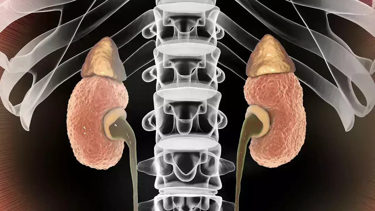
Gestational exposure to NSAIDs associated with childhood chronic kidney disease, finds JAMA study.

Gestational exposure to nonsteroidal anti-inflammatory drugs (NSAIDs) may increase the risk of adverse fetal kidney outcomes. However, details regarding timing, specific NSAIDs, and long-term childhood kidney outcomes are limited. A study was done to evaluate the association between gestational exposure to NSAIDs and the risk of chronic kidney disease (CKD) in childhood. This national cohort study assessed 1 025 255 children born alive in Taiwan from January 1, 2007, to December 31, 2017, with follow-up until December 31, 2021. Children without valid maternal-child linkage and with incomplete birth information were excluded. Data analysis was performed from November 30, 2023, to April 30, 2024. The main outcome was childhood CKD, including congenital anomalies of the kidney and urinary tract and other kidney diseases. Cox proportional hazards regression models with stabilized inverse probability of treatment weighting (weighted hazard ratio [wHR]) and a robust sandwich estimator were used to estimate the relative risk of NSAID exposure in pregnancy, adjusted for newborn characteristics. Results This study included 163 516 singleton-born children (24.0%) whose mothers (mean [SD] age at birth of child, 31.25 [4.92] years) used at least 1 dispensing of an NSAID during pregnancy. Gestational NSAID exposure was significantly associated with a higher risk of childhood CKD (wHR, 1.10; 95% CI, 1.05-1.15). No association was observed between NSAID use and fetal nephrotoxicity in sibling comparisons. Elevated risks were revealed for exposure during the second trimester (wHR, 1.19; 95% CI, 1.11-1.28) and the third trimester (wHR, 1.12; 95% CI, 1.03-1.22) in singleton-born children. Specific NSAID exposures associated with higher CKD risk included indomethacin (wHR, 1.69; 95% CI, 1.10-2.60) and ketorolac (wHR, 1.28; 95% CI, 1.01-1.62) in the first trimester, diclofenac (wHR, 1.27; 95% CI, 1.13-1.42) and mefenamic acid (wHR, 1.29; 95% CI, 1.15-1.46) in the second trimester, and ibuprofen (wHR, 1.34; 95% CI, 1.07-1.68) in the third trimester. In this study, gestational exposure to NSAIDs was not associated with a substantial increase in the risk of childhood CKD when comparing between siblings. However, the findings underscore the need for caution when prescribing NSAIDs during pregnancy, particularly indomethacin and ketorolac in the first trimester, mefenamic acid and diclofenac in the second trimester, and ibuprofen in the third trimester, to ensure the safety of the offspring’s kidneys.

Reference:

Tain Y, Li L, Kuo H, Chen C, Hsu C. Gestational Exposure to Nonsteroidal Anti-Inflammatory Drugs and Risk of Chronic Kidney Disease in Childhood. JAMA Pediatr. Published online December 23, 2024. doi:10.1001/jamapediatrics.2024.4409

Keywords:

Gestational, exposure, NSAIDs, associated, childhood, chronic kidney disease, finds, JAMA, study, Tain Y, Li L, Kuo H, Chen C, Hsu C

Powered by WPeMatico

Micro drops non-inferior to Standard drops for Retinopathy of Prematurity: JAMA

A recent study published in the
journal JAMA Ophthalmology found that substituting micro drops with standard
drops was non-inferior as it reduced the systemic effects in preterm infants
who have retinopathy of prematurity.

Retinopathy of prematurity (ROP)
is one of the most significant risk factors in premature infants who are born
prematurely as it can affect vision. Eyedrops that contain either adrenergic
agonists, muscarinic antagonists, or their combination are used for adequate
pupil dilation. However, they have the risk of causing systemic cardiorespiratory
and gastrointestinal adverse events. Hence, researchers conducted a study to
examine the noninferiority of micro drops of a combined mixture of 1.67%
phenylephrine and 0.33% tropicamide compared to the standard eye drops.

A randomized,
double-masked, noninferiority, crossover design trial was conducted on
premature infants undergoing ROP screening at a tertiary center in Northern
Greece from September 2021 to January 2023. Infants with gestational age below
32 weeks and/or birthweight under 1501 g or infants beyond these thresholds
referred by an attending neonatologist due to comorbidities were considered
eligible for the study. Random allocation was done to prescribe micro drops or
standard drops of the diluted mixture with a 1-week washout period.

The horizontal pupil diameter at
45, 90, and 120 minutes was measured using a customized ruler in 0.5-mm
increments. A predefined noninferiority margin of −0.4 mm was set. Parameters
like heart rate, oxygen saturation, blood pressure measurements at 45, 90, and
120 minutes, 24-hour hypertensive episodes, and 48-hour systemic adverse events
were assessed. Hydrophilic liquid chromatography-tandem mass spectrometry measured
Phenylephrine concentration in peripheral blood within 3 hours post-instillation.

Findings:

  • About 83 infants were randomized with a mean
    [SD] gestational age of 29.7 [2.0] weeks and a mean [SD] birth weight of 1277
    [374] g).
  • Micro drops were superior for mydriatic efficacy
    at 45 minutes and non-inferior at 90 and 120 minutes.
  • After standard drops, lower oxygen saturation
    levels at 45 and 90 minutes and a higher percentage of 24-hour hypertensive
    episodes were observed.
  • A 1-compartment model with first-order
    absorption best described the pharmacokinetic data.

Thus, the researchers concluded
that micro drops of combined 1.67% phenylephrine and 0.33% tropicamide in a
mixture were proven superior regarding mydriatic efficacy at 45 minutes and
non-inferior at 90 and 120 minutes compared with standard drops of the same
regimen administered in infants undergoing ROP screening.

Micro drops were found to be
safe, non-inferior to standard drops, and reduce system absorption. Due to
their diagnostic efficacy, they can be preferable for ophthalmologists and
neonatologists.

Further reading: Seliniotaki AK, Lithoxopoulou
M, Virgiliou C, et al. Efficacy and Safety of Mydriatic Microdrops for
Retinopathy of Prematurity Screening: The MyMiROPS Randomized Clinical
Trial. JAMA Ophthalmol. Published online December 26, 2024.
doi:10.1001/jamaophthalmol.2024.5462

Powered by WPeMatico

Arsenic exposure through drinking water increases kidney cancer risk, finds study

A new study published in the journal of Environmental Pollution found an elevated risk of kidney cancer may be linked to exposure to low levels of drinking water arsenic. Kidney cancer is the 7th most frequent cancer in the United States, with an age-adjusted incidence rate of 17.2 per 100,000 from 2017 to 2021.

Male gender, family history, African American race or ethnicity, obesity, smoking, alcohol use, diabetes mellitus, high blood pressure, severe kidney disease, and chemical exposures like trichloroethylene are risk factors for kidney cancer. One naturally occurring element that is commonly found in groundwater is arsenic. About 2.1 million individuals in over 25 states in the US are exposed to high amounts of arsenic in their drinking water, with levels above the current 10 ppb legal limit. 

The increase of kidney cancer is thought to be caused by arsenic which is a recognized human carcinogen, especially in places where groundwater is high in arsenic. However, nothing is known about the link between kidney cancer and low levels of arsenic in drinking water. In this ecological analysis, this study used data from 240 Texas counties to evaluate the relationship between kidney cancer occurrences and county-level drinking water arsenic levels.

The analysis covered 101,776,294 person-years from 2016 to 2020 and 28,896 cancer diagnoses among people aged ≥20. In addition to geographical determinants spatial Poisson regression models calculated the risk ratio (RR) for incident kidney cancer based on drinking water arsenic levels by controlling for demographic, socioeconomic, and other risk variables. The data from water testing for both private wells and public water systems were used to determine population-weighted drinking water arsenic levels, which were then corrected for the populations supplied by each source.

This study found a dose-response connection (p-trend <0.001) between the medium (1 to 5 ppb) and high (>5 ppb) group counties and the low arsenic level counties (<1 ppb), with the incidence of cancer being 6% and 22% higher, respectively, after controlling for geographical variables and covariates. Furthermore, when drinking water arsenic level was viewed as a continuous variable, the incidence rose by 4% for every doubling of the level.

Overall, in line with other studies showing a link between drinking water exposure and skin, bladder, and lung cancers, this study indicates that even low levels of arsenic exposure may be linked to an increased risk of kidney cancer.

Source:

Hasan, N. T., Han, D., Xu, X., Sansom, G., & Roh, T. (2024). Relationship between low-level arsenic exposure in drinking water and kidney cancer risk in Texas. In Environmental Pollution (Vol. 363, p. 125097). Elsevier BV. https://doi.org/10.1016/j.envpol.2024.125097

Powered by WPeMatico

Signals of inflammation during pregnancy linked to aging and memory changes 50 years later, suggests study

An estimated 13.8 million people in the U.S. will have Alzheimer’s disease (AD) by 2050, two thirds of whom are projected to be women. The brain circuitry underlying memory is widely known to differ based on biological sex, but sex-based drivers of aging and AD are still unclear. A new study by investigators from Mass General Brigham analyzed data from participants who have been followed for over 50 years, starting before their births. Researchers found that maternal immune activity during a critical period of sex-dependent brain development in pregnancy affected the offspring’s long-term memory circuitry and function in childhood and midlife, with different patterns for males and females. Findings are published in Molecular Psychiatry.

“Brain aging is also about brain development, and understanding sex differences in brain development is critical to understanding sex differences in the aging brain,” said corresponding author Jill M. Goldstein, PhD, MPH, founder and executive director of the Innovation Center on Sex Differences in Medicine of Massachusetts General Hospital, a founding member of the Mass General Brigham healthcare system, and Professor of Psychiatry and Medicine at Harvard Medical School. “This paper is a first step toward looking at the fetal origins of Alzheimer’s disease, which, like many chronic diseases, develops across the lifespan and is influenced by early development in a way we might not ordinarily consider.”

This study was based on findings from a cohort established over 60 years ago, which includes the adult offspring of nearly 18,000 pregnancies between 1959 and 1966, followed as part of the New England Family Study (NEFS). The present investigation comprised 204 individuals born during the study who were or were not exposed to an adverse in utero immune environment (i.e., elevated levels of immune markers, such as cytokines IL-6 and TNF-a) and followed into midlife 50 years later. The team used functional brain imaging to examine the impact of this early exposure on brain regions in the memory circuitry that are dense in these cytokine receptors and sex hormone receptors and exhibit sex differences in development and functioning beginning in fetal development.

The researchers found that elevated levels of IL-6 and TNF-a in mothers during pregnancy were linked with sex differences in adverse brain activity in memory circuitry in the offspring later in life, particularly in postmenopausal women. These women also expressed higher markers of a proinflammatory state in midlife. Further, researchers saw evidence of the impact of these immune markers even earlier, in children’s cognitive performance at age seven, underscoring the link between exposures during pregnancy and brain health later in life. Their findings suggest that elevated maternal prenatal immune activity may contribute to the development of heightened immune and stress sensitivity in the offspring, which, the researchers hypothesized, may predispose them to memory disorders, like AD, later in life, in sex-dependent ways.

As participants age, the researchers are continuing to follow them to examine amyloid levels and other measures of AD-related pathology to further explore the association between prenatal immunity and AD. Ongoing goals include understanding the mechanisms by which maternal immune activity influences fetal brain development, identifying biomarkers for future memory dysfunction in early midlife, and understanding how other periods of development, such as puberty, influence the aging brain.

“While prenatal immune activity may affect brain development in the offspring, that does not mean pregnancy is deterministic,” Goldstein said. “Of course, subsequent environmental exposures are key, just as in utero environment is important. Fortunately, the brain is exceptionally adaptable, and we want to understand the cognitive, behavioral, and sex-dependent factors associated with both risk and resilience in order to intervene early and maintain intact memory function as we age.”

Reference:

Goldstein, J.M., Konishi, K., Aroner, S. et al. Prenatal immune origins of brain aging differ by sex. Mol Psychiatry (2024). https://doi.org/10.1038/s41380-024-02798-w

Powered by WPeMatico

Er-YAG Laser Therapy Shows No Significant Benefit for Treating Female Stress Urinary Incontinence: Study

Canada: A recent double-blinded randomized controlled trial published in the American Journal of Obstetrics and Gynecology has explored the effectiveness of Erbium-YAG (Er-YAG) laser vaginal treatment in managing female stress urinary incontinence (SUI).

The researchers found that Er-YAG laser vaginal treatment was not more effective than the sham procedure in curing or improving female stress urinary incontinence after six months. Despite initial hopes that the laser treatment would offer a non-invasive, quick, and effective alternative for managing SUI, the results indicate that it does not provide the expected relief for most patients.

Stress urinary incontinence is a common condition that affects millions of women worldwide, often triggered by physical exertion, sneezing, coughing, or other forms of pressure on the bladder. SUI affects around 40% of adult women and is commonly treated with surgical interventions. While minimally invasive laser vaginal therapy has become increasingly available as a potential treatment, its effectiveness remains uncertain. With this in mind, Patricia LEE, Division of Urogynecology, Dept of Obstetrics and Gynecology, Sunnybrook Health Sciences Centre, University of Toronto, Toronto, Ontario, Canada, and colleagues aimed to evaluate the efficacy of Erbium-Yttrium Aluminum Garnet (Er-YAG) laser therapy as a treatment for stress urinary incontinence.

For this purpose, the researchers conducted a randomized, sham-controlled, double-blinded trial with follow-ups at 6 weeks and 6 months in a single tertiary hospital setting, involving 144 adult women with symptomatic stress urinary incontinence. Participants received two vaginal laser treatments spaced 6 weeks apart and were randomly assigned to either the laser or sham group.

The primary outcome was determined by patients’ self-reported absence of urinary incontinence 6 months after treatment, assessed through question 3 of the International Consultation on Incontinence Questionnaire-Short Form. Secondary outcomes included objective urine loss measurements using 24-hour pad tests, responses to three incontinence symptom questionnaires (Urogenital Distress Inventory-6, Incontinence Impact Questionnaire-7, and International Consultation on Incontinence Questionnaire-Short Form), a quality of life assessment using the King’s Health Questionnaire, and a sexual function evaluation via the Pelvic Organ Prolapse/Urinary Incontinence Sexual Questionnaire.

The following were the key findings of the study:

  • Out of 263 individuals approached, 76 were randomized to the laser treatment group and 68 to the sham treatment group.
  • There was no difference in the reported cure rate between the two groups at the 6-month follow-up, which was the primary outcome:
    • Laser group: 0.013 (1/76).
    • Sham group: 0/68.
  • There was a statistically significant difference at 6 weeks in 1 of the three incontinence questionnaires and 3 of the nine domains of the quality-of-life questionnaire.
  • There were no other significant differences in either subjective or objective measures between the two groups at 6 weeks or 6 months post-treatment.

“After six months, the findings showed that the Er-YAG laser vaginal treatment was not more effective than the sham procedure in curing or significantly improving symptoms of stress urinary incontinence,” the researchers concluded.

Reference:

LEE, P., PERRUZZA, D., EDELL, H., JARVI, S., KIM, K., SULTANA, R., ALAVI, N., KISS, A., CAO, X., GAGNON, L., BODLEY, J., CARR, L., HERSCHORN, S., & KUNG, R. (2024). Double-blinded randomized controlled trial of Erbium-YAG Laser to treat Female Stress Urinary Incontinence. American Journal of Obstetrics and Gynecology. https://doi.org/10.1016/j.ajog.2024.11.021

Powered by WPeMatico

Pancreatic endotherapy improves quality of life in patients with chronic pancreatitis: Study

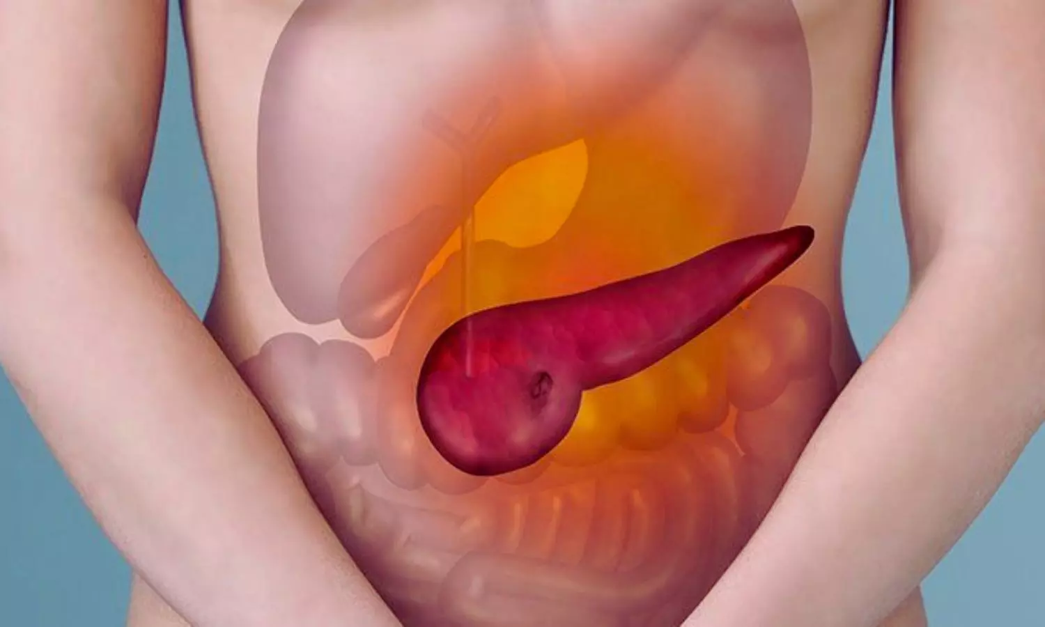
A new study published in the journal of Gastrointestinal Endoscopy showed that for some individuals with chronic pancreatitis (CP), pancreatic endotherapy (PET) can improve quality of life and reduce discomfort. The primary symptom of chronic pancreatitis, pain, is still challenging to successfully manage and can considerably lower a patient’s quality of life. The mechanism of pain is probably influenced by a number of factors, such as inflammation, ductal hypertension, or neuropathic pain resulting from different levels of nervous system sensitization.

The patients with CP-related structural problems, such as pancreatic duct (PD) stones, strictures, stones, or pancreatic fluid collections, such pseudocysts, may benefit from pancreatic endotherapy. Pancreatic duct stones, strictures, and pancreatic fluid collections are among the structural problems associated with chronic pancreatitis that can be managed with pancreatic endotherapy. However, the majority of research on endotherapy has been on technical success outcomes such stricture resolution or stone removal. Thus, this review is to describe in detail how PET affects the quality of life of CP patients.

A validated CP-specific quality of life instrument (PANQOLI) was used in this single-center prospective trial to evaluate quality of life prior to the start of endotherapy. At 1, 3, 6, and 12 months of follow-up, quality of life, pain levels, and opiate use were also assessed. Celiac plexus block (EUS-CPB), EUS-guided pseudocyst drainage, PD stone lithotripsy, and therapeutic PD stent insertion were among the PET modalities. At each follow-up point, quality of life changes before and after PET were compared using a repeated measures mixed effects model.

In all, 120 CP subjects were involved in this investigation. The most common PET modalities were EUS-CPB (32.5%), PD stone lithotripsy (16.7%), and therapeutic PD stenting (49.2%). At 1, 3, 6, and 12 months after completing the PET, there were notable improvements in quality of life scores, with the biggest improvement occurring at 6 months.

At the 1- and 6-month follow-ups, there were notable reductions in pain levels; but, at the 12-month follow-up, there were none. Opiate use decreased at every follow-up point. Despite pancreatic endotherapy has been linked to improvements in quality of life for up to a year after treatment, pain relief was only observed 6 months after treatment which indicates the difficulty of managing pain in this challenging patient population. 

Source:

Han, S., Rueb, N., Nikahd, M., Pessorrusso, F., Wani, S., Edmundowicz, S. A., Duloy, A., Hammad, H. T., & Shah, R. J. (2024). The Impact of Pancreatic Endotherapy on Quality of Life in Chronic Pancreatitis. In Gastrointestinal Endoscopy. Elsevier BV. https://doi.org/10.1016/j.gie.2024.12.002

Powered by WPeMatico

Hypovolemic phlebotomy before major liver surgery cuts need of transfusions by half: Study

Removing 10 per cent of a patient’s blood before major liver surgery and giving it back afterwards reduced transfusions by half, according to a large clinical trial published in The Lancet Gastroenterology & Hepatology. Known as hypovolemic phlebotomy, this practice could save one in every 11 patients having this surgery from needing a transfusion.

“Blood loss is a major concern in liver surgery. Taking out half a litre of blood right before major liver surgery is the best thing we’ve found so far for reducing blood loss and transfusions,” said co-lead author Dr. Guillaume Martel, a surgeon and scientist who holds the Arnie Vered Family Chair in Hepato-Pancreato-Biliary Research at The Ottawa Hospital and University of Ottawa. “It works by lowering the blood pressure in the liver. It’s safe, simple, inexpensive, and should be considered for any liver surgery with a high risk of bleeding.”

Between a quarter to a third of people having major liver surgery will need a transfusion. Cancer is the most common reason for these surgeries and having a transfusion during or shortly after surgery is possibly associated with a higher risk of the cancer coming back.

“Now that we’ve proven removing blood before liver surgery reduces transfusions, we’re spreading the word and teaching our colleagues how to do it,” said co-lead author Dr. François Martin Carrier, an anesthesiologist and critical care medicine specialist at le Centre Hospitalier de l’Université de Montréal who holds the Héma-Québec – Bayer Chair in Transfusion Medicine at Université de Montréal. “Providers find it simple after they’ve done it once, and the impact on surgery is dramatic. It’s now standard of care in the four hospitals that were part of the trial, and other hospitals around the world should start to adopt it after learning of our results.”

Practice-changing trial largest of its kind

In the largest trial of its kind, 446 people having major liver surgery were recruited at four Canadian hospitals between 2018 and 2023 (The Ottawa Hospital, le Centre Hospitalier de l’Université de Montréal, le Centre Hospitalier Universitaire de Sherbrooke, and Vancouver General Hospital).

Once under anesthetic, patients were randomly assigned to receive either hypovolemic phlebotomy or usual care. Only the anesthesiologist knew which patients were in which group.

For patients in the hypovolemic phlebotomy group, the anesthesiologist removed the equivalent of one blood donation (about 450 mL) into a blood bag before surgery. If the patient needed blood during surgery, their blood was used first. Otherwise, it was re-infused before they woke up.

According to hospital blood bank data and patient medical records, 7.6 per cent of those who received hypovolemic phlebotomy (17 of 223) had blood transfusions in the 30 days after surgery compared to 16.1 per cent (36 of 223) of those who received usual care. Hypovolemic phlebotomy caused no more complications than usual care.

Surgeons also said hypovolemic phlebotomy made the surgery easier to do, because there was less blood obscuring the places they needed to cut. Median estimated blood loss was 670 mL with hypovolemic phlebotomy compared to 800 mL with usual care.

“I’m so glad I was picked, and I’m glad it will help other people,” says participant

Rowan Ladd was 44 when she had colon cancer surgery on Christmas Eve 2020. Two years later, the cancer had spread to her liver and she needed another surgery, this time on her liver.

“I signed up for every study I was offered. If I’m going into liver surgery, then why not? I’m a big proponent of research,” says the Ottawa mother of two. “I was interested in this study in particular because they had great results in the pilot trial. You’re told before surgery that the liver is so full of blood vessels that there are risks of major bleeding. I thought it was great that researchers were trying things to reduce those risks.”

During her liver surgery in October 2022, Rowan was randomly selected to have hypovolemic phlebotomy. She found out which group she was in after the surgery. She did not need a blood transfusion. After four days in hospital, she was back home. Two years later, she remains cancer-free.

“I looked at this surgery like it saved my life. I stopped working, I relaxed, I took care of myself. I was unlucky to get cancer, but it woke me up. Now I live life and I really enjoy it where before I was just existing,” she says. “Being part of this trial was a really positive experience, and the team was wonderful. I’m so glad I was picked, and I’m glad it will help other people.”

Saving blood for those who need it most

One blood transfusion in Canada costs at least $500 CAD, mainly in human resources. The blood bags and tubes used for hypovolemic phlebotomy cost less than $30 CAD.

“Blood transfusions can save lives, but if you don’t need one to save your life then it’s better to avoid it. Blood is a precious and limited resource that we need to preserve as much as possible for those who need it most,” said senior author Dr. Dean Fergusson, Deputy Scientific Director, Clinical Research and senior scientist at The Ottawa Hospital and professor at the University of Ottawa.

Dr. Martel’s team previously tested the safety and feasibility of hypovolemic phlebotomy in major liver surgery in a phase 1 trial at The Ottawa Hospital. The procedure is now being tested in liver transplantation, and there may be future interest in testing it for other surgeries with significant blood loss.

Powered by WPeMatico