PGIMER Invites Applications for PhD Program 2024, Know All admission Details Here

Chandigarh- The Postgraduate Institute of Medical Education and Research (PGIMER) is inviting online applications from all the candidates for admission into the PhD Programme for the session 2024. On this, PGMIER has released a prospectus detailing the exam pattern, procedures for applying, fees, etc regarding admission into the PhD Programme 2024.

As per the schedule, the online application registration for admission to PhD Programme 2024 has already begun and the last date for filling the form and payment of online application is 22 November 2024. After the completion of the registration process, the online examination Computer Based Test (CBT) is scheduled to take place on 05 December 2024 and the result to be declared on 16 December 2024. Following this, the counselling is tentatively scheduled to take place on 27 November 2024.

Meanwhile, other details regarding the admission into the PhD Programme 2024 as per the prospectus are mentioned below-

PROCEDURES FOR APPLYING

Fill in the application form online in accordance with the instructions. Please ensure that no column is left blank. Incomplete applications will not be considered and no correspondence will be entertained. Below are some of the important points while filling out the application form-

1 A list of the faculty members who are willing to take PhD students and how many seats are available under them will be displayed on the PGI official website. The faculty member should endorse one or two extra candidates (prospective), than the number of seats advertised under him/her.

2 Applicants for PhD courses who already have fellowship, should choose a department and the faculty member (under whose guidance he/she desires to undergo a Ph.D. program) and indicate the same on the application form. Candidates can apply only in one department.

3 The guide will have the option to accept or not to accept the candidate, who opts to do Ph.D. under that particular faculty member. The candidate may opt. only one faculty member.

4 The candidates carrying their own fellowship from various funding agencies and the Inservice medical faculty of PGI will be exempted from the entrance tests and will appear for counselling directly. However, all the candidates are required to fill out the online application form. Please take a printout of the duly filled Online Application form by logging in with the login ID and password. Affix the same passport-size photograph (which was uploaded in the online form) on it.

INSTRUCTION FOR FILLING THE ONLINE APPLICATION FORM

1 Candidate should fill in the Online Application with utmost care step by step. Candidate should fill in the Online Application form correctly. Incorrectly filled forms may lead to rejection.

2 A candidate seeking admission to the Entrance Examination is required to submit his/her application in the prescribed format.

3 The cost of the Application Form includes the fee for the entrance examination which is nonrefundable and no correspondence in this regard will be entertained.

4 ONLINE REGISTRATION-

i After selecting the online registration, fill in the mandatory details asked for and deposit the prescribed fee through debit/credit card/Net Banking. After submitting fees filled the required information step by step. Follow the instructions carefully.

ii It will be the responsibility of the candidate to ensure that correct details are filled in the Registration Slip. The Institute will not be responsible for any incorrect information/cancellation of candidature/loss or lack of communication etc. due to wrong filled online Application form.

iii No candidate should register more than one application

iv All applicants are required to ensure that Photo/Signature is uploaded according to the instructions. Failure to do so may result in rejection of applications.

v Duplicate applications from any applicant will result in the cancellation of all such applications. No intimation regarding such summary rejections will be provided.

DOCUMENTS

The candidates must upload their self-attested/attested copies of certificates/documents in support of their educational qualifications, marks, date of birth, category, experience etc. If a candidate fails to upload self-attested copies of the requisite documents as above, his/her candidature will be cancelled and he/she will not be allowed to participate in subsequent stages of the selection/admission process.

For Sponsored Candidates and Foreign nationals-

1) Sponsorship Certificate (in the case of sponsored candidate) in the format prescribed in the Prospectus, duly completed and signed by the competent authority.

2 NOC from the Ministry of Health & Family Welfare in case of Foreign National

ONLINE APPLICATION PROCESSING AND EXAMINATION FEES

The General/OBC Category candidates need to pay Rs. 1500/- Plus Transaction charges as applicable and the SC/ST Category candidates need to pay Rs. 1200/- Plus Transaction charges as applicable. The fees should be paid through Debit, Credit Card or Net Banking however the payment through UPI should be avoided.

SUMMARY OF EXAMINATION PATTERN

S.NO

PARTICULARS

PATTERN

1

Duration of Examination.

90 Minutes Part I & II

2

Number of Shifts.

01(One)

3

Timing of Examination (Tentative).

09:00 AM to 10:30 AM (90 Minutes)

4

Location of Examination Centers.

Chandigarh (Tricity) and Delhi (NCR).

5

Language of Paper.

English

6

Type of Examination (CBT).

Objective Type (MCQ)

7

Distribution of Questions.

– Part-I Aptitude Tests Covering General Science, English, Biostatistics and Research Methodology and Mental Ability = 40 Marks.

– Part-II Stream specific (Non-Medical Sciences or; Social & Behavioral Sciences) exam there will be 100 questions of various disciplines and the candidates have to attempt 40 of them.

8

Marking Scheme.

– Correct Answer: One Marks(+): 1

– Incorrect Answer: Minus one-fourth(-): ¼ Marks

– Unanswered/Marked for Review: 0 (Zero)Marks

9

Cut-Off Marks Criteria.

– General/OBC/Spon/FN category: 40 marks

– SC/ST category: 36 marks.

10

Method of resolving ties.

Read concerned section

Also Read: AIIMS Announces Result Of PhD Entrance Exam July 2024 Session

ELIGIBILITY

A Candidate seeking admission to the course of study leading to the award of a Degree of Doctor of Philosophy must possess at least one of the following qualifications-

1 For Medical Sciences- MBBS/MDS/Master of Physio-therapy with minimum 55% aggregate marks or MD/MS in the subject concerned or Diplomate of National Board of Examination. A “Failure” in the examination, “Compartment” or “Re-appear” in the examination will constitute an attempt. Candidates who have obtained MBBS/MD/MS /MDS/Master of Physiotherapy degrees from Medical Colleges not recognized by the Medical Council of India/NMC are not eligible for admission.

2 For Non-Medical/Life Sciences/Social Behavioral Sciences- The candidates with the following qualifications will be eligible-

i The candidates who have passed M.Sc/MA/Masters in Engineering or its equivalent/ examination with at least 60 % marks in the subjects mentioned below: from the colleges/institutes/Universities recognized by the UGC are eligible.

a For Non Medical/Life Sciences– A Postgraduate degree of Master of Science (M.Sc) or Master in Veterinary Science (M.V.Sc.) or M.Sc. (Laboratory Technology) in subjects allied to Medical Sciences such as Respiratory Care, Nuclear Medicine, Forensic Medicine, Anatomy, Physiology, Biochemistry, Biophysics, Human Biology, Molecular Biology, Microbiology, Biotechnology, Immunology, Life Sciences including Botany, Zoology, Genetics, Cell Biology, Pharmacology, Pharmacy, Organic Chemistry, Anthropology & M.Sc (Human Genomics), and ME/M.Tech.

b For Social & Behavioral Sciences- The candidates having Postgraduate degrees in the following subjects are eligible for Social & Behavioural Sciences, Anthropology, Statistics/Biostatistics, Psychology, Sociology, Social Work, Nursing, Nutrition and Child Development.

OR

MA/M.Sc/M.Phil in Health Promotion/Education, Health Management, Epidemiology, Environmental Health/Environmental Sciences and Public Health Nutrition/Applied Nutrition/Food & Nutrition, Health Economics/Applied Economics/Economics, Public Health/Community Health and MPH, Audiology and Speech Therapy.

OR

Post graduation in Law i.e. LLM and its equivalent qualification.

ii The candidates As regards to eligibility of the candidates having their own fellowship with stipends from various funding agencies, they shall be exempted from appearing in the entrance exam. An attested copy of the result/fellowship award letter must be attached.

2 For Sponsored Candidates- Candidates applying for admission as a sponsored/deputed candidate are required to furnish the following certificates/undertaking with his/her application from his/her employer for admission to the course-

i That the candidate concerned is a regular employee of the deputing/sponsoring authority and should have been working for at least three years.

ii That after completion of course/training at PGI, Chandigarh, the candidate will be suitably employed by the deputing/ sponsoring authority to work at least for five years in the speciality in which the training is received by the candidate at PGI, Chandigarh.

iii That no financial implications in the form of emoluments/ stipend etc. will devolve upon PGI, Chandigarh during the entire period of his/her course. Such payment will be the responsibility of the sponsoring authority.

3 For Foreign National- A candidate applying for admission as a Foreign National candidate is required to take the printout of the online application form and furnish the relevant certificates are required to route their application through the Ministry of Health and Family Welfare, Government of India, New Delhi. An advance copy must be submitted at PGIMER, Chandigarh before the last date of receipt of the application, however, applications of such candidates will be processed after receipt of the same through diplomatic channels. These candidates are also required to appear in the entrance examination along with other candidates. A separate merit list of these candidates will be prepared within their own category. There will be another separate merit list for Bhutanese nationals, apart from the list for foreign national seats. Selection of candidates will be made on merit based on their performance in the entrance examination.

SUBMISSION OF APPLICATION BY EMPLOYED CANDIDATES

The candidates in employment applying for Ph.D. Programmes are required to submit their applications through proper channels. They should also sign the undertaking in the downloaded copy of the Registration Form that they have informed their employer about the submission of their application to PGIMER. If any communication is received from their department/office withholding permission for the candidate’s appearing at the entrance examination/admission to the course, the candidature/admission of the candidate will be cancelled, and no further correspondence in this regard will be entertained. (Sponsored candidates for Ph.D. Programmes are required to route their Registration Form through proper channels).

METHOD OF SELECTION

1 Part-I: Method of selection i.e. aptitude test comprises (a) General Science (b) English, (c) Biostatistics & Research Methodology (d) Mental Ability of total of 40 marks. All questions in this part are compulsory with each question carrying one mark.

2 Part-II: Stream Specific (Medical, Non-Medical (Life Science and Social behavioural Sciences), exam. There will be 100 questions of various disciplines and the candidates have to attempt 40 of them.

METHOD OF RESOLVING TIES

If two or more candidates obtained equal marks in the entrance examination, their inter-se-merit for selection shall be determined on the basis of the following criteria-

1 For Medical Candidates-

i A candidate who has made more attempts to pass the various professional MBBS/MD/MS examinations shall rank junior to the candidate who has made lesser attempts.

ii If the attempts made in passing the various MBBS/MD/MS professional examinations are also the same then a candidate who has obtained higher marks in the MBBS examination shall rank senior to a candidate who has obtained lower marks. In case any candidate has not filled up column no. of the application form showing the percentage of aggregate marks in MBBS, he/she will rank junior to other candidates in inter-se-merit.

iii If the attempts made in passing the MBBS/MD/MS professional examination as well as the marks obtained in the MBBS examination are the same, then a candidate senior in age shall rank senior to the candidate who is junior in age.

2 For Non-Medical Candidates-

i A candidate who has made more attempts to pass the M.Sc examination would rank junior to the candidate who has made lesser attempts.

ii If attempts made in passing of M.Sc. examination are also the same then the candidate who has obtained higher marks in the M.Sc. will rank senior to a candidate who obtained lesser marks.

iii If attempts are made to pass the M.Sc. examination and the marks obtained in the MSc examination are also the same then the candidate senior in age shall rank senior to the candidate junior in age.

MERIT LIST & MINIMUM QUALIFYING CRITERIA

Candidates scoring the below-mentioned minimum marks in the aptitude test and speciality-specific theory test (combined) will be eligible to appear in counselling for enrollment to PhD program-

i General/OBC/Spon/FN category – 40 marks.

ii SC/ST category – 36 marks

DECLARATION OF RESULT

The result of PhD programme will be notified on the official website of PGIMER. Results of Individual candidates will not be informed by telephone and candidates are advised not to contact any PGI official from examinations/Sections for such information. However, the individual results can be checked after the completion of the admission process.

DURATION OF COURSE & VIVA VOCE EXAMINATIONS

1 Minimum period of three academic years- Under only exceptional circumstances and on the recommendation of the Doctoral Committee that the candidates’ work has been completed, the period of course can be reduced to two years. The maximum period up to which a candidate can submit his/her thesis is five years. Ordinarily, an extension for the submission of the thesis beyond five years will not be granted unless one year prior to the expiry of the 5 years the Doctoral Committee makes special recommendations for extension giving specific reasons.

2 Viva-Voce Examinations- The candidate should have at least 2(two) publications before the final public (Viva-Voce Examination) defence of his/her.

FEES AND DUES

The following dues are payable to the Institute, by the candidates admitted to the PhD course-

1 Registration fee- Rs. 500/-.

2 Tuition fee- Rs. 350/- per annum.

3 Laboratory fee- Rs. 900/- per annum.

4 Security- Rs. 1000/- (for recovery of breakages or loss of Equipment, balance if any will be refundable on completion of the course.)

5 Amalgamated fund- Rs. 720/- per annum.

6 Examination fee (Viva-Voce)- Rs. 1100/.

AGREEMENT BOND/SURETIES/CONTRACT

Any candidate who joins PhD programme and leaves the course midway will be required to refund the fellowship/ stipend amount if any paid to the candidate in three equal instalments and forfeiture of the security deposited by the candidate. The candidate will also be required to submit two sureties/bank guarantees of equal amount on non-judicial paper both Rs.25/- attested by the Magistrate 1st Class for the period of three/five years at the time of joining the course.

JOINING TIME

Selected candidates must join their respective courses on the prescribed date, as indicated in their admission letters. The selection of those who fail to join by the specified date shall automatically stand cancelled. Under exceptional circumstances, a candidate may be allowed to join late by one month i.e. up to 31st July for the July session and 31st January for the January session every year. The admission for the January session closes on 31st January and for the July Session on 31st July each year.

To view the prospectus, click the link below

Powered by WPeMatico

Increased Diabetes Risk Linked to INSTI Use in HIV Patients, Highlights study

Australia: A recent study published in Clinical Infectious Diseases has shown that the current use of integrase strand transfer inhibitors (INSTIs) compared to protease inhibitors or non-nucleotide reverse transcriptase inhibitors is linked to a higher risk of diabetes mellitus (DM) among individuals with HIV infection.

The prospective study from the RESPOND cohort consortium has revealed concerning findings regarding the use of integrase strand transfer inhibitors in individuals living with HIV. The research indicates that current usage of INSTIs is associated with an increased risk of developing diabetes, a link that appears to be influenced by changes in body mass index (BMI) and other factors.

Given that the use of integrase strand transfer inhibitors is linked to increased BMI and that BMI increases are associated with a higher risk of diabetes mellitus (DM), Dhanushi Rupasinghe, The Australian HIV Observational Database (AHOD), The Kirby Institute, UNSW Sydney, Australia, and colleagues investigated the relationship between INSTI and non-INSTI regimens, changes in BMI, and the risk of developing DM.

To investigate this relationship, the researchers included RESPOND participants who had CD4 counts, HIV RNA levels, and at least two BMI measurements during follow-up. Individuals with a history of diabetes mellitus were excluded. DM was defined based on a random blood glucose level of ≥ 11.1 mmol/L, an HbA1c of ≥ 6.5% (48 mmol/mol), the use of antidiabetic medications, or a clinical diagnosis reported by the site.

The researchers utilized Poisson regression to evaluate the association between the natural logarithm (ln) of time-updated BMI, current use of INSTI versus non-INSTI regimens, and their interactions concerning the risk of developing DM.

The following were the key findings of the study:

  • Among 20,865 people with HIV included, most were male (74%) and White (73%). The baseline median age was 45 years with a median BMI of 24 kg/m2.
  • There were 785 DM diagnoses with a crude rate of 0·73/100 PYFU.
  • Ln(BMI) was strongly associated with DM (adjusted incidence rate ratio (aIRR) 16·54 per log increase).
  • Current INSTI use is associated with increased DM risk (IRR 1·58) in univariate analyses, only partially attenuated when adjusted for variables including ln(BMI) (aIRR 1·48).
  • There was no interaction between ln(BMI), INSTI and non-INSTI use, and DM.

Study limitations include the observational design of BMI assessments, the inability to account for other factors that may have influenced BMI, and the potential for underdiagnosing diabetes.

In the RESPOND study, the current use of INSTIs was linked to a higher risk of diabetes than non-INSTIs; however, this association was somewhat diminished when accounting for changes in BMI and other factors.

The researchers concluded, “The findings indicate that increases in BMI associated with INSTI use are linked to a similar rise in the risk of diabetes as BMI increases from other causes. Therefore, it would be prudent to interpret INSTI-related BMI changes in the context of diabetes risk similarly to other BMI increases.”

Reference:

Rupasinghe, D., Law, M., Zangerle, R., Rauch, A., Tarr, P. E., Greenberg, L., Neesgaard, B., Jaschinski, N., De Wit, S., Wit, F., Monforte, A. D., Fontas, E., Castagna, A., Stecher, M., Brandes, V., Florence, E., Begovac, J., Mussini, C., Sönnerborg, A., . . . Petoumenos, K. Integrase strand transfer inhibitor (INSTI) related changes in BMI and risk of diabetes: A prospective study from the RESPOND cohort consortium. Clinical Infectious Diseases. https://doi.org/10.1093/cid/ciae406

Powered by WPeMatico

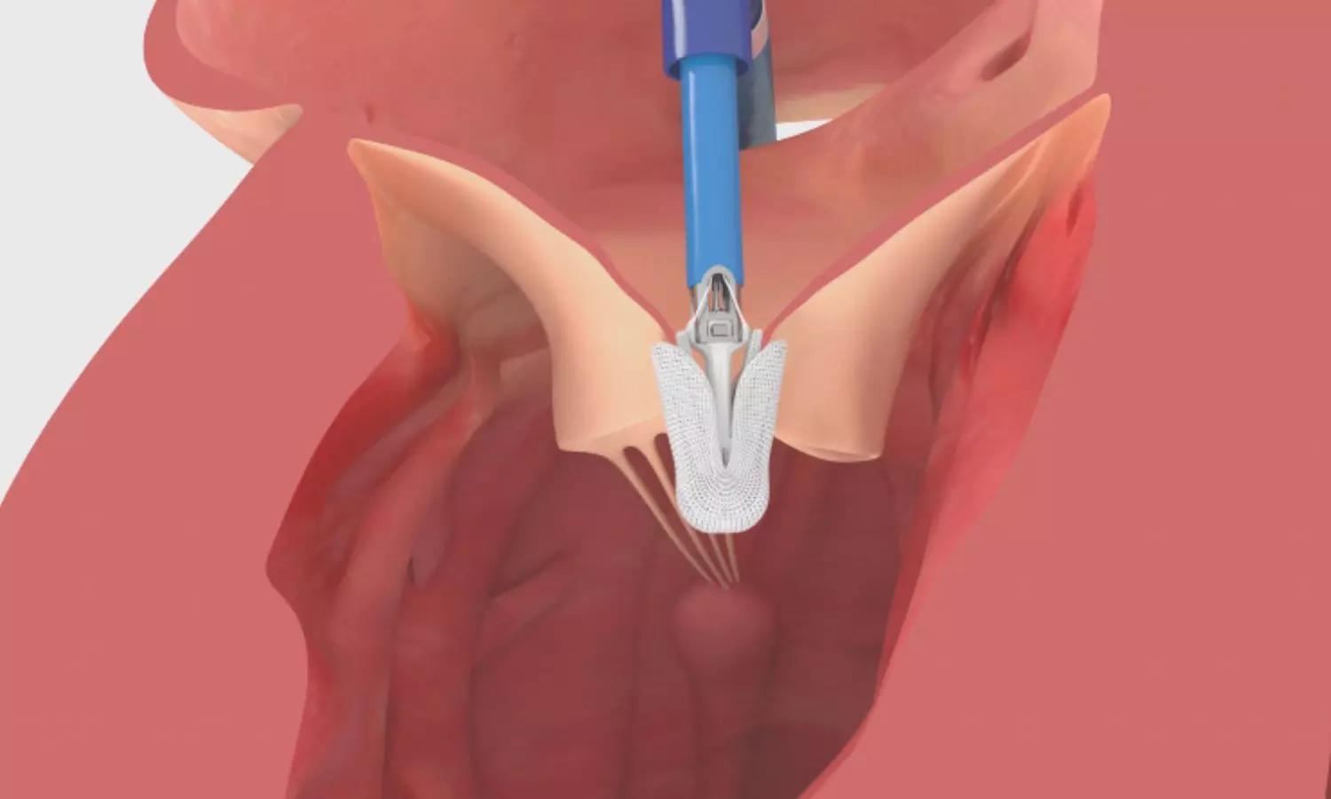
Transcatheter tricuspid valve replacement effective in patients with severe tricuspid regurgitation: Study

A new study published in the New England Journal of Medicine showed that improvements in symptoms and quality of life were the main reasons why transcatheter tricuspid-valve replacement outperformed medical treatment alone for patients with severe tricuspid regurgitation in terms of the key composite outcome.

Even though it has been consistently demonstrated that severe tricuspid regurgitation (TR) is linked to a worse prognosis and a higher death rate, it is still undertreated and treated conservatively. For the following reasons, all patients with severe TR should have access to transcatheter repair and replacement technologies, which are evolving to meet this significant unmet clinical need. A higher risk of mortality and incapacitating symptoms are linked to severe tricuspid regurgitation. Thus, information on the results of percutaneous transcatheter tricuspid valve replacement were required. Therefore, Rebecca Hahn and team conducted this study in order to get an overall effectiveness of percutaneous transcatheter tricuspid-valve replacement in individuals with regurgitation.

This research randomly allocated 400 patients with serious symptomatic tricuspid regurgitation in a 2:1 ratio to either medical therapy alone (control group) or transcatheter tricuspid-valve replacement with medical therapy (valve-replacement group) in this worldwide multicenter study. The following were the hierarchical composite primary outcomes: hospitalization for heart failure, postindex tricuspid-valve intervention, implantation of a right ventricular assist device or heart transplant, all-cause mortality, improvement of at least 10 points on the Kansas City Cardiomyopathy Questionnaire overall summary (KCCQ-OS), improvements of at least one New York Heart Association (NYHA) functional class, and improvement of at least 30 meters on the 6-minute walk distance. By comparing every potential patient pair, beginning with the initial event in the hierarchy, a win ratio was determined for the primary result.

A total of 133 patients were placed in the control group and 267 in the valve replacement group. After a year, the win ratio in favor of replacing the valve was 2.02. In patient pair comparisons, the valve-replacement group outperformed the control group in terms of postindex tricuspid-valve intervention, mortality from any cause, and progress in the KCCQ-OS score, NYHA class, and 6-minute walk length.

The yearly average of hospitalization for heart failure was lower in the valve-replacement group than in the control group. 15.4% of the valve-replacement group and 5.3% of the control group experienced severe bleeding; 17.4% and 2.3%, respectively, had new permanent pacemakers installed. Overall, transcatheter tricuspid valve replacement outperformed medical treatment in patients with severe tricuspid regurgitation.

Source:

Hahn, R. T., Makkar, R., Thourani, V. H., Makar, M., Sharma, R. P., Haeffele, C., Davidson, C. J., Narang, A., O’Neill, B., Lee, J., Yadav, P., Zahr, F., Chadderdon, S., Eleid, M., Pislaru, S., Smith, R., Szerlip, M., Whisenant, B., Sekaran, N. K., … Kodali, S. K. (2024). Transcatheter Valve Replacement in Severe Tricuspid Regurgitation. In New England Journal of Medicine. Massachusetts Medical Society. https://doi.org/10.1056/nejmoa2401918

Powered by WPeMatico

Hourglass-shaped stent could ease severe chest pain from microvascular disease: Study

A study at Mayo Clinic suggests that an hourglass-shaped stent could improve blood flow and ease severe and reoccurring chest pain in people with microvascular disease. Of 30 participants in a phase 2 clinical trial, 76% saw improvement in their day-to-day life. For example, some participants who reported not being able to walk around the block or up a flight of stairs without chest pain were able to do these ordinary physical activities at the end of a 120-day period. Clinical measures of blood flow related to the microvasculature of the heart significantly improved during follow-up, according to findings published in the Journal of the American College of Cardiology: Cardiovascular Interventions.

Microvascular disease is a condition in which tiny blood vessels in the heart are not working properly, resulting in reduced blood flow to the heart. The resulting chest pains, or angina, can be debilitating, limiting a person’s ability to exercise, do household chores or even walk to the mailbox. About 40% of patients receiving a diagnostic coronary angiogram for chest pain do not have blocked arteries that also can cause angina. However, up to 66% of these patients do have coronary microvascular disease, which is more common in women overall and found in people with conditions such as diabetes, high blood pressure and obesity.

For decades, there have been few viable treatment options to improve blood flow through the tiny vessels of the heart. At most, doctors have treated symptoms of angina with several medications and cardiovascular disease prevention methods, including healthy eating, weight loss and regular exercise. The use of a stent could target the issue behind the chest pain-the severe reduction in blood flow affecting the heart muscle.

Unlike tube-shaped stents used to open clogged arteries, the hourglass-shaped stent narrows in the middle. The different design is thought to increase back pressure, redistributing blood flow more fully through small vessels in the heart that were not working at capacity.

“The patients with heart-related microvascular dysfunction in this study had little ability to control their chronic angina, which severely limited their day-to-day activities,” says Amir Lerman, M.D., a cardiologist at Mayo Clinic and senior author of the study. “Beyond reductions in chest pain and being able to comfortably handle more physical activity, the majority of patients in the study also showed a connection between the changes in their coronary flow reserve, which is a measure of maximum blood flow, and changes in their quality-of-life responses on the survey. This points to the link between the physiological measurement and angina symptoms.”

Dr. Lerman notes that more studies are needed to better understand how the reducer stent works and its long-term effects on blood flow. The stent did not improve chest pain symptoms in 20%-30% of the participants, so future research studies will need to better identify which patients respond best to this therapy.

Reference:

David Tryon, Michel T. Corban, Mohamad Alkhouli, Abhiram Prasad, Claire E. Raphael, Charanjit S. Rihal, Guy S. Reeder, Brad Lewis, Diana Albers, Rajiv Gulati, and Amir Lerman, Coronary Sinus Reducer Improves Angina, Quality of Life, and Coronary Flow Reserve in Microvascular Dysfunction, Journal of the American College of Cardiology, DOI: 10.1016/j.jcin.2024.09.018.

Powered by WPeMatico

Ultrasound devise found promising for treating chronic pain, claims research

Pain is a necessary biological signal, but a variety of conditions can cause those signals to go awry. For people with chronic pain, the root is often faulty signals emerging deep within the brain, giving false alarms about a wound that has since healed, a limb that has since been amputated, or other intricate, hard-to-explain scenarios.

Patients with this kind of life-altering pain are constantly looking for new treatment options; now a new device from the University of Utah may represent a practical long-sought solution.

Researchers at the university’s John and Marcia Price College of Engineering and Spencer Fox Eccles School of Medicine have published promising findings about an experimental therapy that has given many participants relief after a single treatment session. They are now recruiting participants for a final round of trials.

At the core of this research is Diadem, a new biomedical device that uses ultrasound to noninvasively stimulate deep brain regions, potentially disrupting the faulty signals that lead to chronic pain.

The findings from a recent clinical trial are published in the journal Pain. This study constitutes a translation of two previous studies, published in Nature Communications Engineering and IEEE Transactions on Biomedical Engineering, which describe the unique features and characteristics of the device and demonstrate its efficacy.

The study was conducted by Jan Kubanek, a professor in Price’s Department of Biomedical Engineering, and Thomas Riis, a postdoctoral researcher in his lab. They collaborated with Akiko Okifuji, professor of Anesthesiology in the School of Medicine, as well as Daniel Feldman, graduate student in the departments of Biomedical Engineering and Psychiatry, and laboratory technician Adam Losser.

The randomized sham-controlled study recruited 20 participants with chronic pain, who each experienced two 40-minute sessions with Diadem, receiving either real or sham ultrasound stimulation. Patients described their pain a day and a week after their sessions, with 60% of the experimental group receiving real treatment reporting a clinical meaningful reduction in symptoms at both points.

“We were not expecting such strong and immediate effects from only one treatment,” Riis said.

“The rapid onset of the pain symptom improvements as well as their sustained nature are intriguing, and open doors for applying these noninvasive treatments to the many patients who are resistant to current treatments,” Kubanek added.

Diadem’s approach is based on neuromodulation, a therapeutic technique that seeks to directly regulate the activity of certain brain circuits. Other neuromodulation approaches are based on electric currents and magnetic fields, but those methods cannot selectively reach the brain structure investigated in the researchers’ recent trial: the anterior cingulate cortex.

After an initial functional MRI scan to map the target region, the researchers adjusted Diadem’s ultrasound emitters to correct for the way the waves deflect off the skull and other brain structures. This procedure was published in Nature Communications Engineering.

The team is now preparing for a Phase 3 clinical trial, the final step before approval from the Food and Drug Administration to use Diadem as a treatment for the general public.

“If you or your relatives suffer from chronic pain that does not respond to treatments, please reach out to us; we need to recruit many participants so that these treatments can be approved for the general public,” Kubanek said. “With your help, we think chronic pain can be effectively silenced. And with new pain treatment options, we can tackle the opioid crisis, too.”

Reference:

Riis, Thomas S.a; Feldman, Daniel A.a,b; Losser, Adam J.a; Okifuji, Akikoc; Kubanek, Jana,*. Noninvasive targeted modulation of pain circuits with focused ultrasonic waves. PAIN ():10.1097/j.pain.0000000000003322, July 30, 2024. | DOI: 10.1097/j.pain.0000000000003322

Powered by WPeMatico

0.01% atropine eye drops slows myopia progression without interfering with exotropia: JAMA

Intermittent exotropia (IXT), characterized by an
intermittent outward deviation of 1 or both eyes, is one of the most common
types of strabismus, particularly in Asian countries. Exotropia and myopia are
commonly coexistent. Myopia has been identified as a risk factor for
concomitant exotropia. It is thought that myopia is associated with a decreased
demand for accommodation and, hence, lower convergence and a predisposition for
developing exotropia. On the other hand, IXT has been regarded as a risk factor
for myopia onset and progression.

In patients with IXT, additional accommodative convergence
is required to maintain binocular vision and ocular alignment, which might
increase accommodative loads and, hence, myopia progression. Alternatively, it
was speculated that increased convergence demand (via convergence
accommodation/ convergence), rather than accommodation, contributes to the
myopia development in IXT.

Increased attention and further investigation seem warranted
in the management of myopia progression in children with IXT, prompting this
evaluation of low-concentration atropine in children with IXT, as previous
clinical trials of atropine for myopia control excluded children with
strabismus. The current 2-year, placebo-controlled, randomized clinical trial
included evaluation of the effects of 0.01% atropine eye drops on accommodative
changes, exotropia conditions, and binocular vision in children with myopia and
IXT (AMIXT) for which we report the 1-year primary results.

This placebo-controlled, double-masked, randomized clinical
trial was conducted from December 2020 to September 2023. Children aged 6 to 12
years with basic-type IXT and myopia of −0.50 to −6.00 diopters (D) after
cycloplegic refraction in both eyes were enrolled. Participants were randomly
assigned in a 2:1 ratio to 0.01% atropine or placebo eye drops administered in
both eyes once at night for 12 months. The primary outcome was change in
cycloplegic spherical equivalent from baseline at 1 year. Secondary outcomes
included change in axial length (AL), accommodative amplitude (AA), exotropia
conditions, and binocular vision at 1 year.

Among 323 screened participants, 300 children were included
in this study. A total of 200 children (66.7%) were in the atropine group, and
100 (33.3%) were in the placebo group. At 1 year, the 0.01% atropine group had
slower spherical equivalent progression (P < .001) and AL elongation (P <
.001) than the placebo group. The mean AA change was −3.06 D vs 0.12 D (P <
.001) in the atropine and placebo groups, respectively. The 0.01% atropine
group had a decrease in near magnitude of exodeviation whereas the placebo
group had an increase (P = .03). In the atropine vs placebo group,
respectively, the incidence of study drug-related photophobia was 6.0% (12 of
200 participants) vs 8.0% (8 of 100 participants; P = .51) and for blurred near
vision was 6.0% (12 of 200 participants) vs 7.0% (7 of 100 participants) (P =
.74).

The AMIXT randomized clinical trial evaluated the efficacy
and safety of 0.01% atropine eye drops on myopia progression, exotropia
conditions, binocular vision, monocular function, and safety parameters in
individuals with myopia and IXT at 1 year. The 0.01% atropine group had slower
myopia progression in both spherical equivalent measurement and AL among
children with myopia and IXT. The 0.01% atropine group did not appear to have
aggravated exotropia conditions, in terms of magnitude of exodeviation and
exotropia control. The 0.01% atropine group did not appear to have worse
binocular vision, supported by assessment of distant stereoacuity, near
stereoacuity, NPC, AC/A, and FVA. In addition, the use of 0.01% atropine
appeared safe with a mild decrease in AA and a mild increase in pupil size;
stability of BCVA, near vision, IOP, and ECD; and low incidence of adverse
events.

In summary, this placebo-controlled, double-masked,
randomized clinical trial established that 0.01% atropine eye drops, although compromising
AA to some extent, appeared effective and safe in slowing myopia progression
without interfering with exotropia conditions or binocular vision in children
with myopia and IXT.

Source: Zijin Wang, MD; Tianxi Li, MD; Xiaoxia Zuo, MD; JAMA
Ophthalmol. doi:10.1001/jamaophthalmol.2024.2295 Published online July 3, 2024.

Powered by WPeMatico

Same-Day Discharge after pediatric laparoscopic appendectomy for non-perforated acute appendicitis on the rise: Study

USA: A recent study using data from the National Surgical Quality Improvement Program Pediatric (NSQIP-P) database has highlighted a significant increase in same-day discharge (SDD) rate following pediatric laparoscopic appendectomy for non-perforated acute appendicitis. The findings suggest that same-day discharge is becoming more common in pediatric surgical practice, and importantly, it does not come at the cost of increased adverse outcomes or readmissions.

“SDD rates are 20–30% lower than those reported in earlier single-center studies, suggesting that a larger proportion of patients may be eligible for same-day discharge than previously anticipated,” the researchers reported in Pediatric Surgery International. 

Laparoscopic appendectomy has long been the gold standard for treating appendicitis in children due to its minimally invasive nature and faster recovery time compared to traditional open surgery. However, allowing patients to go home on the same day of surgery, rather than staying overnight for observation, has been more gradual.

Since the practice patterns surrounding same-day discharge for pediatric laparoscopic appendectomy in cases of non-perforated appendicitis have not been thoroughly studied, P. Benson Ham III, University at Buffalo Division of Pediatric Surgery, Jacobs School of Medicine and Biomedical Sciences, NY, USA, and colleagues aimed to evaluate the current practices for same-day discharge in these patients. Additionally, they seek to assess the associated outcomes of this approach.

For this purpose, the researchers utilized the NSQIP-P retrospective registry to identify patients who underwent laparoscopic appendectomy for non-perforated appendicitis between 2017 and 2021. The annual rate of same-day discharge was calculated, and SDD patients were compared to those discharged on postoperative days 1–2 (non-SDD). A subgroup analysis was conducted using ICD-10 code K35.30, and regression analysis was performed to assess the outcomes.

The study led to the following findings:

  • Total Patients Identified: 67,214 patients were included in the analysis.
  • Same-Day Discharge (SDD) Rate: The SDD rate increased significantly from 33.3% in 2017 to 52.5% in 2021.
  • 30-Day Readmission Rates: SDD patients had lower 30-day readmission rates than non-SDD patients (1.3% versus 2.1%).
  • Sub-group Analysis: Among 7,330 patients, SDD rates increased from 50.7% in 2019 to 60.4% in 2021, with readmission rates again lower for SDD patients (1.3% versus 2.1%).
  • Adverse Events: No increase in adverse events was observed for SDD patients in either analysis.
  • Regression Analysis: Patients with esophageal, gastric, or intestinal diseases had significantly higher odds of early readmission or reoperation (OR 1.85).

“Pediatric laparoscopic appendectomy for non-perforated acute appendicitis continues to evolve, with same-day discharge becoming more widespread. With careful patient selection, the trend of early discharge shows promising outcomes, offering a more efficient and patient-centered approach to appendicitis treatment in children,” the researchers concluded.

Reference:

Woodward, J.M., Brierley, S.F., Bittner, K. et al. Trends and outcomes in pediatric laparoscopic appendectomy: a NSQIP-P analysis of same-day discharge and readmission rates. Pediatr Surg Int 40, 287 (2024).

https://doi.org/10.1007/s00383-024-05869-9

Powered by WPeMatico

MRI-Based MACRO Score Accurately Predicts Macrovascular Causes of Intracerebral Hemorrhage: Study

UK: A new study published in the journal Neurology highlights the effectiveness of the MRI-based MACRO score in predicting macrovascular causes of spontaneous intracerebral hemorrhage (ICH), potentially improving diagnostic accuracy and guiding clinical decision-making.

“The MRI Assessment of the Causes of intracerebral hemorrhage (MACRO) score, developed through a combination of MRI imaging features and clinical factors, demonstrates superior predictive accuracy compared to current CT-based scores in identifying the underlying causes of spontaneous intracerebral hemorrhage,” the researchers wrote. It provides a structured tool to help identify patients whose ICH is likely linked to underlying macrovascular conditions, such as arteriovenous malformations, aneurysms, or cerebral amyloid angiopathy.

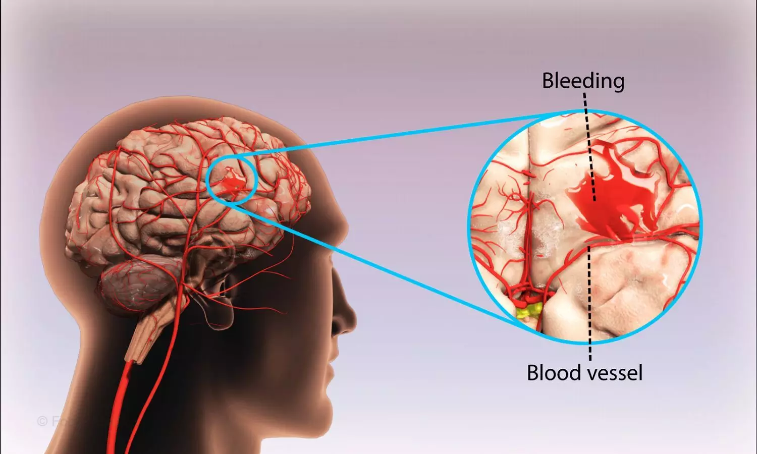
Spontaneous intracerebral hemorrhage, a type of stroke caused by the rupture of a blood vessel in the brain, remains a challenging condition to diagnose and manage due to its varied causes. While the majority of spontaneous ICHs are attributed to cerebral small vessel diseases (SVDs), approximately 1 in 7 to 1 in 10 cases are caused by macrovascular conditions. Identifying the underlying cause is crucial for effective treatment and preventing future bleeding episodes. Traditionally, angiography or digital subtraction angiography (DSA) has been the gold standard for evaluating the vascular origins of ICH, but these methods are invasive and may not always be suitable for every patient.

While MRI offers optimal sensitivity for detecting markers of SVD), it was not incorporated into previous risk stratification scores. To address this gap, Simon Fandler-Höfler, UCL Queen Square Institute of Neurology, United Kingdom, and colleagues aimed to develop and validate a new risk stratification score for macrovascular causes of ICH, incorporating MRI findings to improve diagnostic accuracy and guide further investigations.

For this purpose, the researchers pooled data from two large observational study cohorts (London/United Kingdom and Graz/Austria) consisting of consecutive patients with intracerebral hemorrhage who had undergone brain MRI and at least one angiographic modality within 90 days of symptom onset.

The primary outcome was identifying macrovascular causes of ICH, including conditions such as arteriovenous malformation, dural arteriovenous fistula, aneurysm, cavernoma, or cerebral venous thrombosis, with diagnoses confirmed through neurovascular multidisciplinary meetings. Using lasso logistic regression, the researchers developed the MRI Assessment of the Causes of Intracerebral Hemorrhage (MACRO) score to estimate the probability of a macrovascular etiology.

The following were the key findings of the study:

  • The study included 1,043 patients with intracerebral hemorrhage (ICH), with a mean age of 66 years, 42% of whom were female.
  • Of these patients, 7.5% had a macrovascular cause of ICH.
  • The final MACRO score incorporates age (0–39, 40–69, or ≥70), ICH location (lobar, deep, or infratentorial), and markers of small vessel disease on MRI (≥1 microbleed, ≥1 lacune, presence of cortical superficial siderosis, or white matter hyperintensities based on the Fazekas scale).
  • The MACRO score demonstrated an optimism-adjusted c-statistic of 0.90, outperforming existing CT-based scores.
  • In external validation, the MACRO score achieved a c-statistic of 0.87.
  • MACRO scores ≥6, seen in 59.5% of patients, were associated with a very low risk of a macrovascular cause (0.2%).
  • MACRO scores ≤2, observed in 9% of patients, indicated a high risk of a macrovascular cause (48.9%).

“The MRI-based MACRO score demonstrates strong effectiveness in predicting the likelihood of macrovascular causes of spontaneous intracerebral hemorrhage, offering valuable guidance for further diagnostic investigations,” the researchers wrote.

“However, the study has notable limitations, including its observational design and the fact that digital subtraction angiography was only performed in a small subset of patients,” they concluded.

Reference:

Fandler-Höfler S, Ambler G, Goeldlin MB, Obergottsberger L, Wünsch G, Kneihsl M, Zhang W, Du Y, Locatelli M, Ozkan H, Nash PS, Nistl O, Panteleienko L, Mendel R, Thiankhaw K, Simister RJ, Jäger HR, Enzinger C, Seiffge DJ, Gattringer T, Werring DJ. MRI-Based Prediction of Macrovascular Causes of Intracerebral Hemorrhage: The MACRO Score. Neurology. 2024 Nov 26;103(10):e209950. doi: 10.1212/WNL.0000000000209950. Epub 2024 Oct 31. PMID: 39481069.

Powered by WPeMatico

Increased Dried Fruit Intake May Improve Kidney Function Markers, finds study

Researchers have established that a higher intake of dried fruit was associated with improved markers of kidney function, and effects appeared to be greatest in nonsmokers and nondrinkers. A recent study was published in Frontiers in Nutrition journal conducted by Gao Y. and colleagues. Intake of dried fruits has significant association with markers at lower levels like Blood Urea Nitrogen (BUN), creatinine (CR), uric acid (UA), and cystatin C (CyC) are indicative of good health concerning kidneys.

Millions of people worldwide suffer from chronic kidney disease. Lifestyle factors, including diet, are considered to be involved in the pathogenesis of the disease; however previous studies on dried fruit intake and the health of kidneys have been limited by confounders and bias. Using a technique known as Mendelian randomization (MR), this study aimed to clarify the causal relationship between dried fruit intake and markers of kidney function.

A two-sample MR approach was utilized by the researchers. They sought to investigate whether there was an association, and most importantly, a possible causal one between the consumption of dried fruit and six markers of kidney function that included BUN, creatinine, uric acid, cystatin C, hematuria, and microalbuminuria. Inverse variance weighting was the analytical approach most often used in estimating associations, while other methods-MR Egger and weighted median-were used as supplementary approaches to investigate result robustness. MVMR was conducted with adjustment for smoking and alcohol use to determine if the effect of dried fruit consumption on kidney function may be confounded by smoking and alcohol use.

  • Higher intake of dried fruit was significantly associated with a lower BUN level (β = -0.171, 95% CI: -0.239 to -0.102, p = 1.063 × 10⁻⁶).

  • Positive consumption of dried fruit was linked with lowering creatinine (β = -0.205, 95% CI: -0.311 to -0.099, p = 1.455 × 10⁻⁴).

  • In uric acid as well, a similar association was obtained with β = -0.317, 95% CI: -0.384 to -0.249, p = 4.439 × 10⁻²⁰, that is to say a greater consumption of dried fruits may be associated with the lowering effect of UA.

  • In increased cystatin C, decreased intake of dried fruits is observed with β = -0.323, 95% CI: -0.384 to -0.249, p = 1.074 × 10⁻¹¹.

  • No meaningful association between dried fruit consumption with hematuria or microalbuminuria, which are two of the most common used markers in examinations of renal health,

  • Intake of dried fruit was inversely related to UA and CyC levels, though the association became attenuated; still, it remained significant for both UA (β = -0.296, 95% CI: -0.523 to -0.068, p = 1.094 × 10⁻²) and CyC (β = -0.238, 95% CI: -0.465 to -0.011, p = 4.024 × 10⁻²).

  • For BUN and creatinine, the causal association completely disappeared in smokers and alcoholics (BUN: β = -0.038, 95% CI: -0.215 to 0.138, p = 6.698 × 10⁻¹; CR: β = -0.038, 95% CI: -0.431 to 0.046, p = 1.347 × 10⁻¹).

This study suggests that consumption of dried fruits may be an intervention that improves markers for kidney function in abstainers from smoking and alcohol. Dried fruit can, therefore, be added to a healthy diet for people at risk of kidney disease as a potential additive strategy for preserving renal health and delaying the onset of chronic kidney disease when smoking and alcohol are avoided.

Reference:

Gao Y, Yue X, Zhao W and Yuan F (2024) Association between dried fruit intake and kidney function: research from univariate and multivariate Mendelian randomized studies. Front. Nutr. 11:1440896. doi: 10.3389/fnut.2024.1440896

Powered by WPeMatico

Vitamin D intake during pregnancy may boost bone health of children even at age seven: Study

Children whose mothers took extra vitamin D during pregnancy continue to have stronger bones at age seven, according to new research led by the University of Southampton and University Hospital Southampton (UHS).

Bone density scans revealed that children born to mothers who were given vitamin D supplements during pregnancy have greater bone mineral density in mid-childhood. Their bones contain more calcium and other minerals, making them stronger and less likely to break.

Researchers say the findings, published in The American Journal of Clinical Nutrition, reinforce the importance of vitamin D supplementation in pregnancy as a public health strategy.

Dr Rebecca Moon, NIHR Clinical Lecturer in Child Health at the University of Southampton led the analysis. She said: “Our findings show that the benefits of vitamin D supplementation during pregnancy persist into mid-childhood.

“This early intervention represents an important public health strategy. It strengthens children’s bones and reduces the risk of conditions like osteoporosis and fractures in later life.”

Vitamin D regulates the amount of calcium and phosphate in the body – minerals essential for bones, teeth and muscle health.

In 2009, researchers launched the MAVIDOS study, recruiting over 1000 women from Southampton, Oxford and Sheffield.

During their pregnancy, the women were randomly allocated to two groups: One group took an extra 1,000 International Units per day of vitamin D. The other took a placebo tablet each day. The pregnant women, and the doctors and midwives looking after them, did not know which group they were in.

Previous research assessed the children’s bone health at four years of age and the results showed that the child’s bone mass was greater in children born to mothers who had had vitamin D supplementation during pregnancy compared with those who had not.

In this latest study, the researchers investigated whether the effects on bone health continued into mid-childhood. The team followed up with 454 children aged six to seven. These children were all born to mothers who took part in Southampton.

The results confirmed that the beneficial effect on children’s bones was similar at ages four and six to seven.

Pregnant women in the UK are now routinely advised to take vitamin D supplements.

The Southampton research team are part of the MRC Lifecourse Epidemiology Centre (MRC LEC) and the NIHR Southampton Biomedical Research Centre (BRC).

The MAVIDOS trial has helped the Southampton researchers understand possible mechanisms linking maternal vitamin D supplementation with offspring bone mass. In 2018 they demonstrated that the vitamin D supplementation led to changes in the activity of genes forming part of the vitamin D pathway.

In 2022, they found taking the supplements during pregnancy could substantially reduce the chances of babies up to a year old suffering from atopic eczema.

They also observed that pregnant women given extra vitamin D were more likely to have a spontaneous vaginal delivery, or ‘natural’ delivery.

Professor Nicholas Harvey is Director of the University’s MRC Lifecourse Epidemiology Centre, Professor of Rheumatology and Clinical Epidemiology and project lead.

He said: “These findings add to the important knowledge generated through the MAVIDOS trial.

“We extend our heartfelt thanks to all the mothers and children involved. Their contributions have advanced our understanding of vitamin D supplementation and its role in supporting strong and healthy bones.”

Reference:

Rebecca J Moon, Stefania D’ Angelo, Elizabeth M Curtis, Kate A Ward, Sarah R Crozier, Inez Schoenmakers, M Kassim Javaid, Nicholas J Bishop, Keith M Godfrey, Cyrus Cooper, Nicholas C Harvey, Elaine M Dennison, Richard Eastell, Robert Fraser, Saurabh V Gandhi, Hazel M Inskip, Stephen H Kennedy, Aris T Papageorghiou, Ann Prentice, Pregnancy vitamin D supplementation and offspring bone mineral density in childhood follow-up of a randomized controlled trial, The American Journal of Clinical Nutrition, https://doi.org/10.1016/j.ajcnut.2024.09.014.

Powered by WPeMatico