Pleural effusion in HF patients indicates poor QoL and worse outcomes: Study

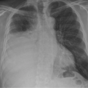
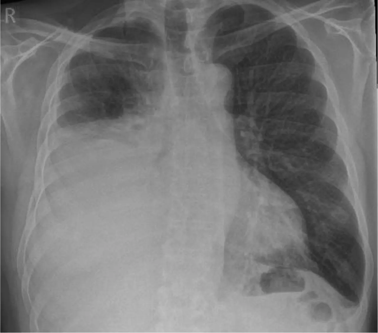
Researchers have established that pleural effusion (PE), which is a common complication of acute decompensated heart failure (ADHF), severely impacts the quality of life (QOL) in patients. Fluid accumulation in the lungs is often observed among ADHF patients, also known as pleural effusion. A recently published study was conducted at a university hospital in Mexico, investigating the relationship between PE diagnosed by thoracic ultrasound and the QOL of patients with a reduced ejection fraction. The study was conducted by Rendon-Ramirez and colleagues and was published in the journal Heart & Lung.

This was a prospective, longitudinal study with patients admitted for ADHF and low ejection fraction. The diagnosis of PE was assessed with TUS within 48 hours after admission. QoL of the patients was measured according to the MLHFQ. The patients were of an average age of 51.24 years (±16.942 years), and 30 males and 10 females participated. The participants were divided into two groups based on the presence of PE: those with PE (n=25, 62.5%) and those without (n=15, 37.5%).

  • The study found that the presence of PE was associated with much lower scores on QoL. The patients with PE had an MLHFQ score of around 40, significantly more than the average score of 12 patients who were without PE, meaning the severity of QoL was reduced greatly (p < 0.001).

  • Volume pleural fluid had correlation with lower QoL scores. Patients who have larger PEs as diagnosed by having more intercostal spaces, the QoL of patients was poorer (p < 0.001).

The presence of pleural effusion in patients with acute decompensated heart failure and a reduced ejection fraction is highly associated with a reduced quality of life. This study supports the potential benefits of early identification and management of PE as part of a comprehensive heart failure care plan. By addressing this condition, healthcare providers could help alleviate some of the QoL burdens on heart failure patients.

Reference:

Rendón-Ramírez, E. J., Fraga-Enríquez, V. M., Colunga-Pedraza, P. R., Nañez-Terreros, H., Flores-Ramírez, R., Canseco, F., Porcel, J. M., & Carrizales-Sepúlveda, E. F. (2024). Pleural effusions identified by thoracic ultrasound predict poor quality of life in patients with acute decompensated heart failure. Heart & Lung: The Journal of Critical Care, 68, 279–283. https://doi.org/10.1016/j.hrtlng.2024.08.008

Powered by WPeMatico

3% of NICU Patients using Central Venous Catheters may develop Deep Vein Thrombosis: Study

Central venous catheters are frequently used in babies in neonatal intensive care units (NICUs)—providing key nutrition, fluids, and medications. But while these catheters are critical for survival, they can also be linked to risks such as deep vein thrombosis.

Thromboses can lead to a host of issues, including long-term swelling of the affected limb, pulmonary embolism, longer hospital stays, and mortality. And yet, little is understood about how frequently these events occur in NICU patients—and whether certain catheter characteristics might increase that risk.

Now, a new study at Children’s Hospital Los Angeles is shedding light on the answers to these questions. The team’s preliminary data recently found that about 3% of central venous catheters placed in NICU babies were affected by blood clots.

Researchers also examined the impact of specific catheter and patient characteristics on thrombosis risk. Results were presented by lead author Molly Easterlin, MD, MS, at the 2024 Pediatric Academic Societies meeting in Toronto.

“Our data shows that catheter-related thrombosis is relatively common in NICU patients and may be linked to specific catheter and patient risk factors,” says Dr. Easterlin, a neonatologist in the Fetal and Neonatal Institute at Children’s Hospital Los Angeles.

Key findings

Although prior studies have examined the issue of central venous catheters in NICU patients, that literature often lacks details of the actual catheters used and where they were placed, Dr. Easterlin says.

“This detailed granular data can be hard to come by in existing large data sets,” she notes. “Even mining your own electronic medical record data can be difficult because you traditionally have to rely on chart review to pull out variables for thousands of patients.”

To solve this problem, Dr. Easterlin and her colleagues partnered with CHLA data science experts. The team used natural language processing—a form of machine learning—to create a database of every central venous catheter placed in a NICU patient from January 2017 to December 2023.

That database includes about 3,900 central venous catheters that were placed in approximately 2,100 unique patients in the Level IV Steven & Alexandra Cohen Foundation Newborn and Infant Critical Care Unit (NICCU) at CHLA.

In addition to finding a 3.6% rate of deep vein thrombosis among those catheters, the team found that:

Catheters placed in the femoral vein or lower extremity blood vessels were more likely to be associated with clots.

Peripherally inserted central catheters (PICCs) were linked to thrombosis events more frequently than other catheter types, such as umbilical catheters.

Catheters with two lumens had a higher risk of clotting than those with one lumen.

Central lines affected by thrombosis were in place for a longer amount of time than those not linked to clotting.

Integrating data to improve care

Dr. Easterlin notes that a multidisciplinary approach is key to the study, which is ongoing. In addition to neonatologists and data science experts, the team includes Joseph Miller, MD, MS, Director of Interventional Radiology at CHLA and senior author on the study.

“Dr. Miller’s team places these central lines in patients and sees many of the complications when they arise, so he brings a different perspective to the issue of thrombosis in these patients,” she explains. “This work would also not be possible without the incredible expertise of our Data Science team, Mohit Mehra and Xinwen (Melody) Li, in extracting this information from electronic medical records.”

The team is now working to develop a real-time dashboard that could be integrated into the electronic medical record. The dashboard would show the characteristics of every central line placed in a patient—including type of catheter, where it was placed, number of lumens, and more.

“Our goal is to be able to use this database in real time to provide patient-specific clinical guidance,” Dr. Easterlin explains. “The goal is to better centralize all this information and help guide decision-making that can hopefully reduce these complications in patients.”

Study authors, all from CHLA, were Molly Easterlin, MD, MS; Mohit Mehra, MS; Xinwen Li, MS; Srikumar Nair, MD; Philippe Friedlich, MD, MSEpi, MBA; and Joseph Miller, MD, MS.

Powered by WPeMatico

People with cardiovascular-kidney metabolic syndrome may land up with CVD much earlier, reveals research

People with chronic kidney disease, Type 2 diabetes or both were predicted to have elevated cardiovascular disease (CVD) risk 8 to 28 years sooner than someone without those conditions, according to a preliminary study to be presented at the American Heart Association’s Scientific Sessions 2024. 

Current guidelines on CVD prevention define CVD risk as elevated if the chance of having a heart attack or stroke in the next 10 years is 7.5% or higher. Chronic kidney disease and Type 2 diabetes increase risk and are two of the four components of cardiovascular-kidney-metabolic (CKM) syndrome, which the American Heart Association defines as the interplay of cardiovascular disease, kidney disease and the metabolic disorders Type 2 diabetes and obesity. This study aimed to better understand the effects of (CKM) syndrome on CVD risk.

Researchers created risk profiles to simulate men and women with and without chronic kidney disease and/or Type 2 diabetes at each age from 30 to 79 years. They used the American Heart Association Predicting Risk of cardiovascular disease EVENTs (PREVENT™) calculator to determine at what age someone with each risk profile would be expected to have elevated CVD risk.

The risk profiles were based on data from the National Health and Nutrition Examination Survey 2011-2020. Chronic kidney disease was defined as estimated glomerular filtration rate (eGFR) of 44.5, which indicates stage 3 kidney disease. Type 2 diabetes was indicated as a “yes” response to the PREVENT calculator prompt, “Any history of diabetes.” Profiles without CKM syndrome were derived from average blood pressure, cholesterol and eGFR levels representing survey participants who did not have diabetes, were not taking medicines to lower blood pressure or cholesterol and were non-smokers.

According to the American Heart Association, nearly half of all U.S. adults are living with some form of CVD, and one in three has at least three risk factors that contribute to CKM syndrome. Identifying who is at greatest risk earlier can improve primary prevention and reduce the risk of premature death from CVD.

“Our findings help to interpret the combination of risk factors that will lead to a high predicted CVD risk and at what age they have an impact on risk,” said lead study author Vaishnavi Krishnan, B.S., a researcher at Northwestern University in Chicago and a medical student at Boston University School of Medicine in Boston. “For example, if someone has borderline-elevated levels of blood pressure, glucose and/or impaired kidney function, but they don’t yet have hypertension or diabetes or chronic kidney disease, their risk may not be recognized. Understanding how age interacts with risk factor levels is important to optimize CKM health.”

Without CKM syndrome, the expected age to reach elevated CVD risk was 68 years for women and 63 for men. However, with CKM components added to the simulated patient profile, the same risk level was predicted to occur at a much younger age:

For adults with stage 3 chronic kidney disease, predicted 10-year CVD risk was elevated at age 60 for women and 55 for men. This is 8 years younger than expected for men and women without CKM syndrome.

For adults with Type 2 diabetes, predicted 10-year risk of CVD was elevated at age 59 for women and 52 for men. This is 9 years younger for women and 11 years younger for men compared to those without CKM syndrome.

For adults who had both Type 2 diabetes and stage 3 chronic kidney disease, predicted 10-year risk of CVD was elevated at age 42 for women and 35 for men. This is 26 years younger for women and 28 years younger for men compared to those without CKM syndrome.

“A strength of this study is that it spans nearly the full adult life course and examines how risk factor levels due to CKM conditions of chronic kidney disease and Type 2 diabetes may impact predicted CVD risk,” Krishnan said. “However, a limitation of this study is that these are predicted risk calculations based on a simulated population.”

“This is an early step in the process of understanding how a risk model works,” said Sadiya S. Khan, M.D., M.Sc., co-author of the study, the Magerstadt Professor of Cardiovascular Epidemiology and an associate professor of cardiology, medical social sciences and preventive medicine (epidemiology) at Northwestern School of Medicine in Chicago, and chair of the writing group for the PREVENT equations. “Future work and guidelines are needed to determine how to use the PREVENT equations and what risk thresholds should be used in a clinical setting.”

To address the complex health threat of CKM syndrome, the American Heart Association, celebrating 100 years of lifesaving service as a global force for healthier lives for all, has launched a four-year Cardiovascular-Kidney-Metabolic (CKM) Health Initiative, supported by Novo Nordisk and Boehringer Ingelheim. The initiative will track CKM measures in the Association’s Get With The Guidelines® and outpatient data registries; improve coordination among CVD, kidney and diabetes specialists; and promote best practices, including guideline-directed treatment. This work will help assess gaps in clinical care, identify areas for future research, and implement guidelines and screening recommendations to provide clear and definitive advice for CKM syndrome treatment. 

Powered by WPeMatico

Canagliflozin Enhances Iron Utilization and Hemoglobin Levels in Diabetic CKD Patients, CREDENCE Trial Reveals

Australia: Recent findings from the CREDENCE trial have shed light on the significant role of Canagliflozin, a sodium-glucose co-transporter 2 (SGLT2) inhibitor, in iron metabolism among patients with type 2 diabetes and chronic kidney disease (CKD).

The study, published in Nephrology Dialysis Transplantation, revealed that treatment with canagliflozin led to an increase in total iron binding capacity (TIBC) while decreasing ferritin levels. These changes suggest a shift in iron metabolism, indicating that Canagliflozin may enhance iron utilization in the body.

“Canagliflozin was linked to enhanced iron utilization and elevated hemoglobin levels in adults with type 2 diabetes and chronic kidney disease,” the researchers wrote.

Iron deficiency is a common complication in individuals with type 2 diabetes and CKD, often leading to anemia and exacerbating the overall health of patients. Research involving patients with heart failure has shown that sodium-glucose cotransporter 2 (SGLT2) inhibitors can boost iron utilization and promote erythropoiesis. In a post hoc analysis of the CREDENCE trial, Hiddo J L Heerspink, The George Institute for Global Health, UNSW Sydney, Sydney, Australia, and colleagues assessed the impact of canagliflozin on iron metabolism in CKD patients. They investigated how iron deficiency influenced the drug’s effects on hemoglobin levels and cardiorenal outcomes.

For this purpose, the researchers measured serum iron, total iron binding capacity (TIBC), transferrin saturation (TSAT), and ferritin levels at baseline and after 12 months. They used analysis of covariance to assess the effects of canagliflozin compared to placebo on these iron markers. Additionally, mixed-effect and Cox regression models were employed to evaluate how baseline iron deficiency, defined as TSAT <20%, influenced the impact of canagliflozin on hemoglobin levels and cardiorenal outcomes.

The study revealed the following findings:

  • Of the 4,401 participants randomized in the CREDENCE trial, 2,416 had their iron markers measured at baseline, with 924 identified as iron deficient.
  • Compared to placebo, Canagliflozin resulted in a 2.1% increase in TIBC and an 11.5% reduction in ferritin levels, without significantly impacting serum iron or transferrin saturation (TSAT).
  • Over the trial period, Canagliflozin increased hemoglobin by 7.3 g/L for patients with iron deficiency and 6.7 g/L for those without iron deficiency, with no significant interaction effect.
  • The relative impact of Canagliflozin on the primary outcome—defined as a doubling of serum creatinine, kidney failure, or death from cardiovascular disease or kidney failure—was consistent across both groups, with a hazard ratio of 0.70 and no significant interaction effect.
  • Similar consistency was observed for other cardiovascular and mortality outcomes.

According to the authors, iron deficiency is highly prevalent among patients with type 2 diabetes and chronic kidney disease.

“Their findings indicate that Canagliflozin increases total iron binding capacity and decreases ferritin levels in this population, suggesting enhanced iron utilization. Additionally, the treatment is associated with improved hemoglobin levels and favorable clinical outcomes, regardless of whether patients have iron deficiency,” the authors concluded.

Reference:

Koshino, A., Heerspink, H. J., Jongs, N., Badve, S. V., Arnott, C., Neal, B., Jardine, M., Mahaffey, K. W., Pollock, C., Perkovic, V., Hansen, M. K., Bakker, S. J., Wada, T., & Neuen, B. L. Canagliflozin and iron metabolism in the CREDENCE trial. Nephrology Dialysis Transplantation. https://doi.org/10.1093/ndt/gfae198

Powered by WPeMatico

Persistent Menopausal Symptoms Linked to Higher Diabetes Risk, reveals study

New research from the Study of Women’s Health Across the Nation (SWAN), found that women who regularly suffer with hot flashes and night sweats during the menopause phase may have a higher possibility of developing type 2 diabetes (T2DM). This study was conducted by Monique H. and colleagues. The study was recently published in JAMA Network Open.

The SWAN study was a long-term prospective study of 2761 midlife women from a diverse racial and ethnic backgrounds of the United States. At baseline, the women studied were at intermediate levels between being premenopausal and being at an early stage of perimenopause and followed for as long as up to 13 annual visits. Vasomotor symptoms (VMS) frequency was determined based on participants’ self-reports regarding frequency of hot flashes and night sweats and significantly varies in the population across studies. Women already diagnosed with diabetes, using hormone therapies, or lacking adequate follow-up data were excluded for the analysis, thereby providing a clearly defined cohort that could be followed with respect to VMS trajectories and risk of incident diabetes.

The number of VMS was assessed at each follow-up visit in the SWAN study, and diabetes status was determined by use of medication for antidiabetic use, repeated high fasting glucose (≥126 mg/dL), or self-reported diabetes with high fasting glucose. The discrete-time hazard models were used to calculate hazard ratios (HRs) of diabetes associated with different frequencies and patterns of VMS. Other covariates tested were demographics and lifestyle factors such as age, race/ethnicity, education, BMI, physical activity, smoking, alcohol intake, and menopausal stage.

  • At baseline, 28% of women reported VMS 1-5 days/wk, 10% reported VMS 6 or more days/wk, and 62% reported VMS 0 days/wk.

  • At study completion, 338 of the 2768 participants (12.2%) had developed diabetes.

  • The incidence of frequent VMS was associated with an increased risk of diabetes (45%; HR, 1.45; 95% CI, 1.11-1.95), and women reporting infrequent VMS were at a 30% higher risk (HR, 1.30; 95% CI, 1.00-1.70) compared with women without VMS.

  • The four major patterns of VMS trajectories across participants were classified into four categories: consistently low VMS probability (26%), persistently high VMS probability (31%), early onset with a gradual decline (25%), and late onset with a gradual increase (19%).

  • Women with the persistently high VMS patterns had a 50% higher risk of diabetes compared with women with consistently low VMS probability (HR, 1.50; 95% CI, 1.12-2.02).

The persistence of VMS during menopause may thus be considered a marker of the risk of T2DM, thereby identifying the at-risk population for diabetes prevention. These findings further suggest to health care providers the necessity of considering VMS patterns while making assessments regarding metabolic health in menopausal women and further reinforce the need for targeted preventive care and lifestyle management among women with persistent VMS.

Reference:

Hedderson, M. M., Liu, E. F., Lee, C., El Khoudary, S. R., Gold, E. B., Derby, C. A., & Thurston, R. C. (2024). Vasomotor symptom trajectories and risk of incident diabetes. JAMA Network Open, 7(10), e2443546. https://doi.org/10.1001/jamanetworkopen.2024.43546

Powered by WPeMatico

Pulp canal obliteration and calcification more common in females on opposite side of proximal caries: Study

Recent study has found that proximal caries in mandibular molars are associated with significant calcification, or pulp canal obliteration (PCO), in the contralateral root canal. The new study of CBCT scans done recently looked into the prevalence and pattern of PCO in the molars of patients with proximal caries, showing that the amount of calcification typically occurs on the opposite side of the root canal on which caries are visible. The study was conducted by Ganesh R. and colleagues and was published in the Journal of Endodontics.

Dental caries remain a very common oral health problem, generally resulting in pulp tissue inflammation and other sequelae. Proximal caries, those occurring between two adjacent teeth, may often present a therapeutic challenge due to the limited mobility of the affected tooth. In mandibular molars, these caries are often accompanied by PCO – a calcification process that might hinder blood supply, impair root health, and complicate root canal procedures. The present study aimed to determine how the location of proximal caries relates to the site and degree of PCO and any differences among age, gender, and position of caries.

A total of 1,491 CBCT scans of dental patients aged 18 to 49 years old from the period of 2002 to 2022 were analyzed. Out of these, 328 teeth showing visible proximal caries were chosen using the defined inclusion criteria for more thorough examination. The studied teeth for both calibrated examiners were scored for PCO presence and severity along the sagittal and coronal planes. Comparisons of data were conducted on the basis of alterations in presence and PCO severity with respect to age, sex, and caries location by independent sample t-test.

  • Among patients with proximal caries, PCO was observed in 47% of cases, underscoring its common occurrence in caries-affected mandibular molars.

  • Proximal caries in the mesial (front) position correlated with greater PCO in the distal root canal (73.4%). Conversely, distal caries led to increased PCO in the mesial canal, affecting 77.3% of cases.

  • The mean level of intraradicular calcification was significantly higher in the distal canal compared to the mesial canal (p< 0.001).

  • PCO occurrence was significantly more common in female patients than males, with females showing a higher likelihood of developing PCO (p=0.003).

The study validated that proximal caries in mandibular molars are observed to have a high incidence of calcification on the contralateral side of the root canal. The prevalence of this calcification was more prominent at the distal side of the canals. This may lead to improving the accuracy of diagnosis and consequently the outcome for patients in dental practice.

Reference:

Jadhav, G. R., & Mittal, P. (2024). CBCT scan-based assessment of the correlation between the location of caries and pulp canal obliteration: an aid to treatment planning. Journal of Endodontics. https://doi.org/10.1016/j.joen.2024.10.005

Powered by WPeMatico

Fasting blood glucose levels may predict prognosis of recanalization procedures like thrombectomy and thrombolysis: Study

A new retrospective study published in the Frontiers in Neurology highlighted the relationship between mean fasting blood glucose (MFBG) levels after the reperfusion of acute large vessel occlusion (ALVO) and patient prognosis. The analysis focused on fasting blood glucose as a significant indicator of patient outcomes following recanalization procedures such as mechanical thrombectomy and bridging thrombolysis.

The study explored the predictive factors influencing functional outcomes at 90 days post-reperfusion. The patients were divided into 2 groups based on their modified Rankin Scale (mRS) scores where one was a good prognosis group (mRS 0–2) and the other a poor prognosis group (mRS 3–6). Out of the total 2,056 participants across 3 major stroke centers in China, 1,370 patients were classified in the good prognosis group, while 686 fell into the poor prognosis category.

This research observed significant differences in age, preoperative mRS scores, diabetes history, and mean fasting blood glucose (MFBG) levels between the two groups (p < 0.001). The patients in the poor prognosis group expressed higher MFBG levels within 72 hours after recanalization. The data analysis demonstrated that these MFBG levels served as independent predictors for stroke outcomes, along with other factors such as age, preoperative functional status, and a history of diabetes.

Through both univariate and multivariate logistic regression analyses, MFBG stood out as a robust predictor of functional recovery which showed a higher predictive value when compared to other variables. The area under the receiver operating characteristic (ROC) curve for MFBG was 0.644 by indicating a noteworthy level of predictive accuracy. This metric surpassed the predictive capacities of age and preoperative mRS that emphasized the critical nature of post-recanalization blood glucose management.

The results underline the importance of closely monitoring fasting blood glucose levels in ALVO patients following vascular reperfusion. Elevated MFBG within the first 72 hours after recanalization may serve as an early warning for clinicians in guiding a more tailored post-procedural care and potentially improving long-term recovery outcomes.

Given the AUC value of 0.644 for MFBG, the clinicians may consider prioritizing glucose level monitoring as part of the early post-procedural evaluation to better stratify risk and implement timely interventions. While age, preoperative health, and diabetes history are important considerations, the monitoring and management of MFBG levels post-reperfusion emerge as pivotal elements in the pursuit of improved patient outcomes following ALVO recanalization.

Reference:

Luo, B., Xiang, Y., Meng, F., Wang, Y., Zhang, Z., Ren, H., & Ma, L. (2024). Impact of fasting blood glucose on prognosis after acute large vessel occlusion reperfusion: results from a multicenter analysis. In Frontiers in Neurology (Vol. 15). Frontiers Media SA. https://doi.org/10.3389/fneur.2024.1422851

Powered by WPeMatico

Arthrex Internal Brace viable option for open reduction and internal fixation of Lisfranc ligamentous injuries: Study

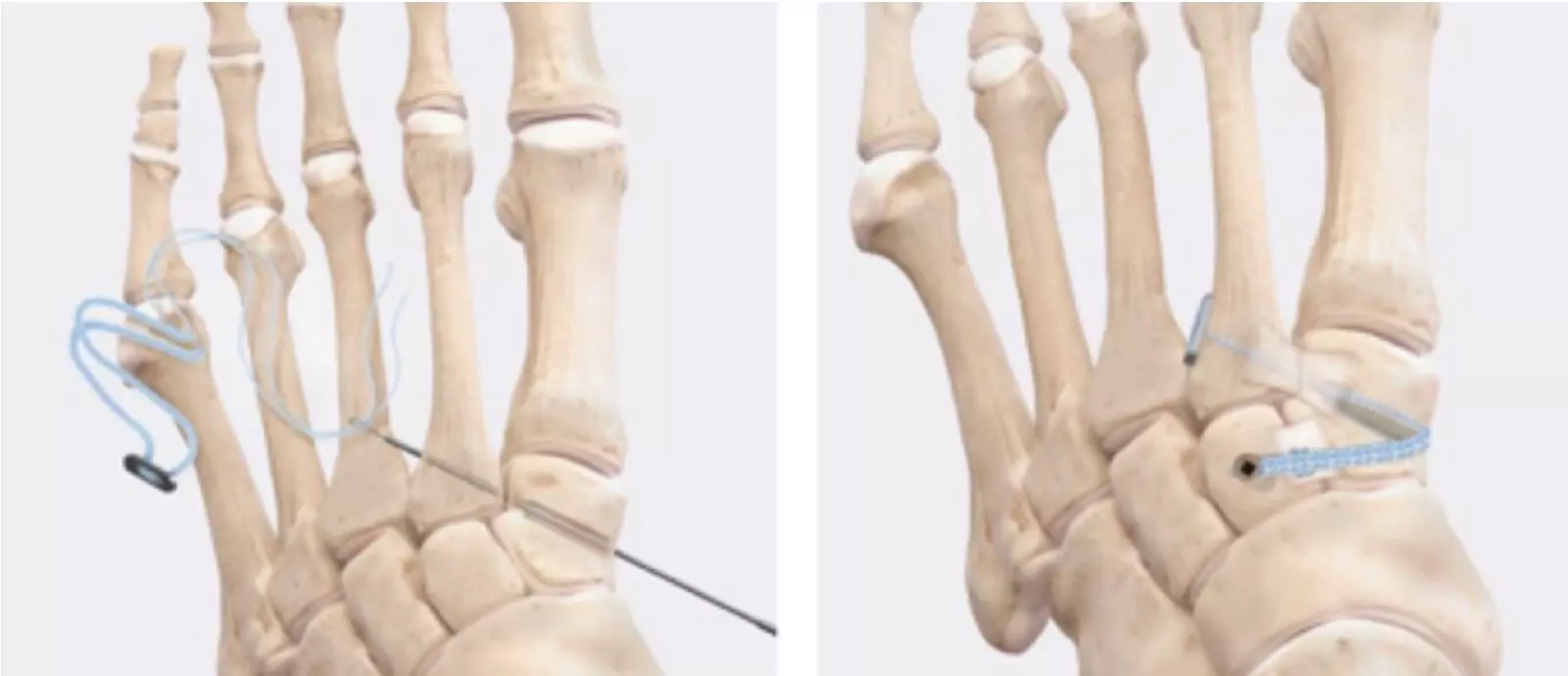
The treatment of Lisfranc injuries continues to evolve with time. Meloria Hoskins et al conducted a study to report early outcomes of patients with Lisfranc ligamentous injuries treated with the Arthrex InternalBrace, which has benefits to other previously described techniques. The article has been published in Indian journal of Orthopedics.

The authors retrospectively identified 15 adult patients with Lisfranc injuries that were treated via open reduction internal fixation with the Arthrex InternalBrace at two level one trauma centres in USA. Demographic data, mechanism of injury, and concomitant foot injuries were recorded. Outcomes were assessed by return-to-work or sport and time to weight-bearing. Secondary complications or revision surgeries were noted.

Key findings of the study were:

• Of the 15 patients identified, 8 were male and 7 were female.

• Ten out of fifteen patients were white and there was only one smoker in the group.

• Average age at time of surgery was 35.2 with a range of 18–66 years old.

• Eight patients had isolated Lisfranc ligamentous injuries and seven had additional intercuneiform instability, which required a supplemental limb of the fixation device.

• The most common mechanism of injury was a cutting/pivoting maneuver (n = 5) followed by fall (n = 4).

• The mean radiographic follow-up time was 7.3 months.

• The average time to weight-bearing as tolerated was 6.6 weeks (± 2.2).

• The average time to return-to-work/sport as tolerated was 14.1 weeks (± 3.6).

• Only two minor complications were noted at follow-up but no major complications or revision surgeries occurred.

The authors concluded that – “our retrospective review from two large academic hospitals demonstrated that for a ligamentous Lisfranc injury, the InternalBrace and additional flexible fixation limb for intercuneiform instability is a reasonable option. Our study demonstrated that with an average of 7-month follow-up, there were zero failures, all persons returned to work/play in less than 15 weeks, and unrestricted weight-bearing began at 6.6 weeks on average. This is the first paper to our knowledge that has reported outcomes of this technique in a non-biomechanical setting. Flexible suture button fixation for Lisfranc injuries continues to be recognized as a viable and stable fixation method but a future prospective, comparative study is necessary to determine if this specific InternalBrace technique is comparable or superior to other techniques..”

Further reading:

Early Outcomes of Lisfranc Injuries Treated with Arthrex InternalBrace: A Case Series

Meloria Hoskins et al

Indian Journal of Orthopaedics (2024) 58:257–262

https://doi.org/10.1007/s43465-024-01097-4

Powered by WPeMatico

Cancer immunotherapy may cause heart inflammation in some patients, reveals research

Some patients being treated with immune checkpoint inhibitors, a type of cancer immunotherapy, develop a dangerous form of heart inflammation called myocarditis. Researchers led by physicians and scientists at the Broad Institute of MIT and Harvard and Massachusetts General Hospital (MGH), a founding member of the Mass General Brigham healthcare system, have now uncovered the immune basis of this inflammation. The team identified changes in specific types of immune and stromal cells in the heart that underlie myocarditis and pinpointed factors in the blood that may indicate whether a patient’s myocarditis is likely to lead to death.

Appearing in Nature, the results are among the earliest translational findings to come from the Severe Immunotherapy Complications (SIC) Service and Clinical-Translational Research Effort, which is based at Mass General Cancer Center and includes Broad researchers. Launched in 2017, this is a first-of-its-kind program in North America focused on improving the diagnosis, treatment, and understanding of serious immunotherapy complications, which can affect nearly every organ system. The team focused on myocarditis as one of their first research projects because despite being one of the rarer complications from immune checkpoint inhibitors (ICIs), it is the most deadly.

Importantly, these findings provide the first evidence for an immune reaction in the heart that is distinct from the immune response at the tumor, suggesting that targeted treatments might be able to address myocarditis while allowing patients to continue receiving potentially life-saving anti-tumor immunotherapy. The results also highlight possible therapeutic targets that bolster the rationale behind an ongoing clinical trial recently launched at MGH that is testing a drug for this kind of heart inflammation.

Roughly 1 percent of patients treated with an ICI-more than 2,000 individuals a year in the US-will develop myocarditis, and this number goes up to nearly 2 percent among patients treated with certain immunotherapy drugs in combination. Myocarditis leads to dangerous cardiac events such as arrhythmia and heart failure in 50 percent of cases, and about a third who develop the condition will die from it, despite current treatments. In addition, treatments and supportive care approaches used for other forms of myocarditis, such as viral myocarditis, don’t work for this type.

“We don’t have great solutions now to help these patients, so we try everything to shut down the immune system and reverse myocarditis, but that’s an imprecise approach that comes with its own risks,” said study co-senior author Alexandra-Chloé Villani, an institute member at the Broad, an investigator in the Krantz Family for Cancer Research and the Center for Immunology and Inflammatory Diseases at MGH, and an assistant professor of Medicine at Harvard Medical School who leads the translational research endeavors related to the SIC Service at MGH. “Our results provide a more detailed picture of what’s happening in the heart and suggest intriguing new ways forward to improve patient care.”

“Myocarditis from immune checkpoint inhibitors is a major hurdle for us clinically,” said co-senior author Kerry Reynolds, the clinical director of inpatient oncology at MGH, director of the SIC Service, and an assistant professor of medicine at Harvard Medical School. “This study is a game-changer, paving the way to unearthing the roots of these complications. We are incredibly grateful to each and every patient who partnered with us, all those involved in their clinical care, and the exceptional team in our lab who made this research possible.”

“This work provides a biological foundation for testing more targeted therapies for myocarditis due to an immune checkpoint inhibitor. This paper is a major step forward as we need to improve our understanding of this toxicity, and this will lead to improved outcomes,” said co-senior author Tomas Neilan, an associate professor of medicine at Harvard Medical School and director of the Cardio-Oncology Program and co-director of the Cardiovascular Imaging Research Center at Mass General.

Benefits and risks

Approximately one-third of patients with cancer in the United States are eligible to receive the revolutionary drugs known as immune checkpoint inhibitors (ICIs), which are part of the immunotherapy class of medicines that take the brakes off the body’s immune system so that it can fight cancer.

The threat of serious complications and the challenge of how to manage them is growing as more patients undergo ICI treatment each year. More than 230,000 patients in the US were treated with ICIs in 2020, and that number has likely grown since then as the FDA has approved more than 80 indications for these medicines. Most patients taking one or more ICI drugs will develop at least one form of toxicity and, depending upon the drug given, ten percent to more than 50 percent will develop a severe complication. The complications can be difficult to halt or reverse, even if the treatment is stopped, and patients can develop life-threatening organ inflammation after a single dose. Doctors currently don’t have effective targeted treatments, so they often have to stop the anti-tumor therapy or give large amounts of steroids, which have their own undesired side effects such as lowering the efficacy of the ICI anti-tumor treatment.

One of the more feared complications of immunotherapy, checkpoint myocarditis is significantly more dangerous for patients than myocarditis from other causes and it’s unclear why. “Since we first started seeing checkpoint myocarditis less than a decade ago, it’s largely been a black box,” said co-first author Daniel Zlotoff, a cardiologist and assistant in medicine at MGH and postdoctoral fellow in the Villani lab. “Only now are we starting to answer the fundamental biological questions, which we hope will shed light on the optimal treatments to make it more tolerable and improve outcomes for patients.”

In the new study, the researchers collected blood from individuals who developed myocarditis while on ICI therapy and consented to be part of the study, along with paired heart and tumor tissue from some. As patients underwent diagnostic procedures at the SIC Service, or after they succumbed to the illness, samples were taken and rapidly sent to the lab, where the research team performed single-cell RNA sequencing analysis along with microscopy, proteomic analysis, and T-cell receptor sequencing to identify cells involved in driving and sustaining the inflammatory processes associated with myocarditis.

In the heart tissue of patients, the team observed the upregulation of molecular pathways that help recruit and retain immune cells involved in inflammation. They also saw an increase in abundance of several immune cell subsets, as well as an increase in abundances of certain cellular groupings composed of specific cytotoxic T cells, conventional dendritic cells (cDCs), and inflammatory fibroblasts that were found together in the hearts of patients with active disease. In the blood, they found reductions in plasmacytoid dendritic cells, cDCs, and B lineage cells along with increased numbers of other mononuclear phagocytes.

The team also analyzed the T-cell receptor, a unique protein complex that binds and responds to foreign particles known as antigens. T-cell receptors abundant in the affected heart tissue were distinct from those seen in tumors, a result that is different from findings by other researchers which suggested that the immune responses in a patient’s heart and tumor were the same. The team also found no evidence for T-cell receptors recognizing the α-myosin protein, which was previously reported to be a pivotal antigen driving checkpoint myocarditis. These results suggest that the T-cell receptors most abundant in affected heart tissue recognize undetermined antigens. In future work, the researchers hope to identify the particular antigens at play in the heart and the tumor and discern whether they are normal proteins, mutated tumor proteins, foreign particles like viruses, or something novel.

“Because the responses in the tumor and the heart are different, it makes us hopeful that we can someday disentangle the two and treat them separately,” said co-first author Steven Blum, an oncologist at MGH and postdoctoral fellow in the Villani lab. “We’re especially grateful to the patients who are willing to participate. Ultimately, it’s the biggest gift that a patient can give to research.” The researchers acknowledge that the result was only possible with crucial contributions from MGH and Broad members who lead the Rapid Autopsy Program, developed by Dejan Juric, and the hospital’s pathology team, notably James Stone.

The pattern of T cell subtypes in the blood also indicated which individuals were more likely to succumb to myocarditis, suggesting that a blood-based measurement could one day be used to flag patients who are at increased risk and should be monitored closely or avoid immunotherapy altogether. They also found T cells in the peripheral blood that originated in the heart and correlated with severity of disease. The findings open the door to developing a diagnostic blood test that could replace invasive heart biopsies for patients suspected of having myocarditis.

The work also lends support to an ongoing clinical trial (ATRIUM, NCT05335928) based at MGH exploring the use of an arthritis drug, abatacept, to control myocarditis in these patients. “We always want better patient outcomes, but we need hard evidence from clinical trials on how to resolve the inflammation while preserving anti-tumor responses,” said Reynolds. “These cell maps help guide us to what we should be studying in clinical trials.”

By treating and studying complications across different organ systems, the researchers hope to find both distinct and shared mechanisms that can shed light on adverse events that affect diverse parts of the body in these patients, often simultaneously. The researchers are also working to bring together other institutions that share the goal of improving immunotherapy and cancer patient care, and are providing guidance for similar efforts elsewhere.

“It’s important to remember that immunotherapy drugs are miracle life-saving medicines, and patients should not be afraid of them,” said Villani. “We just need to make them work better so that we can maximize their anti-tumor treatment benefit while minimizing the risk of adverse events.”

Reference:

Blum, S.M., Zlotoff, D.A., Smith, N.P. et al. Immune responses in checkpoint myocarditis across heart, blood and tumour. Nature (2024). https://doi.org/10.1038/s41586-024-08105-5.
Powered by WPeMatico

Transcranial magnetic stimulation Found Effective in Reducing Auditory Hallucinations in Schizophrenia: Study

Researchers have found that repetitive transcranial magnetic stimulation (rTMS) can efficiently reduce the symptoms of auditory verbal hallucination in schizophrenic patients. It was concluded on the basis of a randomized controlled trial where scores for the hallucination severity showed a highly significant reduction in patients treated with rTMS compared to sham-treated patients. The study was conducted by HuA Q. and colleagues published in JAMA Network Open.

Schizophrenia is a mental disorder characterized by auditory verbal hallucination whereby the drugs and prescribed doses are usually unable to be treated. Despite that these symptoms may be significantly reduced by pharmacologic treatments, they are very often accompanied by severe disturbances of metabolism as well as by movement disorders. This study aimed at evaluating the efficacy of rTMS for the treatment of auditory hallucinations and if precise neuronavigation will improve the outcome of the treatment.

Between September 2016 and August 2021, 66 participants with schizophrenia and prominent auditory verbal hallucinations were recruited from the Anhui Mental Health Center to participate in this six-week, double-blind, randomized clinical trial. Of the 66 recruited participants, 62 completed the two-week treatment phase; 53% of whom were women, with an average age of 27.4 years.

Participants were recruited into an active rTMS treatment group (n = 32) and a sham control group (n = 30). The active treatment consisted of three daily rTMS sessions over two weeks using a 70-mm figure-of-8 air-cooled coil, while the sham device just released sound and vibration but no current. This was crucial: the use of neuronavigation will optimize coil placement during rTMS sessions to target precisely the brain region associated with the generation of auditory hallucinations.

  • Compared with those receiving sham rTMS, significantly more of the patients in the active rTMS group showed a decrease in scores on the AHRS at week 2 (-5.96 points; 95% CI = 3.42 to 8.50, P < 0.001). At week 6, the difference was even larger, at -7.89 points (95% CI = 4.77 to 11.01, P < 0.001).

  • At six weeks, RTerapy was sustained and at both week 2 and week 6, there were significant time-by-group interactions for AHRS scores (P<0.001 for both time points).

  • Higher strength of the electric field due to TMS was associated with greater reductions in hallucination severity, P=0.002, implying that treatment accuracy contributes to this effect.

Based on this study’s findings, rTMS with neuronavigation significantly reduces auditory verbal hallucinations in patients with schizophrenia. This advance would be promising in symptom management in schizophrenia, however, provided that the technique becomes more accessible for broader clinical utility.

Reference:

Hua, Q., Wang, L., He, K., Sun, J., Xu, W., Zhang, L., Tian, Y., Wang, K., & Ji, G.-J. (2024). Repetitive transcranial magnetic stimulation for auditory verbal hallucinations in schizophrenia: A randomized clinical trial. JAMA Network Open, 7(11), e2444215. https://doi.org/10.1001/jamanetworkopen.2024.44215

Powered by WPeMatico