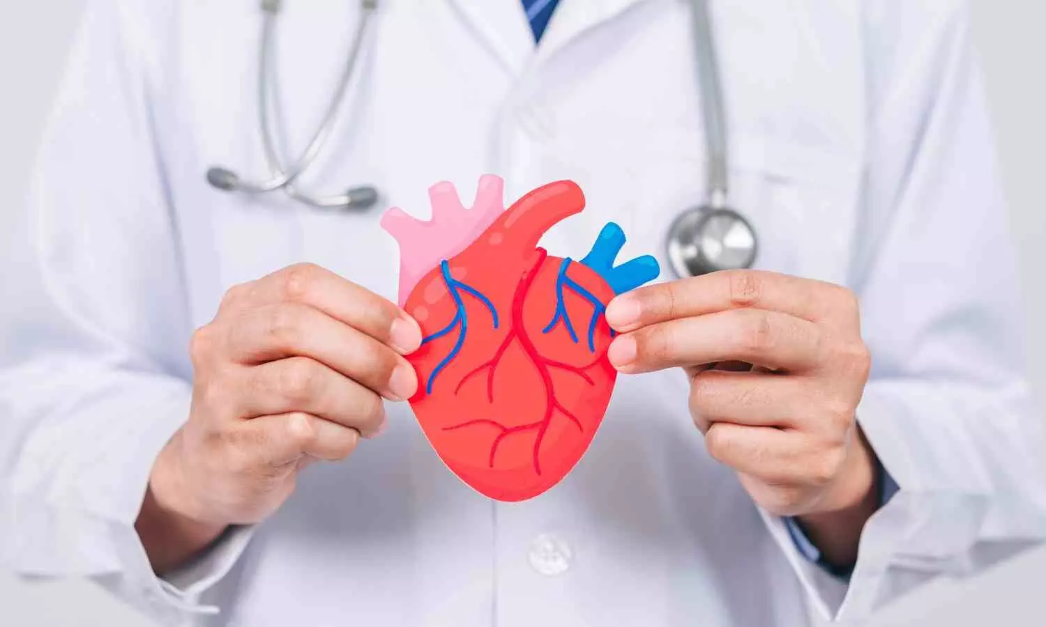
Reconditioned pacemakers worked as well as new ones, finds study

A randomized trial that compared previously used and new pacemakers in patients found the reconditioned devices were as safe and effective as new pacemakers, potentially offering affordable options for patients in low-and middle-income countries.

This was the finding of late-breaking science presented today at the American Heart Association’s Scientific Sessions 2024. The meeting, Nov. 16-18, 2024, in Chicago, is a premier global exchange of the latest scientific advancements, research and evidence-based clinical practice updates in cardiovascular science.

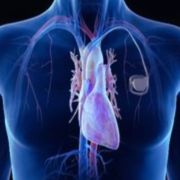
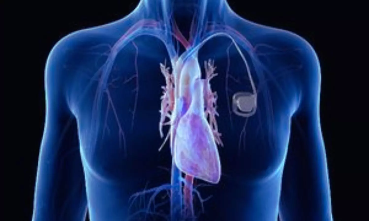
Reusing pacemakers is prohibited in the U.S. However, the U.S. Food & Drug Administration allows re-sterilized devices to be exported for reuse. Pacemakers were recovered from deceased patients and patients undergoing surgeries that required the removal of their existing devices. Pacemakers with at least six years of battery life and proper electrical function were sterilized for reuse.

The “My Heart Your Heart” (MHYH) study compared the function of refurbished pacemakers to new devices in a randomized trial of nearly 300 adults in seven countries.

“Access to pacemakers is limited in many low- and middle-income countries due to the relatively high cost of the devices,” explained lead study author Thomas Crawford, M.D., a professor of internal medicine in the division of cardiovascular disease at the University of Michigan Medical School in Ann Arbor, Michigan. “In some high-income countries, as many as 1,000 people per million population may receive a pacemaker annually. In low-income countries, it could be 3 per million population or fewer who get pacemakers each year.”

Previous research has suggested that refurbished pacemakers could be safely implanted. However, those studies were retrospective, noting trends from previously collected information. This is the first prospective, randomized study that included patient outcomes related to function and infection with reconditioned devices, with follow-up information collected up to 90 days after implantation.

“With reconditioned pacemakers, there is worry about whether the device will cause infection and whether it will function properly,” Crawford said. “The 90-day endpoint of the My Heart Your Heart study addresses the most immediate concern of infection because most of the infections related to the implantation procedure occur within that period.”

The trial assessed outcomes in patients evaluated in the hospital at two weeks after pacemakers were implanted and for up to 90 days afterward.

The analysis found:

Five cases of infection at the implant site (device pocket infections) required implant removal: three of which were among the patients who had received new devices and two patients in the group that had received reconditioned pacemakers.

A superficial skin infection responsive to antibiotics occurred in one patient with a new device. This device did not have to be removed, and the infection cleared.

Five patients with new pacemakers required surgery to move or replace the device’s leads after pacemaker implantation, compared to six patients with reconditioned devices. Lead dislodgement is a known complication of a standard pacemaker implantation procedure.

No device malfunctions were reported among any patients.

Three deaths unrelated to the device implantation occurred in the reconditioned group and none in the new pacemaker group.

The researchers concluded that reconditioned pacemakers were comparable to new pacemakers in terms of safety and effectiveness up to 90 days after implantation; however, longer-term follow-up is necessary.

“Our study shows pacemaker recycling is green, good for the environment and can save the lives of people in other countries who can’t afford a new device,” Crawford said. “However, longer-term follow-up will be necessary to confirm the safety and efficacy of reconditioned devices, including whether they continue to work as expected and whether the batteries depreciate at the expected rate.”

Powered by WPeMatico

Coronary calcium scores effectively predict risk of heart attack and overall mortality in both women and men: Study

Coronary artery calcium (CAC) scores have become a non-invasive way for physicians to easily determine how much plaque has built up inside a patient’s coronary arteries, but the question has been how accurate the score is in identifying women, as well as men, who are at high risk for a heart attack or death.

Now, a major new study by researchers at Intermountain Health in Salt Lake City finds coronary artery calcium scores are not only highly effective in identifying those at risk for future heart attacks, but also for death, and risk prediction was found to be as good in women as in men.

Results from the Intermountain Health study found that in addition to predicting risk of coronary death and non-fatal heart attacks, coronary artery calcium scores also correlated to all-cause mortality. Researchers found that people with a score of zero were three times less likely to die from any cause.

“The coronary artery calcium score appears to be an excellent and accurate indicator of health and overall prognosis, even beyond heart disease,” said Jeffrey L. Anderson, principal investigator of the study and distinguished research physician at Intermountain Health.

Findings from the Intermountain Health study were presented at the American Heart Association National Scientific Sessions 2024 in Chicago on Monday, Nov. 18, 2024.

Coronary artery calcium testing is becoming more common in heart healthcare. These tests are non-invasive, use as little radiation as a mammogram, and are relatively cheap, especially compared to PET stress tests, coronary CT angiograms, or coronary angiography.

For the Intermountain Health study, researchers reviewed the electronic health records of 19,495 women and 20,523 men who had undergone PET/CT scans because their physicians suspected they had a risk of heart disease, but who hadn’t yet had a heart event, such as a heart attack.

Of these patients, 7,967 had a CAC score of zero, meaning they had no calcified plaque in their coronary arteries. Of this group, women on average were older than men (60.5 years for women, 53.8 for men), which correlates to how women often develop heart disease later in life than men, Dr. Anderson said.

Researchers then followed up on patients about two years later. They found that a zero CAC score predicted a low risk for coronary death or nonfatal heart attacks in both men and women. CAC scores of zero were more frequently observed in women despite having an older average age.

Intermountain researchers also found that those with a zero CAC had three times lower risk of all-cause death or heart attack in both sexes.

While it’s obvious why a CAC score of zero would be a good indicator of heart health, its ability to predict all-cause mortality is novel and deserves additional study.

“We will be exploring this further to understand better why a zero-calcium score is such a good signal to overall health,” said Dr. Anderson.

Reference:

New study finds prognostic value of coronary calcium scores effective in predicting risk of heart attack and overall mortality in both women and men, Intermountain Healthcare, Meeting: AHA Scientific Sessions 2024.

Powered by WPeMatico

Topical human platelet extract and vitamin C both help rejuvenate skin on the dorsal hands: Study

A new study published in the journal of Plastic and Reconstructive Surgery showed that after using the human platelet extract (HPE) and vitamin C  twice a day for 12 to 26 weeks were able to revitalize the skin on the dorsal hands. Traditionally, the face has been the primary target of topical skin-rejuvenating therapies, while the hands and other visible body parts have received less attention. Skin photoaging can be corrected using a variety of efficient therapies.

Topical human platelet extract (HPE) and topical L-ascorbic acid (vitamin C), a well-known antioxidant, are new off-the-shelf cosmetic ingredients that have demonstrated promising outcomes in recent clinical trials. HPE is a leukocyte-depleted allogeneic product made from pooled, apheresed platelets imported from the United States. In order to determine how topical HPE (plated) intense serum (Rion Aesthetics) and vitamin C (C E Ferulic with 15% L-ascorbic acid) affected the skin renewal of dorsal hands after 12 to 26 weeks of twice-daily use, Saranya Wyles and her team carried out this study.

This prospective, long-term study used photographic documentation was used at baseline, 6, 12, and 26-week evaluations to evaluate typical aging-related skin issues. The primary finding of this study was found that at 12 weeks following twice-daily topical application, topical HPE was not inferior to topical vitamin C for improving the fractional areas of wrinkles, brown spots, and luminosity on the dorsal hands. Despite their different function, vitamin C and HPE were both useful skin-rejuvenation therapies.

While vitamin C serves as an antioxidant and free radical seeker, protecting cells from oxidative stress, HPE offers other benefits for skin rejuvenation, including angiogenesis, collagen formation, and inflammatory management. Both compounds in a topical serum, brighten and level out skin tone while reducing photoaging symptoms including wrinkles, erythema, and brown spots when applied consistently as part of a skin care regimen. Only 50% of the overall sample population completed the research between 12 and 26 weeks, and there were few darker skin types (8% Fitzpatrick IV or V, 0 type VI), as well as a limited number of men. Overall, after 12 to 26 weeks of twice-daily topical use, HPE is equally effective as vitamin C to rejuvenate the skin on the dorsal hands.

Source:

Wyles, S., Jankov, L. E., Copeland, K., Bucky, L. P., Paradise, C., & Behfar, A. (2023). A Comparative Study of Two Topical Treatments for Photoaging of the Hands. In Plastic & Reconstructive Surgery (Vol. 154, Issue 5, pp. 978–984). Ovid Technologies (Wolters Kluwer Health). https://doi.org/10.1097/prs.0000000000011240

Powered by WPeMatico

New oral drug derived from Oxytocin May calm abdominal pain in IBS patients, reveals research

University of Queensland researchers have developed a new class of oral painkillers to suppress chronic abdominal pain that is based on the peptide hormone oxytocin that drives childbirth contractions.

Associate Professor Markus Muttenthaler from UQ’s Institute for Molecular Bioscience led a team that has changed the chemical structure of oxytocin to make it gut-stable after earlier work revealed the hormone could treat abdominal pain.

Dr Muttenthaler said there was an urgent need for new treatments for the chronic pain caused by gastrointestinal disorders such as irritable bowel syndrome (IBS) and irritable bowel disease (IBD).

“This pain affects up to 15 per cent of adults in their lifetime, and all we have are anti-inflammatories and opioids which can cause side effects and addiction,” Dr Muttenthaler said.

“Our research focuses on peptides that are highly potent and selective molecules and have few side effects. However, nearly all peptide drugs must be injected as they are rapidly digested in the gut.

“We have now developed a way of making peptides gut-stable so they can be given orally.

“This is a new and highly promising approach to treating gut disorders.”

Oxytocin is a peptide hormone produced in the brain which is known as the ‘bonding hormone’ or the ‘love molecule’ due to its effects on relationship building, empathy and trust.

Oxytocin is also the key hormone that induces uterine contractions during labour and facilitates milk release during breastfeeding.

“We identified the parts of oxytocin that are rapidly broken down by gut enzymes and used medicinal chemistry to render them gut-stable while ensuring that the new molecule was still able to activate the oxytocin receptor,” Dr Muttenthaler said.

“We now have a new class of molecules that are potently active but do not degrade in the stomach or intestine, meaning they can be taken orally.”

The research indicates that the new molecules work in the colon and do not need to cross the gut barrier into the bloodstream to suppress abdominal pain.

“This is a very safe therapeutic approach as it reduces the risk of side effects in the rest of the body, a problem with many other systemic drugs,” Dr Muttenthaler said.

“This is an exciting new mode of action to prevent pain.

“We are currently looking for investors to accelerate pre-clinical studies, with the goal of taking it into the clinic.

“Now that we have perfected making peptides stable, we are looking at other gut drugs to improve treatment options for gastrointestinal disorders, an unmet medical need.”

Reference:

Thomas Kremsmayr, Gudrun Schober, Matthias Kaltenböck, Bradley L. Hoare, Stuart M. Brierley, Markus Muttenthaler, Oxytocin Analogues for the Oral Treatment of Abdominal Pain, Angewandte Chemie,  https://doi.org/10.1002/anie.202415333

Powered by WPeMatico

Severe Anemia Elevates Risk of PPH During Elective Labor Induction, reveals research

According to researchers, pregnant women with severe anemia stand at a more substantial risk of experiencing postpartum hemorrhage (PPH) if labor is induced electively during late pregnancy. A new study was recently published in the Journal of Gynecology and Obstetrics conducted by Tuck S. and colleagues.

The most important cause of maternal mortality in many parts of the world is PPH. Anemia in pregnancy is a known risk factor for PPH, though the ways in which anemia severity and methods of labor induction/augmentation interact to influence risk for PPH has not been well understood. This study examines in detail how those factors interact to raise risk for PPH and provides insights that can guide clinical decision-making in obstetric care.

This prospective cohort study enrolled 9,420 pregnant women across India in 13 hospitals. Hemoglobin at ≥28 weeks of gestation was assessed and blood loss after delivery during follow-up. PPH was defined as blood loss ≥500 mL for vaginal births or ≥1,000 mL for cesarean sections. Anemia severity was defined by the following:

  • No/Mild: Hemoglobin ≥10 g/dL

  • Moderate: Hemoglobin 7 to 9.9 g/dL

  • Severe: Hemoglobin <7 g/dL

The indications of labor induction/augmentation were categorized as either clinically indicated or elective, and multivariable Poisson regression was used to compute associations between these factors and PPH, adjusting for confounders.

Anemia Severity and Risk of PPH:

  • Patients with severe anemia (hemoglobin <7 g/dL) were at strongly increased risk of PPH.

  • Associations were not significant at no/mild or moderate anemia levels.

Impact of Labor Induction/Augmentation:

  • Elective labor induction significantly increased the risk for PPH in women with severe anemia (adjusted risk ratio, 3.44; 95% CI: 1.29–9.18).

  • Clinically indicated inductions were not associated with a significant increase in the risk for PPH in this group (adjusted risk ratio, 1.22; 95% CI: 0.42–3.55).

Interaction Between Factors:

  • A significant interaction was found between anemia severity and type of labor induction/augmentation regarding the risk for PPH (P = 0.003).

Labor management should be stratified according to the severity of anemia. Induction/augmentation of labour is safe in most women with anemia, but elective procedures should be avoided in severe cases to minimize the risk of PPH. These findings stress the need for individualised obstetric care for better maternal outcomes.

Reference:

Cheng, T. S., Zahir, F., Solomi, C., V., Verma, A., Rao, S., Choudhury, S. S., Deka, G., Mahanta, P., Kakoty, S., Medhi, R., Chhabra, S., Rani, A., Bora, A., Roy, I., Minz, B., Bharti, O. K., Deka, R., Opondo, C., Churchill, D., … Nair, M. (2024). Does induction or augmentation of labor increase the risk of postpartum hemorrhage in pregnant women with anemia? A multicenter prospective cohort study in India. International Journal of Gynaecology and Obstetrics: The Official Organ of the International Federation of Gynaecology and Obstetrics. https://doi.org/10.1002/ijgo.16008

Powered by WPeMatico

Nasal spray version of bumetanide has potential to help treat heart failure, reveals study

A new nasal spray form of the medication bumetanide may reduce the tissue swelling caused by heart failure as effectively as the standard oral and intravenous formulations of the medication, according to late-breaking science presented today at the American Heart Association’sScientific Sessions 2024. This research is simultaneously published today in the American Heart Association journal Circulation.

Heart failure occurs when the heart does not pump blood as well as it should, resulting in decreased blood flow to organs and fluid buildup in the lungs and other tissues. Lifestyle changes, including controlling obesity, smoking cessation, being physically active, controlling high blood pressure and controlling blood glucose levels can help prevent the development of heart failure.

Medications used to treat heart failure include diuretics, which reduce tissue swelling and can be administered orally or intravenously. Bumetanide is one of the standard diuretics administered orally or intravenously to reduce tissue swelling from heart, kidney or liver disease by removing excess salt and water through the urine.

In this clinical trial called RSQ-777-02, researchers explored a new nasal spray form of bumetanide in healthy adults. They compared its absorption and ability to reduce swelling to those of oral and intravenous bumetanide among 68 adults who did not have heart failure or risk factors for heart failure at the time of enrollment.

“In patients with heart failure, the body’s ability to absorb oral medications in the stomach and gut often decreases as fluid overload builds up (this is called diuretic resistance) and, thus, oral medications are often least effective when the body needs them most,” said study presenting author Daniel Bensimhon, M.D., medical director of the Advanced Heart Failure/Mechanical Circulatory Support Program at Cone Health in Greensboro, North Carolina. “Having a diuretic that does not rely on gut absorption to be effective may be a very important tool to help patients with heart failure and other conditions without having to rely on intravenous administration of these medications, which can only be done at hospitals and in heart failure clinics.”

The study found:

  • The nasal spray was absorbed effectively and was safe, meaning that side effects were consistent with the other versions of administration and that it had fewer treatment-emergent adverse events compared to the oral version.
  • Compared with the oral and IV versions of bumetanide, the nasal spray formulation led to a similar amount of urine output.
  • Compared with the oral version, people treated with the nasal spray bumetanide reached a similar blood concentration, and the medication was absorbed 33% faster. The absorption rate of the IV form was faster than both the oral and nasal versions, however, the onset of sodium excretion through urine was faster with the nasal version, the authors noted. Previous research has suggested urinary sodium as a biomarker of diuretic response in heart failure.
  • Study participants received all three forms of bumetanide in various order. The nasal and IV forms of bumetanide were absorbed at more reliable rates than the oral form for each study participant – a measure known as intrasubject variability. The nasal and IV forms had a variability in absorption of 27%, compared to >40% for the oral form, suggesting that the nasal and IV forms are more stable routes of dosing, the authors noted. This would be an advantage for the nasal spray over the oral form for self-administration of the medication at home, they added.

“We were surprised by how fast the nasal spray worked and by how variable the absorption of the oral drug was even in healthy subjects,” Bensimhon said. “Patients who require diuretic treatment for swelling associated with chronic heart failure and liver disease may now have a new, self-administered option, particularly when they are unable to take their oral medication or it no longer works.”

The study’s primary limitation is that participants were healthy and did not have heart failure or risk factors for developing heart failure at the time of enrollment. Now that safety and tolerability have been established in healthy adults, the authors said they will be conducting future studies to evaluate the bioavailability and clinical effectiveness of intranasal bumetanide in patients with heart failure.

“We think this will be a valuable tool for treating heart failure by promoting care at home and potentially reducing the need for costly hospital admissions and readmissions,” Bensimhon said. “Keeping patients at home is good for the patient and our health systems.”

Study background and details:

  • The RSQ-777-02 clinical trial was conducted at Orange County Research Center in Irvine, California, from December 2023 to April 2024.
  • The study included 68 healthy adults, ages 18 to 55 years old, who did not have heart failure or risk factors for developing heart failure at the time of enrollment.
  • 66.2% of participants identified as men; 33.8% as women. 60.3% of participants self-identified as white adults, 27.9% as Black adults, 10.3% as Asian adults and 1.5% self-identified as adults of “other” race. 32.4% identified as Hispanic or Latino ethnicity and 67.6% as not Hispanic or Latino ethnicity.
  • The researchers administered nasal, oral and intravenous bumetanide. Each participant received all three forms of bumetanide in a varied order.
  • Study participants were monitored on-site for 10 days.

Reference:

Nasal spray version of common diuretic has potential to help treat heart failure, American Heart Association, Meeting: AHA Scientific Sessions 2024.

Powered by WPeMatico

Transscleral Cyclophotocoagulation effectively lowers IOP among glaucoma patients:Study

Glaucoma encompasses a heterogeneous group of eye diseases
associated with progressive loss of optic nerve fibers, with consequent loss of
corresponding visual field. Several risk factors have been identified for the
development and progression of this condition, but intraocular pressure (IOP)
is still the primary modifiable risk factor. IOP-lowering can be achieved with
eye drops or laser trabeculoplasty (both currently considered to be first line
options in most glaucoma cases), or surgery, which is mostly reserved for
patients who progress despite non-surgical maximal tolerated treatment or if
there is a very advanced disease stage or glaucoma type where non-surgical
treatment is not expected to be able to lower IOP to the levels that are expected
to stop disease progression. The majority of surgeries performed are filtering
surgeries and aim to lower IOP through an increased outflow, but there are also
options that aim for a reduction of aqueous humour (AH) production, such as the
cyclodestructive or cyclomodulating procedures, which are still usually used as
a last resource or when other types of surgery are deemed very likely to be
unsuccessful.

In cyclodestructive procedures, unlike other surgeries, the
IOP lowering effect is obtained by ablation of the ciliary body with subsequent
reduction of AH production. The most widely used technique is Laser Cyclophotocoagulation
(CPC), namely the Diode Laser Transscleral Cyclophotocoagulation (TSCPC).
During TSCPC, a semiconductor diode laser (810 nm) is transmitted through the
overlying sclera and absorbed by melanin in the ciliary processes, leading to
selective thermal coagulation of the ciliary body. Despite the simplicity of
the procedure (comparing to incisional filtering surgery), cycloablation is
feared among some ophthalmologists due to the risk of persistent hypotony,
phthisis bulbi and loss of visual acuity. The risk of hypotony with TSCPC can
be as high as 18% (namely in cases of neovascular glaucoma (NVG). Moreover, the
reported success rates are highly variable (36.7–94.4%), possibly due to
heterogeneity in methodologies regarding the definitions of patient population,
energy settings, success and follow-up duration. It is also important to
acknowledge that the majority of the studies only established an upper IOP
limit for the definition of success, therefore not excluding patients with
hypotony.

Despite the increasing evidence regarding TSCPC, according
to the latest version of the European Glaucoma Society Guidelines, refractory
glaucoma (glaucoma for which the target intraocular pressure (IOP) has not been
achieved despite maximum tolerated medical treatments and/or conventional,
properly performed filtration surgery) or expected incisional surgery failure
remain the only formal indications for TSCPC use.

There is a need for more evidence concerning this procedure,
in order to try to expand its indications. Therefore, this study intended to
provide more data regarding the efficacy and safety of TSCPC, based on a
Portuguese population with glaucoma or ocular hypertension treated at a
tertiary centre, with a 2-year follow-up post-procedure.

Ribeiro et al carried a retrospective review of the records
of all adult patients who underwent their first TSCPC treatment between 2014
and 2019 at Unidade Local de Saúde de São João, Porto, Portugal. Data regarding
intraocular pressure (IOP), best corrected visual acuity, number of
IOP-lowering medications, use of oral acetazolamide, retreatments and
complications during a 2-year period following TSCPC were registered. The
primary outcome was overall success at 2 years, defined as IOP≥ 6 and ≤21 mmHg,
with at least 20% IOP reduction from baseline, with or without IOP-lowering
medications (qualified and complete success, respectively), without the
development of phthisis bulbi or loss of light perception due to glaucoma and
no further glaucoma procedures except TSCPC retreatment.

Ninety-six eyes from 96 patients were included, mean age was
63 (±14) years. Mean IOP at baseline was 39.1 (±13.3) mmHg. Mean IOP reduction
at 2 years was 18.5 (42.9%) mmHg (±16.0, min −16.0, max 56.0) (p < 0.001)
and a significant reduction in the number of IOP-lowering medications and use
of oral acetazolamide was observed. IOP reduction at 2 years was positively
correlated with baseline IOP (r=0.682; p < 0.001). Overall success (including
complete and qualified) was achieved in 42 patients (43.8%), with 34 (35.4%)
presenting qualified success. Neovascular glaucoma (NVG) was the predominant
diagnosis (n = 30, 31.3%), with a higher mean baseline IOP of 46.3 mmHg (±11.8,
min 21.0, max 70.0) and a larger mean IOP reduction at 2 years of 24.7 (51.0%)
mmHg (±16.4, min −2.0, max 55.0). Thirteen patients (13.5%) developed
persistent hypotony, eight of which converted to phthisis bulbi, of which half
had NVG.

The results of the study demonstrated a significant effect
of TSCPC in reducing both IOP and the need for IOP-lowering topical and/or oral
medications in the management of patients with glaucoma. The overall success
rate was 37.5% at 12 months and 43.8% at 24 months. The majority (23 patients,
64%) of patients with overall success after 12 months also achieved success at
24 months without the need of additional TSCPC, suggesting a sustained effect
of this procedure between the first and second year. Nonetheless, the rate of
complications observed is not negligible, with 13 (13.5%) of the patients
developing persistent hypotony, and of these, 8 evolved to phthisis bulbi.
Regarding BCVA, 26.3% of patients with vision (light perception, hand motion
and counting fingers) progressed to no light perception.

In conclusion, the study shows that TSCPC can be an
effective IOP-lowering procedure, demonstrating a stronger effect when
preoperative IOP is highest. Authors confirmed that there is a wide variability
in the effect and a considerable amount of postoperative complications, such as
persistent hypotony and phthisis bulbi, which are more pronounced in certain
glaucoma types such as NVG. More studies are required, ideally with less
advanced cases, to establish appropriate energy levels, duration and extension,
based on each patient’s clinical characteristics, thus allowing for higher
success rates, more predictability and less complications.

Source: Ribeiro et al; Clinical Ophthalmology 2024:18

https://doi.org/10.2147/OPTH.S473788

Powered by WPeMatico

Sensitive assessment tools like Teate Depression Inventory may accurately diagnose Antenatal Depression, study finds

Antenatal depression presents as feelings of sadness, sleep problems, disorganized behavior, irritability, and restlessness during pregnancy. If not properly identified, it remains untreated and has links to increased suicide risk, higher chances of postpartum depression, preeclampsia, premature birth, low birth weight, impaired mother-child interactions, and severe obstetric complications. Recent findings highlight the crucial need to prevent depression risk during pregnancy. Recent study examined the predictive validity of socio-demographic and psychological factors in the development of antenatal depression (AD) in a sample of 247 Italian pregnant women. AD is characterized by low mood, insomnia, disorganized behavior, irritability, and agitation during pregnancy. If left untreated, AD can have serious consequences, including increased risk of suicide, postpartum depression, preeclampsia, preterm birth, low birth weight, poor mother-child interactions, and other adverse obstetric outcomes.

Assessment of Depressive Symptoms

The researchers assessed depressive symptoms using the Edinburgh Postnatal Depression Scale (EPDS) and the Teate Depression Inventory (TDI), a new measure designed to better differentiate between depressive symptoms and normal pregnancy-related somatic changes. They also collected data on demographic factors like age, marital status, employment, and education level.

Results and Predictive Risk Factors

The results showed that only the psychological factors, as measured by the TDI, were significant predictive risk factors for AD. There were no significant associations between AD and the demographic variables examined. Specifically, higher scores on the TDI were associated with higher scores on the EPDS, indicating more severe depressive symptoms. In contrast, the demographic factors did not significantly predict EPDS scores.

Implications and Recommendations

These findings suggest that screening for and treating depressive symptoms during pregnancy, rather than focusing on socio-demographic risk factors, may be the most effective approach for preventing the negative consequences of AD. Early identification and intervention for depressive symptoms in pregnancy could help reduce the risk of postpartum depression and associated complications. The researchers recommend using reliable, sensitive assessment tools like the TDI to accurately diagnose and monitor AD. Further research with larger, more diverse samples is needed to confirm and extend these results.

Key Points

1. The study examined the predictive validity of socio-demographic and psychological factors in the development of antenatal depression (AD) in a sample of 247 Italian pregnant women.

2. AD is characterized by low mood, insomnia, disorganized behavior, irritability, and agitation during pregnancy, and can have serious consequences if left untreated.

3. The researchers assessed depressive symptoms using the Edinburgh Postnatal Depression Scale (EPDS) and the Teate Depression Inventory (TDI), and also collected data on demographic factors.

4. The results showed that only the psychological factors, as measured by the TDI, were significant predictive risk factors for AD, while the demographic variables did not significantly predict EPDS scores.

5. The findings suggest that screening for and treating depressive symptoms during pregnancy, rather than focusing on socio-demographic risk factors, may be the most effective approach for preventing the negative consequences of AD.

6. The researchers recommend using reliable, sensitive assessment tools like the TDI to accurately diagnose and monitor AD, and call for further research with larger, more diverse samples to confirm and extend these results.

Reference –

Sergi, M.R., Saggino, A., Balsamo, M. et al. Risk factors of the antenatal depression in a sample of Italian pregnant women: a preliminary study. BMC Pregnancy Childbirth 24, 689 (2024). https://doi.org/10.1186/s12884-024-06704-8.

Powered by WPeMatico

Lung ultrasound effective as point-of-care investigation to diagnose childhood pneumonia, reveals research

Lung ultrasound is effective as a point-of-care investigation to diagnose childhood pneumonia, reveals research published in Lung India.

Lung ultrasound is emerging as a rapid, simple and safe alternative for diagnosing pneumonia since it has a higher sensitivity than X-rays and lower radiation exposure than computerized tomography. This is a prospective observational study done at a tertiary care centre in Chennai to study the diagnostic utility of lung ultrasound in pneumonia. Children aged 1 month to 12 years who were admitted to the hospital with complaints of cough, fever and/or breathing difficulty and on examination had tachypnea and/or chest indrawing were included in the study. All children underwent chest X-rays which was a standard hospital protocol. At admission, an independent investigator who was blinded to the clinical and radiological features of the child performed lung ultrasound. In all children, the final diagnosis of pneumonia was made by another independent expert paediatrician on the basis of the clinical features and chest X-ray. The test characteristics of ultrasound and chest X-ray were compared against this gold standard of physician-diagnosed pneumonia. Results: Out of the 252 children studied, 225 (89.3%) had pneumonia while the rest 27 (10.7%) had other diagnoses. Among the 225 children with pneumonia, 223 (99.1%) were detected by ultrasound while 157 (69.8%) were detected by chest X-ray. All the test characteristics such as sensitivity, specificity, positive and negative predictive values of ultrasound were higher than those of chest X-ray. The sensitivity and specificity of ultrasound to diagnose pneumonia were 99.11% and 81.48%, respectively, while the sensitivity and specificity of X-ray for the same were 69.77% and 74.07%, respectively. Overall diagnostic accuracy for chest ultrasonography was 97.22% (94.36% to 98.88%), whereas for chest radiography, it was found to be 70.24% (64.18% to 75.81%).While both modalities were able to diagnose pneumonia significantly, ultrasound had better strength of association (Cramer’s V value = 0.849) than X-ray to the final diagnosis. Lung ultrasound can be employed as a point-of-care investigation to diagnose pneumonia in suspected cases and can even replace chest X-ray in such circumstances.

Reference:

Vaitheeswaran, Gayathri, et al. “Point-of-care Lung Ultrasound in the Diagnosis of Childhood Pneumonia.” Lung India : Official Organ of Indian Chest Society, vol. 41, no. 6, 2024, pp. 411-415.

Keywords:

Lung, ultrasound, effective, point-of-care, investigation, diagnose, childhood, pneumonia, reveals, research, lung India, Vaitheeswaran, Gayathri

Powered by WPeMatico

Routine imaging in postoperative follow-up of patients with pancreatic ductal adenocarcinoma may help improve overalls survival: JAMA

Recent research has shown that, in comparison to symptom-based follow-up, routine imaging in follow-up after pancreatic resection for pancreatic ductal adenocarcinoma (PDAC) is associated with survival and a higher rate of treatment targeted towards recurrence. The study was conducted by Paul C. and colleagues published in JAMA Surgery journal.

PDAC is considered one of the most aggressive forms of malignancy with high recurrence following surgical resection. The overall objective of this research was to examine how these follow-up interventions impacted overall survival and chances of access to recurrence-specific treatments in patients with recurrent PDAC.

This was a prospective, cross-sectional trial conducted in 13 countries and 33 E-AHPBA centers across the years 2020 to 2021, recruiting patients with PDAC. A total of 333 participants, with a mean age of 65 years (SD, 11 years), and 184 male patients (55%), had follow-up after pancreatic resection assessed. Two approaches to the strategy of follow-up were compared: symptomatic follow-up without routine imaging and routine imaging follow-up. The overall survival was analyzed through Kaplan-Meier estimates. Logistic and Cox proportional hazard models were used to control the confounding factors.

Key Findings

  • Of 333 patients with PDAC recurrence, routine imaging follow-up was done in 71% (235 patients), and the follow-up was based on symptoms in 29% of the patients (98 patients).

  • Median overall survival was significantly longer for the routine imaging group, at 28 months (95% CI, 24-30 months), versus 23 months (95% CI, 19-29 months) in the symptomatic follow-up group (P = 0.01).

  • Routine imaging follow-up was associated with a higher likelihood of receiving recurrence-focused treatment (adjusted odds ratio, 2.57; 95% CI, 1.22-5.41; P =0.01).

  • In contrast, overall survival was prolonged among patients who had routine imaging at regular intervals, with an adjusted hazard ratio of 0.75 (95% CI, 0.56-0.99; P = 0.04).

Routine imaging post-resection of PDAC was independently associated with greater recurrence-focused treatment and longer survival, and an overall survival rate of 28 months in patients monitored by routine imaging versus symptomatic follow-up at 23 months. Specific imaging protocols must be further explored as well as their cost-effectiveness, and then uniform, evidence-based guidelines for follow-up care have to be developed.

Reference:

Andel, P. C. M., van Goor, I. W. J. M., Augustinus, S., Berrevoet, F., Besselink, M. G., Bhojwani, R., Boggi, U., Bouwense, S. A. W., Cirkel, G. A., van Dam, J. L., Djanani, A., Dorcaratto, D., Dreyer, S., den Dulk, M., Frigerio, I., Ghorbani, P., Goetz, M. R., Groot Koerkamp, B., Gryspeerdt, F., … Wellner, U. (2024). Routine imaging or symptomatic follow-up after resection of pancreatic adenocarcinoma. JAMA Surgery. https://doi.org/10.1001/jamasurg.2024.5024

Powered by WPeMatico