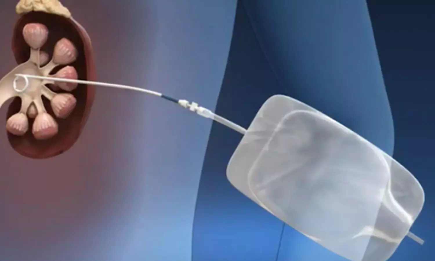
CT-Defined Coronary Artery Calcification may predict overall survival and major CV events in Lung Cancer patients: Study

A recent study published in the
journal Academic Radiology found that coronary artery calcification can be used
as a diagnostic tool for estimating overall survival and predicting the major
cardiovascular events in individuals with lung cancer.

Lung cancer is the leading cause
of increased morbidity and mortality globally. Computed Tomography (CT) scan is
used for diagnosing, staging, and assessing the prognosis of lung cancer.
Literature shows that Coronary artery calcification (CAC) can be used to diagnose
and quantify v=cardiovascular diseases using a CT scan. Agatston score is used
to quantify CAC based on the cardiac-gated CT images. Previous studies
showed that artificial intelligence algorithms can calculate CAC scores in oncology
patients. As there is ambiguity in using the CAC score in lung cancer,
researchers have conducted a systematic review to establish the effect of the CAC
score on overall survival (OS) in lung cancer patients.

Literature databases like the
MEDLINE library, Google Scholar, and SCOPUS databases were screened for papers
analyzing the association between CAC and overall survival in lung cancer
patients up to June 2024. The study included lung cancer patients in whom CT
can define CAC for the overall survival of major adverse cardiac events. The
primary endpoint of the systematic review was overall survival (OS) presented
as hazard ratio for CAC with a reported 95% confidence interval and p-value in
univariable and multivariable analyses.

Findings:

  • The included studies comprised 2292 patients
    undergoing curative treatment.
  • The pooled hazard ratio for the association
    between CAC score and OS was HR= 1.42 (95% CI=(1.19; 1.69),
    p < 0.0001) in the univariable analysis and HR= 1.56 in the
    multivariable analysis.
  • A higher CAC score was associated with poor
    overall survival.
  • The pooled odds ratio for the association
    between CAC score and major cardiovascular events was OR= 1.97.
  • A higher CAC score was found to be strongly associated
    with an increased likelihood of MACE

Thus, the study concluded that the
CAC score can be used as a good predictive tool for overall survival and the occurrence
of major cardiovascular events. A CT-defined CAC score significantly influences
overall survival and strongly predicts major adverse cardiovascular events. Researchers
emphasized adding CAC to radiological reporting in lung cancer patients to
assess the prognosis. The study highlights the potential outcomes that can be
obtained by multidisciplinary care by promoting cardioprotective interventions
along with oncological care for Lung cancer.

Further reading: Meyer HJ, Wienke
A, Surov A. CT-Defined Coronary Artery Calcification as a Prognostic Marker for
Overall Survival in Lung Cancer: A Systematic Review and
Meta-analysis. Acad Radiol. Published online November 18, 2024.
doi:10.1016/j.acra.2024.10.046

Powered by WPeMatico

Addition of mid-section loop ligation enhances hemostatic efficacy of intrauterine balloon tamponade for treating PPH: Study

Researchers have demonstrated that intrauterine balloon tamponade combined with mid-section loop ligation significantly improves the management of postpartum hemorrhage (PPH), especially in patients who have undergone cesarean section. A recent study was conducted by Liu W. and colleagues published in the journal Archives of Gynecology and Obstetrics.

Postpartum hemorrhage is a leading cause of maternal morbidity and mortality worldwide. Intrauterine balloon tamponade applies internal uterine pressure, while uterine compression sutures like mid-section loop ligation exert external pressure to control bleeding. This study aimed to evaluate the hemostatic and perioperative outcomes of combining these techniques in PPH management.

The study analyzed 74 pregnancies complicated with PPH after CS at a single hospital between February 2021 and May 2022.

Patients were divided into two groups:

  • Study group (30 cases): Intrauterine balloon tamponade combined with mid-section loop ligation.

  • Control group (44 cases): Treated with intrauterine balloon tamponade alone. Important endpoints such as surgical time, blood loss, perioperative outcomes, hospital stay, and uterine recovery were compared between groups.

Surgery Time:

  • The study group had a significantly shorter procedural time compared to the control group.

Blood Loss:

  • The study group had reduced postoperative bleeding, suggesting better hemostatic control.

Hospital Stay:

  • No statistical difference was observed in the hospital stay between the groups.

Uterine Recovery:

  • Both groups achieved similar results on uterine recovery; hence, combining techniques has no undesirable effects.

This study shows that combining mid-section loop ligation with intrauterine balloon tamponade significantly enhances hemostatic efficacy, cuts surgical time and minimises post-operative blood loss. Such a finding gives a strong basis for trying this combined approach within a clinical setting, thereby providing an opportunity to improve patients’ outcomes for postpartum hemorrhage.

Reference:

Liu, W., Sha, Y., Yang, X., Yan, X., Yang, L., Li, J., Tang, Y., & Yu, J. (2024). Intrauterine balloon tamponing combined with mid-section loop ligation for postpartum hemorrhage: a retrospective analysis. Archives of Gynecology and Obstetrics. https://doi.org/10.1007/s00404-024-07824-4

Powered by WPeMatico

Combination of surgery and embolization for treatment of subdural hematomas reduces recurrence: NEJM

A dramatic, threefold reduction in repeat operations in patients surgically treated for chronic subdural hematoma was achieved when the artery supplying the brain covering was blocked, according to results of a national clinical trial led by neurosurgeons at the University at Buffalo and Weill Cornell Medicine that was published in the New England Journal of Medicine.

“We are changing the way that we are treating this very common disease,” says Jason M. Davies, MD, PhD, corresponding author and associate professor of neurosurgery in the Jacobs School of Medicine and Biomedical Sciences at UB. “We are changing subdural hematoma from being a disease that commonly requires multiple surgeries to a disease that can be better treated with a simple, minimally invasive procedure that produces better outcomes.”

Led by Davies and Jared Knopman, MD, associate professor of neurological surgery at Weill Cornell Medicine, the EMBOLISE trial followed 400 participants at 39 community and academic hospitals with chronic/subacute subdural hematoma, 197 of which were randomized to the treatment group and 203 to the control group. Their average age was 72. Of the 400 patients, 40 were enrolled through UBMD Neurosurgery and treated through the Gates Vascular Institute.

“The problem we wanted to address was whether or not we could change the way we treat this disease that’s very common in the elderly,” says Davies. “Our study found a threefold reduction in the rates of recurrence. So, for every hundred patients who undergo treatment for this disease, we’ve reduced the number of recurrences from about 11 to about four.”

Common in older adults

Subdural hematomas are collections of blood and fluid that accumulate between the surface of the brain and the protective covering around it, called the dura. They typically result from head injuries, and there are two types: acute and chronic. Acute subdural hematomas result from traumatic injuries, such as car accidents, and are the most serious type.

The study dealt only with chronic subdural hematomas, which can result either from a head injury or because of normal aging. As we age, the brain atrophies so that veins that connect the surface of the brain to the dura may become stretched and leaky, leading to a subdural hematoma. And even if someone experiences a mild fall from which they immediately recover, that fall can jar the brain and cause a hematoma to form.

Complications from blood thinners

Already quite common in older adults, Davies notes that by 2030, chronic subdural hematomas are expected to be the most common cranial neurosurgical disease in the world. What complicates the picture for older patients is that they often have other medical problems, some of which require them to be on blood thinners.

“A patient with a chronic subdural hematoma who is on a blood thinner presents an even more complicated picture,” says Davies. “If you are on a blood thinner, once you start bleeding, you’re not going to stop.”

Symptoms of a chronic subdural hematoma can come on slowly over days or weeks, sometimes after a fall that at first didn’t seem serious. Symptoms include weakness, numbness, headaches, nausea, confusion or dizziness.

“These hematomas often get bigger over time and can be very irritating to the brain, exerting more pressure and preventing the brain from functioning properly,” says Davies.

Although some chronic subdural hematomas may heal on their own, many older adults with chronic subdural hematomas will require surgical drainage of the blood and fluid through craniotomy, which involves drilling a small hole into the skull.

“Right now, the problem is that in up to 20% of these patients who require surgery, the hematoma will come back, so they have to undergo another craniotomy,” says Davies. “Every time they go back to the OR, it’s going to cause that patient more pain and suffering. It’s no fun to have to get a hole in the head. It increases the risk of infection and contributes to an increase in morbidity and mortality, in addition to higher health care costs. So we really want to reduce the number of times they go back to the OR.”

In the study, the 197 patients who received the intervention underwent a nonsurgical procedure called middle meningeal artery embolization, aimed at reducing the blood supply to inflamed vascular membranes in the dura. Blocking or essentially gluing shut the artery in the brain that is bleeding resulted in a far lower rate of hematoma recurrence.

To do it, the investigators used Onyx, made by Medtronic, a liquid embolic agent that is used to occlude blood vessels. The liquid is administered non-surgically through the endovascular system via catheterization in the patient’s wrist or groin, another important advantage.

Davies says the next phase in the trial involves patients who do not require surgery initially, assessing whether embolization of the middle meningeal artery can avoid the need for surgery in the first place.

“In addition to demonstrating the role that the middle meningeal artery plays in the formation and recurrence of subdural hematomas, we have discovered an entirely new facet about the brain that has gone unknown and untreated for decades,” Knopman added.

“The publication of the EMBOLISE trial results in the New England Journal of Medicine is a testament to the dedication and expertise of the neurosurgeons at the Jacobs School,” says Allison Brashear, MD, vice president for health sciences at UB and dean of the Jacobs School. “This significant advancement in treating chronic subdural hematoma, particularly for our elderly population, underscores our commitment to improving patient outcomes through innovative research and clinical excellence.”

Powered by WPeMatico

Cetuximab outperforms durvalumab in treatment of head and neck cancers: Lancet

Head and neck cancers are the seventh most common type of cancer worldwide, according to the 2020 World Cancer Report. Smokers and drinkers, as well as those with HPV infections, are disproportionately affected. The chemotherapy drug cisplatin, when administered alongside radiation therapy, is the gold standard for treating these malignancies. However, more than 30% of patients, including some older adults and those with pre-existing kidney disease or hearing loss, cannot take cisplatin due to severe side effects.

The monoclonal antibody cetuximab, while not as effective as cisplatin, is often used as an alternative in these patients. However, there is currently no consensus on the standard of care for this population. Durvalumab, an immune checkpoint inhibitor, has shown promise for treating a wide range of cancers, and has been proposed as a potentially safer and more effective option than cetuximab, according to Loren Mell, MD, professor and vice chair of clinical and translational research at University of California San Diego School of Medicine Department of Radiation Medicine and Applied Sciences, and co-leader of the UC San Diego Moores Cancer Center Head/Neck Disease Team.

In a clinical trial, Mell and cancer researchers from two dozen other institutions compared the effectiveness of durvalumab and cetuximab-in combination with radiation therapy-in 186 patients with advanced squamous cell carcinoma of the head or neck who were ineligible for cisplatin. They report in The Lancet Oncology that contrary to expectations, cetuximab performed better than durvalumab, with a similar rate of adverse events. As a result, the researchers discontinued the study early. The results were published on November 14, 2024.

“We found that the probability of being alive and free of disease at two years was approximately 64% for cetuximab versus 51% for durvalumab, indicating no evidence of a benefit of durvalumab over cetuximab,” said Mell. “We had a lot of reasons to be optimistic about durvalumab, but it turned out to be potentially worse than the standard.”

Monoclonal antibodies like cetuximab bind to proteins on the surface of cancer cells, halting their growth and proliferation. In contrast, durvalumab, an immune checkpoint inhibitor, blocks a protein on cancer cells called PDL1, which allows tumors to thrive, allowing the immune system’s T cells to attack cancer cells directly. Mell says there is some evidence that durvalumab could still have a place in treating a very specific subset of patients with highly immunoreactive tumors, but more research is needed.

The results have significant implications for the treatment of head and neck cancers. The study-the first of its kind conducted in North America-demonstrated a higher success rate than any previous study of cetuximab in this patient population, and Mell said the drug is likely to become the new standard of care for these patients. The study also revealed that cetuximab showed similar efficacy whether or not a patient’s cancer was associated with HPV.

“Our study helps reinforce that radiation with cetuximab is a very good alternative for patients who cannot get standard cisplatin,” said Mell. “Historically, this has been an underserved group, so our trial sought to establish an evidence base for this rather unique population that skews very much towards older individuals and those with competing health problems that have in many cases been expressly excluded from clinical trials based on those comorbidities.”

Mell says research is in progress to compare cetuximab against competing standard alternatives, such as carboplatin and paclitaxel combination therapy, with new trials under development. In addition, ongoing trials continue to test novel therapeutic agents in conjunction with radiotherapy as an alternative to radiation with cetuximab.

Reference:

Mell, Loren K et al., Radiotherapy with cetuximab or durvalumab for locoregionally advanced head and neck cancer in patients with a contraindication to cisplatin (NRG-HN004): an open-label, multicentre, parallel-group, randomised, phase 2/3 trial, The Lancet Oncology,DOI:10.1016/S1470-2045(24)00507-2.

Powered by WPeMatico

Preterm birth associated with increased mortality risk into adulthood, study finds

According to a new study from researchers at Wake Forest University School of Medicine and The Hospital for Sick Children (SickKids) in Toronto, being born preterm is associated with an increased risk of death from birth until the third and fourth decades of life.

The study appears online today in JAMA Network Open.

About 10% of all births worldwide are classified as preterm, which occurs when a baby is born before 37 weeks gestation. Globally, preterm birth is the leading cause of infant mortality and the second leading cause of death for children under the age of 5.

“Understanding the long-term effects of preterm birth can help us develop preventative strategies and identify interventions to improve the health of individuals who are born preterm,” said Asma M. Ahmed, M.D., Ph.D., assistant professor of epidemiology and prevention at Wake Forest University School of Medicine, former post-doctoral fellow at SickKids, and the study’s lead author.

Ahmed said while the majority of people who are born preterm survive into adulthood, there’s growing evidence that highlights an increased risk of death throughout life.

“We wanted to know whether people born prematurely have a higher risk of short-term and long-term mortality compared to individuals who were born full term,” Ahmed said.

In the first known population-based study of preterm birth in North America, Ahmed and team analyzed a cohort of nearly 5 million live births in Canada of which 6.9% were preterm. The participants were born between 1983 and 1996, and were tracked through 2019, providing a follow-up period ranging from 23 to 36 years.

The preterm births were categorized into gestational age subcategories: 24-27 weeks, 28-31 weeks, 32-33 weeks and 34-36 weeks and compared with the full-term births, 37-41 weeks.

The researchers found that preterm birth is associated with increased risk of death across all age groups up to the age of 36 years, with the highest risks occurring from birth to infancy, 0-11 months, and early childhood, 1-5 years.

“We also found that the risk of mortality is higher with a lower gestational age at birth, with those born before 28 weeks facing the highest risks,” Ahmed said.

The study also identified increased risks of mortality associated with several causes such as respiratory, circulatory and digestive system disorders; nervous system, endocrine and infectious diseases; cancers; and congenital malformations.

“These findings suggest that preterm birth should be recognized as a significant risk factor for mortality,” Ahmed said. “The risk remains until the third and fourth decades of life. While clinical care during the neonatal period is critical, long-term follow-up care and monitoring for people born preterm is crucial to help mitigate the risks.”

Ahmed said additional research is needed in other populations, especially in low-income and middle-income countries where preterm birth rates are highest. She also highlighted the importance of research that identifies underlying factors contributing to the increased mortality risk after preterm birth.

Reference:

Ahmed AM, Grandi SM, Pullenayegum E, et al. Short-Term and Long-Term Mortality Risk After Preterm Birth. JAMA Netw Open. 2024;7(11):e2445871. doi:10.1001/jamanetworkopen.2024.45871

Powered by WPeMatico

Metabolic and bariatric surgery safe and effective for patients with severe obesity, finds study

A team of researchers led by Pennington Biomedical Research Center’s Dr. Florina Corpodean confirmed through a data analysis that metabolic and bariatric surgery is largely safe and effective for patients who are experiencing severe obesity. In the recent study “BMI ≥ 70: A Multi-Center Institutional Experience of the Safety and Efficacy of Metabolic and Bariatric Surgery Intervention,” published in Obesity Surgery: The Journal of Metabolic Surgery and Allied Care, researchers affirmed that, following surgery-based obesity treatment, patients in this category had an overall rate of serious postoperative complications at 30 days as low as 0.7%, an acceptable surgical risk.

With more than 40 percent of the U.S. population experiencing obesity, health care providers have an increased likelihood of seeing patients with a body mass index, or BMI, exceeding 70 kg/m2. The researchers analyzed the data of 84 patients with excess obesity who received metabolic or bariatric surgery, discerning that while these patients generally had increased likelihood of visits to emergency department, they exhibited low rates of complications at 30 days post operation.

“This research aimed to survey the data from a population that is understudied, but we are proud to have contributed to this necessary data analysis,” said Dr. Corpodean, who is a postdoctoral researcher and surgical research fellow at Pennington Biomedical and who works in the Metamor Metabolic Institute. “Patients with this level of obesity represent a growing demographic, and one that requires nuanced care. The good news is, though these patients may be considered high risk due to their BMIs, bariatric and metabolic surgery remain largely safe for these patients.”

The researchers conducted an analysis of the data, reviewing it for demographics, postoperative outcomes, and changes in BMI and weight. Weight loss was evaluated at different intervals: 30 days, 6 months, and 1 year. They also evaluated rates of emergency department visits, readmissions and reoperation in the first year following the surgery.

“As the prevalence of obesity continues on its current trajectory, doctors, surgeons and researchers are more and more likely to work with patients with BMI’s above 70 kg/m2,” said Dr. Philip Schauer, Director of the Metamor Institute. “Our analysis of the data demonstrates the BMI levels of these patients do not warrant hesitations for performing bariatric and metabolic surgery as a method of treatment to treat them. At Metamor, we see it as our mission to provide this kind of clarity, and we welcome other institutions to pursue future studies with this expanding demographic.”

As of now, there is no accepted maximum BMI value considered to be prohibitive for those in need of bariatric surgery, but it is commonly accepted that an increased BMI translates to increased surgical risk. The researchers’ findings indicated that patients in the demographic show robust weight loss following their metabolic or bariatric surgery, with results durable up to one year following the operation. Findings further showed that, though these patients exhibited high rates of emergency department visits, they demonstrated low rates of complications and reoperations within the first year following surgery.

“Addressing the leading health epidemic of our time is the mission of Pennington Biomedical, and our researchers at the Metamor Institute are delivering on that mission through studies and analysis like this,” said Dr. John Kirwan, Executive Director of Pennington Biomedical. “Though we encourage further research, this study and others like it offer hope to those who are suffering from the deleterious health effects of obesity. Researchers, patients and surgeons can have an increased sense of confidence that metabolic surgery is safe and effective in treating obesity.”

Reference:

Corpodean F, Kachmar M, Popiv I, LaPenna KB, Lenhart D, Cook M, Albaugh VL, Schauer PR. BMI ≥ 70: A Multi-Center Institutional Experience of the Safety and Efficacy of Metabolic and Bariatric Surgery Intervention. Obes Surg. 2024 Sep;34(9):3165-3172. doi: 10.1007/s11695-024-07419-7. 

Powered by WPeMatico
Habitual Fish Oil Use Reduces Risk of Kidney Stones in Individuals with Genetic Risk, reveals study

A groundbreaking study found that
habitual usage of fish oil was associated with a lower risk of new-onset kidney
stones in individuals having a low or intermediate genetic risk of kidney
stones. The study results were published in the Journal of Clinical Lipidology.

Kidney stones or renal stones are
common, expensive, and painful diseases affecting several people worldwide. The
presence of renal stones significantly increases the risk of metabolic,
cardiovascular, and inflammatory diseases. Evidence shows that inflammation and hyperlipidemia
play a vital role in the pathogenesis of renal stones. Hence, anti-inflammatory
and lipid-lowering treatments or supplements are necessary to prevent the
development of renal stones. Previous research shows that fish oil supplementation
has potential health benefits as it can reduce blood pressure and triglycerides
levels as well as pose anti-inflammatory effects. Hence, researchers from Nanfang
Hospital conducted a study to assess the association between habitual fish oil
use and new-onset kidney stones in participants with different levels of
genetic risks of kidney stones.

A prospective cohort study was
carried out by recruiting approximately 500,000 participants aged 37–73 years
from the UK biobank. Individuals with kidney stones at baseline as determined
by self-report or hospitalization records and used fish oil and having
information on the kidney stone’s genetic risk score were included in the
study. Fish oil usage was determined through a food frequency questionnaire
(FFQ) and a series of 24-hour dietary recalls. Based on the fish
oil usage individuals were categorized into constant nonusers, occasional
users, modest users, and moderate-high users. Participants were categorized
into three groups low risk, moderate risk, and high risk based on the genetic
risk categories. The main outcome was the new-onset kidney stones.

Findings:

  • During a median follow-up of 12.0 years, 5,637
    cases of kidney stones were documented.
  • The study found that high genetic risks of
    kidney stones had a higher prevalence of developing new-onset kidney stones.
  • The habitual use of fish oil was associated with
    a lower risk of new-onset kidney stones when compared with those who did not
    use it in participants with low or intermediate genetic risks.
  • However, the same was not reflected in those
    with high genetic risks of kidney stones.
  • Individuals with low to intermediate risks have a
    lower risk of kidney stones with frequent fish oil use while modestly constant
    users have the most significant reduction in the risk.

Thus, the study concluded that habitual
fish oil use was associated with a lower risk of new-onset kidney stones in
participants with low or intermediate genetic risk of kidney stones. This study
reinforces the importance of fish oil intake against the development of
gene-less-determined kidney stones.

Further reading: Gan X, Liu M, He
P, et al. Habitual fish oil supplementation, genetic susceptibility of kidney
stones and the risk of new-onset kidney stones. J Clin Lipidol.
2024;18(1):e116-e124. doi:10.1016/j.jacl.2023.11.013

Powered by WPeMatico
Early Intravitreal Triamcinolone Acetonide Injection Improves Visual Outcomes in Open Globe Injury: Study

Researchers have found that early intravitreal injection of triamcinolone acetonide (TA) significantly reduces the severity of traumatic proliferative vitreoretinopathy (TPVR) and improves functional outcomes of patients with open globe injury (OGI). A recent study was published in The British Journal Of Ophthalmology by Guo H. and colleagues. This approach is novel and provides a hint toward better outcomes in patients with severe ocular trauma.

A prospective single-center randomized controlled trial was conducted on patients presenting with globe rupture injuries in zone III. Patients were then randomly assigned into one of two groups: 34 patients received a 0.1 mL intravitreal injection of TA at the end of the emergency surgery, while 34 patients in the control group received 0.1 mL of balanced salt solution. The allocation ratio was 1:1 to balance the two arms in this study. The primary outcome measure was the severity of TPVR during the performance of vitrectomy, 10±3 days after the initial surgery. The secondary outcomes measured included the improvement in VA; the anatomical retinal and macular attachment rates; the recurrence of PVR; and the side effects noted six months following vitrectomy.

There were a number of key differences in outcomes between the TA-treated versus the control groups:

  • There was significantly less serious TPVR in the TA group when compared to controls during vitrectomy (p=0.028).

  • During TPVR scoring, the mean in the TA group was 9.30 ± 0.82 and that of the control group was 6.44 ± 1.06. This increase in the TA group was statistically significant upon comparison (p=0.036).

  • The improvement in visual acuity was statistically significant after six months, showing 92% in the TA group versus 63.64% in the control group, with p=0.008, which may be indicative of early visual recovery from severe injury to the eye following injection of TA.

  • Overall attachment rate in the TA group was 88% while in the control group it was 63.64% with a p value of 0.049. This shows that the most vital factor in maintaining the vision, namely the retinal attachment is very much dependent on the treatment with TA.

  • There was no significant difference between the two groups regarding macular repositioning and PVR recurrence, with p-values of 0.215 and 0.191, respectively.

  • Temporary elevation of intraocular pressure occurred in one eye in the TA group after the initial surgery, but there were no major side effects reported. This therefore suggests that intravitreal TA is generally safe to use in patients with OGI.

Early intravitreal injection of triamcinolone acetonide has been documented to reduce traumatic proliferative vitreoretinopathy, improve the attachment of the retina, and consequently improve the visual outcome in patients suffering from open globe injury. The current study emphasizes the role of TA as a treatment modality in complication suppression post-OGI and states that long-term studies regarding its benefit in reducing the recurrence of PVR and ensuring better visual recovery are required. These findings may form newer aspects of treatment protocols in the management of severe ocular trauma and ensure better patient outcomes.

Reference:

Guo, H., Yu, J., He, T., Chen, S., Sun, Z., Zhang, J., Sun, Z., Yang, W., Yao, B., Yang, X., Liu, Y., Zhang, M., Meng, Y., Yang, L., & Yan, H. (2024). Early use of intravitreal triamcinolone to inhibit traumatic proliferative vitreoretinopathy: a randomised clinical trial. The British Journal of Ophthalmology, 108(8), 1161–1167. https://doi.org/10.1136/bjo-2023-32431

Powered by WPeMatico

Early BMI Management Linked to Better Lung Health in Adulthood, finds study

Researchers have found that having a normal body mass index during childhood might protect one later on from lung health issues. According to results from a Swedish BAMSE birth cohort, the researchers were following participants’ track of BMI from birth until 24 years and identified that specific BMI trajectories had an impact on lung function outcomes as young adults. The study was conducted by Gang Wang and colleagues which was published in The European Respiratory Journal.

So far, only a few studies investigated the effects that trajectories of growth in BMI have on lung health over the lifecycle. To this end, a long-term dataset from the BMI cohort known as BAMSE study was followed from birth to young adulthood. Six BMI development groups were identified and differing trajectories in weight progression with associated lung function outcomes at ages 8, 16, and 24 years could be observed. This approach provided a holistic view on different BMI patterns and their impacts on respiratory health outcomes at these developmental stages.

In the present prospective study, researchers applied latent class mixture modeling to classify BMI z-scores for 3,204 individuals with four or more records of BMI from birth through age 24. Lung function was assessed at ages 8, 16, and 24 by pre-bronchodilator (BD) spirometry. On entry at age 24, they added analysis with post-BD spirometry, multiple-breath nitrogen washout for lung clearance index (LCI) and urinary metabolomics.

  • Accelerating Increasing BMI Group showed significant worsening in both pre- and post-BD FEV1/FVC ratio z scores (β=-0.26, 95% CI [-0.44, −0.08] and -0.22, [-0.39, -0.05], respectively), as well as a higher LCI score of 0.30 (95% CI [0.22, 0.42]) at age 24.

  • Persistent High BMI Group had decreased growth of both FEV1 and FVC z scores between ages 16 and 24 (-0.24, [-0.42, −0.05] and −0.27, [-0.45, −0.01], respectively) and increased LCI at age 24 (0.20, [0.03, 0.39]) to represent that impaired lung function growth and respiratory efficiency.

  • The Accelerated Resolving BMI Group had fewer impairments to lung function, signifying that stabilizing BMI after rises might have muted adverse impacts on control of respiration.

  • The Persistent Low BMI Group had lower FEV1 and FVC from age 8 through 24 years of age, signifying lower lung capacity and lung function growth with age.

  • Metabonomic correlations and implications on lung health

  • Associations were demonstrated between histidine-related metabolites and both pre- and post-BD FEV1 (using a hypergeometric FDR of 0.008 and <0.001, respectively).

In conclusion, this study suggests that monitoring growth patterns in BMI during childhood and adolescence can become in itself a critical factor in supporting long-term respiratory health. The achievement of a normal BMI trajectory through early intervention may reduce the risk of lung function impairments, therefore reinforcing the critical importance of early, proactive health management during childhood.

Reference:

Wang, G., Hallberg, J., Merid, S. K., Kumar, A., Klevebro, S., Habchi, B., Chaleckis, R., Wheelock, C. E., Hernandez-Pacheco, N., Ekström, S., Janson, C., Kull, I., Bergström, A., & Melén, E. (2024). Body mass index trajectories from birth to early adulthood and lung function development. The European Respiratory Journal: Official Journal of the European Society for Clinical Respiratory Physiology, 2400298. https://doi.org/10.1183/13993003.00298-2024

Powered by WPeMatico

AI Enhances Carotid Plaque Detection in Dental Imaging, Paving Way for Early Atherosclerosis Diagnosis: Study

Netherlands: In recent years, advancements in artificial intelligence (AI) have begun transforming various fields of medicine, and dentistry is no exception. A recent study has revealed that AI can potentially enhance the detection of carotid artery calcifications (CACs) that might be overlooked in panoramic radiographs (PRs).

“Integrating AI models into dental imaging to support dental professionals in detecting CACs on panoramic radiographs could greatly improve the early identification of carotid artery atherosclerosis, thereby enhancing its clinical management,” the researchers wrote in the Journal of Dentistry.

Carotid artery atherosclerosis, a condition where fatty deposits build up in the carotid arteries, is often asymptomatic until it leads to life-threatening consequences. Early identification of CACs is critical for managing the disease and preventing complications. However, detecting these calcifications in panoramic radiographs—an imaging tool primarily used for dental assessments—can be challenging. Traditional methods depend heavily on the clinician’s experience and expertise, and small or subtle calcifications may go unnoticed.

The researchers note that panoramic radiographs can incidentally reveal atherosclerosis, or carotid artery calcification (CAC), in 3–15% of patients examined. However, due to limited training in recognizing these calcifications, dental professionals often miss such diagnoses.

Against the above background, Shankeeth Vinayahalingam, Department of Oral and Maxillofacial Surgery, Radboud University Medical Center, Nijmegen 6500 HB, the Netherlands, and colleagues aimed to detect carotid artery calcification on panoramic radiographs using an AI model powered by a vision transformer.

For this purpose, the researchers collected 6,404 PRs from a single hospital and screened them for CAC using electronic medical records. The CAC regions were manually annotated with bounding boxes by an oral radiologist and then reviewed and refined by three experienced clinicians to reach a consensus. An artificial intelligence model combining Faster R-CNN and Swin Transformer was trained and evaluated on a dataset of 185 PRs with CAC and 185 PRs without CAC.

To assess the performance of this AI model, the researchers compared its diagnostic accuracy with previously reported results from AI models based on convolutional neural networks (CNNs). The models were quantitatively evaluated using several metrics, including precision, F1-score, recall, area under the curve (AUC), and average precision (AP).

The findings of the study are as follows:

  • The AI model based on Faster R-CNN and Swin Transformer achieved a precision of 0.895.
  • The model demonstrated a recall of 0.881.
  • The F1-score was 0.888, indicating balanced performance.
  • The area under the curve (AUC) was 0.950, reflecting strong overall diagnostic ability.
  • The model achieved an average precision (AP) of 0.942.

“The Faster R-CNN and Swin Transformer model demonstrated strong diagnostic performance and hold promise as a solid foundation for the further advancement of automated detection of carotid artery calcification on panoramic radiographs,” the researchers wrote.

“The detection performance of this newly developed and validated model showed significant improvement over previously reported models,” they concluded.

Reference:

Vinayahalingam, S., Van Nistelrooij, N., Xi, T., Heiland, M., Bressem, K., Rendenbach, C., Flügge, T., & Gaudin, R. (2024). Detection of carotid plaques on panoramic radiographs using deep learning. Journal of Dentistry, 151, 105432. https://doi.org/10.1016/j.jdent.2024.105432

Powered by WPeMatico