Less than 50% of many prenatal supplements have the adequate amount of choline and iodine, reveals study

New research reveals many prenatal vitamins don’t contain enough of the nutrients that are essential for a healthy pregnancy, while others contain harmful levels of toxic metals.

The study published in The American Journal of Clinical Nutrition checked the amounts of choline and iodine in nonprescription and prescription prenatal vitamins. The research also checked for toxic metals like arsenic, lead and cadmium.

“During pregnancy, many women rely on prenatal vitamins and minerals to support their health and their baby’s development. Among the most crucial nutrients for fetal development are choline and iodine. However, some prenatal vitamins may not contain the exact amounts listed on the label and some may not contain any choline or iodine,” said the study’s first author Laura Borgelt, PharmD, MBA, professor at the University of Colorado Skaggs School of Pharmacy and Pharmaceutical Sciences at CU Anschutz. “Our study aims to help women better understand the nutrient content in prenatal supplements, empowering them to make more informed choices and select the best options for their health and their baby’s well-being.”

The researchers tested a sample of 47 different prenatal vitamins (32 nonprescription and 15 prescription products) bought from online and local stores where people commonly shop. They then measured the actual amounts of choline and iodine in their lab versus what was on the label and also checked for arsenic, lead and cadmium. They compared their findings with official safety standards within 20% of the claimed amount.

The Food and Nutrition Board of the Institute of Medicine recommends dietary reference intakes for choline at 450 mg/day during pregnancy and 550 mg/day during lactation, with a tolerable upper limit of 3,500 mg/day. For iodine, the recommended dietary reference intake for females aged 19 and older is 150 mcg/day, increasing to 220 mcg/day during pregnancy and 290 mcg/day during lactation. The tolerable upper limit for iodine is 1,100 mcg/day.

Additionally, the United States Pharmacopeia has established purity standards for pharmaceuticals, including limits for harmful substances: arsenic (2.5 mcg per oral daily dose), cadmium (0.5 mcg per oral daily dose) and lead (0.5 mcg per oral daily dose).

The researchers found most prenatal vitamins do not list choline, and many of those that do, don’t contain the correct amount. Only 12 listed the choline content, which is about 26% and only five products (42%) had the right amount of choline as promised on the label.

When checking for iodine, they found most prenatal vitamins contain less than advertised, and very few provided the correct amount, with 53% of products listing iodine content, but only four (16%) products contained the claimed amount of iodine on the label.

They also found some products contained levels of heavy metals that were higher than expected. Specifically, seven products had too much arsenic, two had too much lead and 13 had too much cadmium, all above the purity limits set by the U.S. Pharmacopeia. Exposure to these heavy metals in pregnancy has been associated with adverse birth outcomes.

“We’re one of the first studies to measure the actual amounts of choline and iodine in a large sample of prenatal supplements. The presence of contaminants, especially cadmium, was also concerning. Our findings highlight a significant gap between what’s listed on the labels and what’s actually in the products, underscoring the urgent need for stronger regulatory oversight in this area,” Borgelt adds.

The authors mention that although there’s a need for more oversight in ensuring supplements have enough of each ingredient, prenatal supplements are still important to take during pregnancy. They recommend double-checking ingredients or working with a doctor or healthcare professional to choose the prenatal supplement.

Reference:

Laura M. Borgelt, Michael Armstrong, Stephen Brindley, Jared M. Brown, Nichole Reisdorph, Carol A. Stamm. Content of Selected Nutrients and Potential Contaminants in Prenatal Multivitamins and Minerals: an Observational Study. The American Journal of Clinical Nutrition, 2024; DOI: 10.1016/j.ajcnut.2024.11.014.

Powered by WPeMatico

Females Heal More Quickly than Males after Muscle Injuries, finds research

Females may begin to recover twice as fast as males when it comes to muscle injuries, according to a new study in mice. The findings support the idea of forming individualized treatment plans based on a person’s biological sex. The research will be presented at the 2024 Integrative Physiology of Exercise conference, Nov. 20–22, in State College, Pennsylvania.

Muscle injuries affect more than 65 million people each year in the U.S. Severe muscle injuries can leave people with a less than 100% recovery rate. Personalized medicine could help the recovery process based on the amount of inflammation and the development of scar tissue (fibrosis) that occurs during a person’s healing. A new study explored the possibility that inflammation and fibrosis levels are dependent on biological sex.

Researchers looked at muscle function repair in the calf muscle of male and female mice. This included the ability of the muscle to contract and produce force as well as muscle fatigue, inflammation and fibrosis. The female mice had significant improvement in force production and contraction compared to the males and healed 50% to 60% faster than the males in these areas. The females showed less inflammation and fibrosis overall, with sex hormones potentially playing a role in muscle repair, the research team explained.

“By understanding the difference between males and females in the healing process, medical professionals can give personalized treatment plans,” study authors Matthew Kostek, PhD, and Siyu Liu, said in a statement. “Based on the healing process difference in the time course, exercise treatment could begin earlier or be more intense in one [sex] or may have more benefits in one sex.”

Powered by WPeMatico

Single intraoperative steroid injection improves outcomes of idiopathic subglottic stenosis: Study

A recent retrospective study published in The Laryngoscope journal found the patients with single intraoperative steroid injection experience shorter surgery-free interval in idiopathic subglottic stenosis (iSGS). This research was conducted over an 11-year period from January 1, 2012 to December 1, 2023 to evaluate how different treatment strategies impacted the surgery-free interval (SFI), which represents the duration between surgical interventions.

The study encompassed a total of 305 procedures involving 104 patients to compare the outcomes among different treatment protocols. The patients were divided into 4 main groups based on their treatment as endoscopic dilation (ED) alone, ED with a single intraoperative corticosteroid injection, ED followed by a single in-office intralesional steroid injection (ILSI), and ED with serial in-office steroid injection (SILSI) procedures postoperatively.

  • Endoscopic Dilation Alone: This group consisted of 55 procedures and had a median SFI of 658 days (IQR: 595 days). It emerged as the treatment associated with the longest interval between surgeries.
  • ED with Single Intraoperative Steroid Injection: This was the largest subgroup, with 102 procedures recorded. The median SFI here dropped notably to 395 days (IQR: 296 days) by indicating a statistically significant decrease in the surgery-free period when compared to ED alone (p = 0.001).
  • ED Followed by Single Postoperative ILSI: This approach was applied in 27 procedures and resulted in a median SFI of 533 days (IQR: 351 days). Although longer than the intraoperative-only group, it still did not surpass the outcomes of ED alone.
  • ED with Serial SILSI Procedures: This method comprised of 15 procedures and showed a median SFI of 585 days (IQR: 338 days). Despite the introduction of regular, serial steroid injections, the increase in SFI was not significantly different from the ED-alone group.

The study highlighted a critical insight, where the use of a single intraoperative steroid injection correlated with a significantly reduced SFI when compared to using endoscopic dilation alone. This outcome points to the potential limitations of single-use steroid injections in extending the time between surgical interventions.

The patients managed with ED followed by serial SILSI procedures did not experience significant improvements in SFI when compared to the ones who only underwent dilation. This finding suggests that while serial steroid injections might offer some benefits, their impact may not be substantial enough to alter the surgical interval significantly within this patient population. Overall, the outcomes of this research reinforces the importance of treatment selection in managing iSGS by emphasizing that while steroids are a common adjunct to surgical procedures, their long-term efficacy in extending SFI warrants further exploration.

Reference:

Ramazani, F., Bosch, J. D., & Randall, D. R. (2024). Comparing Intraoperative and In‐Office Steroid Injections for Management of Subglottic Stenosis. In The Laryngoscope. Wiley. https://doi.org/10.1002/lary.31903

Powered by WPeMatico

People with migraine at high risk of depression during pandemic, reveals research

 A recent longitudinal study from the University of Toronto reveals the mental health consequences of the COVID-19 pandemic on older adults living with migraine.

Using a sample of more than 2,000 older adults with migraine from the Canadian Longitudinal Study on Aging, researchers examined changes in depression status among this population during the pandemic. More than 1 in 7 older adults with migraine experienced depression for the first time during the COVID-19 pandemic, while approximately 1 in 2 with a previous history of depression experienced a recurrence during this period.

“People living with migraine are already known to be highly vulnerable to adverse mental health outcomes, such as depression,” said senior author Esme Fuller-Thomson, a professor at the University of Toronto’s Factor-Inwentash Faculty of Social Work (FIFSW) and director of the Institute for Life Course & Aging (ILCA). “Considering the increases in stressors during the pandemic, such as disruptions to medication and healthcare access, we wanted to understand how this may have impacted the mental health of those with migraine.”

The researchers looked at risk factors for depression to better understand the subpopulations of people with migraine that had the highest risk of pandemic-related depression.

Increased family conflict during the pandemic was associated with a five-fold risk of incident depression and a three-fold risk of recurrent depression.

“Many families experienced increased discord and conflict during periods of lockdown, which were coupled by declines in access to important coping mechanisms that can mitigate family stress, such as physical activity outside and time spent socializing with friends. This likely impacted the mental health challenges observed during the pandemic,” said co-author Andie MacNeil, PhD student at FIFSW.

Other pandemic-related stressors that were associated with incident depression among older adults with migraine included experiencing difficulty accessing healthcare.

“Access to comprehensive healthcare was already recognized as a major challenge for people with migraine prior to the pandemic,” said co-author Aneisha Taunque, a research assistant at the Institute for Life Course and Aging. “We know access to healthcare worsened during the pandemic, which may have exacerbated mental health challenges among this population.”

Increased time spent caregiving and/or challenges linked to caregiving were also associated with incident depression, which, like difficulties in accessing health care, approximately doubled the risk of incident depression.

“Caregiving responsibilities can be extremely challenging for people living with debilitating pain, such as that caused by migraine — and many people reported increases in their caregiving responsibilities during the pandemic,” said co-author Ying Jiang, Senior Epidemiologist at the Public Health Agency of Canada.

Other contributors to the study include Sarah Leo, a recent MSW graduate from FIFSW, Dr. Grace Li, who was a research associate with ILCA, and Margaret de Groh, a retired Scientific Manager at the Public Health Agency of Canada.

The authors emphasized the importance of ongoing research to examine depression among people with migraine.

“It will be important to identify if the observed increases were a temporary occurrence due to pandemic-related stressors, or if these trends have persisted after the pandemic,” said Fuller-Thomson. “This knowledge can help inform targeted screening and intervention for people with migraine.”

Reference:

MacNeil A, Taunque A, Leo SN, Li G, de Groh M, Jiang Y, Fuller-Thomson E. The Mental Health Toll of the COVID-19 Pandemic on Older Adults with Migraine: A Prospective Analysis of Depression Using the Canadian Longitudinal Study on Aging. J Pain Res. 2024;17:3845-3866 https://doi.org/10.2147/JPR.S469798

Powered by WPeMatico

Bone Mineral Density Declines More Rapidly in Adults With Cystic Fibrosis, finds study

Bone mineral density declines more rapidly in adults with cystic fibrosis, according to a study published in the Journal of Bone and Mineral Research.

Improved survival in people with cystic fibrosis (pwCF) presents new complexities of care, including CF-related bone disease, a common complication in older pwCF. The trajectory of bone loss with age in this population remains unclear. The objective of this study was to estimate the average rate of change in bone mineral density (BMD) in adults with CF.

This retrospective study included adults with CF, aged 25-48 years, followed between January 2000 and December 2021. Subjects with at least one dual-energy X-ray absorptiometry (DXA) scan were included. Scans obtained post-transplantation, after the initiation of bisphosphonates or cystic fibrosis transmembrane conductance regulator (CFTR) modulator therapy were excluded. The primary outcome was BMD (g/cm2) at the lumbar spine (LS) and femoral neck (FN). A linear mixed-effects model with both random intercept and random slope terms was used to estimate the average annual change in BMD. Results: A total of 1502 DXA scans in 500 adults (average age 28.4y) were included. There was a statistically significant annual decline in BMD of -0.008 gm/cm2/year (95% CI -0.009, -0.007) at the FN and -0.006 gm/cm2/year (95% CI -0.007, -0.004) at the LS. Relative to BMD at age 25, there was a -18.8% decline at the FN by age 48 years and a -11% decline at the LS. Pancreatic insufficient (PI) subjects had a faster rate of decline in BMD compared to pancreatic sufficient (PS) subjects. After adjusting for markers of disease severity, the annual rate of decline remained significant. Individuals with CF experience bone loss at an age when it is not anticipated, thereby entering early adulthood, where further bone loss is inevitable especially with the decrease in estrogen during menopause, with suboptimal BMD. As the CF population ages, it will become very important to consider interventions to maximize bone health.

Reference:

Jad R, Ma X, Stanojevic S, et al. Longitudinal changes in bone mineral density in adults with cystic fibrosis. J Bone Miner Res. Published online September 2, 2024. doi:10.1093/jbmr/zjae139.

Keywords:

Bone, Mineral, Density, Declines, More, Rapidly, Adult, Cystic, Fibrosis, find, study, Jad R, Ma X, Stanojevic S, Journal of Bone and Mineral Research, Adults; Bone disease; Cystic fibrosis; Rate of decline in bone mineral density.

Powered by WPeMatico
Photon-counting detector CT is an Accurate Alternative to Quantify Liver Fat, finds study

A new study published in the journal of Radiology found that counting photons when evaluating liver fat in individuals with fatty liver disease, computed tomography (CT) may be used instead of magnetic resonance imaging (MRI). Photon-counting detector (PCD) CT provides a consistent CT value and might overcome the restriction of traditional energy-integrating detector CT in precisely measuring liver fat because of protocol-induced CT value variations. To improve the accuracy of fat measurement across different PCD CT procedures in relation to MRI proton density fat fraction (PDFF), Huimin Lin and colleagues carried out this investigation in order to create and validate a universal CT to MRI fat conversion formula.

The viability of fat measurement in phantoms with different nominal fat fractions was assessed in this prospective investigation. Between September 2023 and March 2024, 157 persons with suspected metabolic dysfunction–associated steatotic liver disease (MASLD) and 500 asymptomatic subjects were recruited. 6 groups with varying CT protocols (90, 120, or 140 kVp tube voltage and standard or reduced radiation dosage) were randomly allocated to the participants.

The training cohort consisted of 51% (53 of 104) of the subjects in the 120-kVp standard-dose asymptomatic group, whereas the validation cohort consisted of the remaining asymptomatic patients. To estimate the CT-derived fat fraction (CTFF), a method for quantifying fat from CT to MRI was developed using the training cohort. The asymptomatic validation cohort, subcohorts stratified by radiation dosage, tube voltage, and body mass index, as well as the MASLD cohort, were used to assess CTFF agreement with PDFF and its error. Further analysis was done on the factors affecting CTFF error.

Excellent agreement between CTFF and the nominal fat fraction was observed in the phantoms (intraclass correlation coefficient: 0.98; mean bias: 0.2%). There were a total of 122 MASLD participants and 412 asymptomatic participants in all. The following formula was developed to convert CT to MRI fat: MRI PDFF (%) = −0.58· CT (HU) + 43.1. CTFF and PDFF showed excellent agreement in all comparisons (mean bias values < 1%). Tube voltage, radiation dosage, body mass index, and PDFF had no effect on CTFF error. The MASLD cohort similarly showed agreement between CTFF and PDFF (mean bias, -0.2%). Overall, across several protocols, the standardized CT value from PCD CT demonstrated a strong and impressive agreement with MRI PDFF, and it might be a reliable substitute for measuring liver fat.

Source:

Lin, H., Xu, X., Deng, R., Xu, Z., Cai, X., Dong, H., Yan, F., & Weintraub, E. (2024). Photon-counting Detector CT for Liver Fat Quantification: Validation across Protocols in Metabolic Dysfunction–associated Steatotic Liver Disease. In K. Fowler (Ed.), Radiology (Vol. 312, Issue 3). Radiological Society of North America (RSNA). https://doi.org/10.1148/radiol.240038

Powered by WPeMatico
Opioid use above 90 total standardized doses closely associated with increased dementia risk: JAMA

A new study published in the Journal of American Medical Association showed that fewer than 90 total standardized doses (TSDs) of opioids were not substantially linked to an increased risk of dementia. With over 10 million new cases reported each year and a fast rising incidence worldwide, dementia is becoming a more serious public health concern. Between 2000 and 2019, the usage of opioids quadrupled more than usual throughout the globe. Although opioids have been investigated as a possible risk factor for dementia, there is little data linking long-term noncancer opioid usage and the sole use of mild opioids to an increased risk of dementia. Thus, Nelsan Pourhadi and colleagues carried out this investigation to evaluate the relationship between the risk of age-related all-cause dementia and the cumulative noncancer usage of opioids.

In this population-based cohort nested case-control research, 1,872 854 people without a history of dementia, opioid addiction, cancer, or opioid use in terminal illness were included. Every person with dementia during follow-up was incidence-density matched to 5 controls who did not have dementia. The period of statistical analysis was August 2023 to March 2024. The total amount of opioid exposure was calculated using filled prescriptions from 1995 to 2020. The main findings of this study were adjusted incidence rate ratios (IRRs) for the relationships between opioids and dementia, which were obtained using conditional logistic regression.

A total of 93,638 of the 1,872 854 participants in the trial who had no prior history of dementia, opioid addiction, cancer, or opioid use in terminal illness acquired all-cause dementia during follow-up. These participants were matched to 468 190 control participants. The risk of dementia was not consistently linked to the use of opioids up to 90 total standardized doses. Increased IRRs of dementia occurring before the age of 90 years were found for opioid exposure above 90 TSDs. These IRRs ranged from 1.29 for 91 to 200 TSDs to 1.59 for higher than 500 TSDs for age-band 60 to 69 years at dementia diagnoses.

For the age group of 70 to 79 years, the corresponding IRRs were 1.16 to 1.49, and for the age group of 80 to 89 years, they were 1.08 to 1.21. The sensitivity tests confirmed correlations between mild opioids and persistent non-cancer pain. Overall, while some models revealed a statistically significant positive correlation with 31 to 90 TSDs, this investigation found that cumulative opioid exposure below 90 TSDs was not consistently linked to an elevated risk of dementia.

Reference:

Pourhadi, N., Janbek, J., Gasse, C., Laursen, T. M., Waldemar, G., & Jensen-Dahm, C. (2024). Opioids and Dementia in the Danish Population. In JAMA Network Open (Vol. 7, Issue 11, p. e2445904). American Medical Association (AMA). https://doi.org/10.1001/jamanetworkopen.2024.45904

Powered by WPeMatico
Nutrition aligning with guidelines during pregnancy associated with better infant growth outcomes, finds NIH study

Expectant mothers who maintain a diet that meets USDA dietary guidelines during pregnancy may be more likely to have infants with healthy birthweights, steadier growth patterns, and potentially a reduced risk of obesity later in childhood, according to a new study funded by the Environmental influences on Child Health Outcomes (ECHO) Program at the National Institutes of Health.

The research, involving more than 2,800 mother-child pairs across eight ECHO Cohort Study Sites, suggests that following a healthy prenatal diet in line with the USDA Dietary Guidelines for Americans could have long-term positive effects on infant growth up to 24 months.

This study found that eating a healthy diet during pregnancy was linked with a lower chance of extremely rapid infant growth,” said Assiamira Ferrara, MD, PhD, of Kaiser Permanente Northern California Division of Research. Dr. Ferrara noted that rapid growth from birth to 24 months is a strong predictor of obesity later in life.

The study used two dietary measures-the Healthy Eating Index (HEI) and the Empirical Dietary Inflammatory Pattern (EDIP)-to look at maternal eating patterns. The HEI measures overall diet quality. The EDIP score measures how likely a diet is to cause inflammation in the body. Diets with high EDIP scores have been linked to increased levels of inflammation.

Key findings from the research showed that:

  • Higher HEI scores, reflecting healthier diets, were associated with a 12% reduced chance of infants being born large for gestational age (LGA), alongside lower rates of rapid growth up to 6 and 24 months.
  • Lower EDIP scores, indicating diets with reduced inflammatory potential, were associated with a 24% higher chance of LGA and had mixed effects on growth: slower from birth to 6 months but faster growth between birth and 12 months. This association was less clear and warrants further study.

To assess rapid growth, the study relied on a measurement called the weight-for-length z-score (WLZ) a tool used to track an infant’s growth. It compares a baby’s weight to its length and tells you how far a baby’s weight is from the average weight of babies of the same length. Rapid growth was defined as a significant increase in WLZ scores from birth to 6, 12, or 24 months. Babies whose WLZ scores jumped more than expected moved to a higher percentile on the growth chart, meaning they weighed more relative to their peers of the same length.

“The findings support a role for a balanced prenatal diet that aligns with the USDA Dietary Guidelines in promoting healthy birthweights and balanced growth through early childhood,” said Monique Hedderson, PhD, of Kaiser Permanente Northern California Division of Research. “This suggests the need for programs to help improve pregnant people’s access to healthy food and interventions to support healthy eating during pregnancy.”

Researchers said that the findings represent an opportunity for an early obesity prevention strategy. More research is needed to learn how low-inflammatory diets during pregnancy might benefit fetal and infant growth.

Reference:

Hedderson MM, Schuh HB, Knapp EA, et al. Prenatal Diet and Infant Growth From Birth to Age 24 Months. JAMA Netw Open. 2024;7(11):e2445771. doi:10.1001/jamanetworkopen.2024.45771.

Powered by WPeMatico

Early detection, intensive treatment critical for high-risk patients with Kawasaki Disease, reveals research

Advances in cardiac imaging techniques and risk categorization have led to improvements in diagnosis, initial treatment and long-term management of patients with Kawasaki Disease, according to a new scientific statement published today in the American Heart Association’s flagship, peer-reviewed journal Circulation.

The new statement, “Update on Diagnosis and Management of Kawasaki Disease,” summarizes the data published since the 2017 American Heart Association Scientific Statement on Kawasaki Disease related to diagnostic criteria, risk scores and treatment options for children and adults with this condition.

Statement highlights include:

Kawasaki Disease is a rare but serious illness primarily affecting children younger than five years old. The disease can cause inflamed blood vessels throughout the body, and it is the leading cause of acquired (not congenital) heart disease in children throughout the developed world.

Although the cause of Kawasaki Disease is still unknown, however, there is a strong suspicion that the cause is infectious but no single infectious agent has been implicated. Health care professionals diagnose Kawasaki Disease based on a set of well-established symptoms, including prolonged fever, rash, reddened eyes and swelling of the hands and feet. Without timely treatment, one in four children can develop coronary artery dilation and/or coronary artery aneurysms.

Health care professionals use coronary artery Z-scores, a measurement that compares the coronary artery diameter of a child with Kawasaki Disease to the expected diameter of coronary arteries in healthy children of similar age, sex and body size to evaluate coronary abnormalities and classify risk. Children with Z-scores greater than 2.5 are at increased risk for developing coronary artery aneurysms. Although a Z-score alone should not determine the damage to coronary arteries, the statement highlights the need for consistent Z-score equation over time to ensure a patient’s risk classification does not change.

Early identification of children with Kawasaki Disease at risk of developing coronary artery aneurysms remains a challenge in a multi­ethnic population and it is critical to diagnose Kawasaki Disease early for optimal management. A new risk scoring system specific to children in North America combines factors like age younger than six months, Asian race, elevated coronary artery Z-scores of at least 2 and higher C-reactive protein levels (a substance in the blood that is elevated when there’s inflammation in the body). These advancements help clinicians identify high-risk patients who might require more intensive initial treatment to potentially reduce coronary artery complications​.

The statement suggests echocardiograms at regular time intervals, with a higher frequency in patients with coronary artery Z-scores greater than 2.5, to track the progression of any coronary artery abnormalities during hospitalization or after discharge. Echocardiographic, or heart ultrasound, techniques have improved since 2017 to better detect coronary artery dilation or aneurysms, which can help patients avoid more serious and/or long-term heart problems.

Intravenous immunoglobulin (IVIG), a mixture of antibodies and proteins to help fight infections, remains the standard of care for patients with Kawasaki Disease. Studies have shown, however, that dosing adjustments based on lean body mass in patients with obesity can help reduce the risks of complications.

The statement reevaluates the use of aspirin during the acute phase of Kawasaki Disease. Recent studies have shown using low- or medium-dose aspirin may be just as effective as high-dose aspirin. Ongoing trials are comparing different doses to confirm these results.

Recent studies have found adding other therapies, such as corticosteroids or infliximab, can help children with Kawasaki Disease who are at high risk for IVIG resistance. These new options may be effective in preventing coronary artery complications and offer health care professionals more options in managing treatment-resistant cases.

For children with Kawasaki Disease who have larger coronary aneurysms, blood-thinning medications like aspirin (typically 81 mg daily) combined with anticoagulants, such as warfarin or low molecular weight heparin, may help prevent dangerous blood clots. Recent evidence indicates that using direct oral anticoagulants in the treatment of patients with large coronary artery aneurysms may be more effective and safer, require less monitoring and have fewer side effects, however, further research is needed to confirm these findings.

The outbreak of COVID-19 saw an increase in a related condition called Multisystem Inflammatory Syndrome in Children (MIS-C). MIS-C became a diagnostic challenge since many symptoms including fever, rash and heart-related issues, overlap with symptoms of Kawasaki Disease. New data have helped differentiate the two conditions by identifying additional symptoms of MIS-C that include gastrointestinal issues, a low number of platelets that could cause bruising or bleeding, and a lower than normal number of white blood cells. In contrast, coronary artery involvement remains a hallmark of Kawasaki Disease, helping health care professionals make more accurate diagnoses. Machine learning algorithms have also been developed that may help clinicians distinguish between Kawasaki Disease and MIS-C.

All medical centers with patients with Kawasaki Disease and giant coronary artery aneurysms need to have a multidisciplinary heart team and a protocol in place to address major adverse cardiac events.

This updated scientific statement highlights the need for a formal transition program to ensure continuity of care for adolescents and adults with a history of Kawasaki Disease. Patients with giant aneurysms remain at high risk for heart attack and require lifelong monitoring and care. Further research is needed to optimize the timing and methods used to monitor long-term heart health in patients with the disease.

Individuals with Kawasaki Disease who plan on becoming pregnant are at high risk for adverse cardiac events and require care from obstetricians who have knowledge of Kawasaki Disease in tandem with consultations with a cardiologist.

This scientific statement was prepared by the volunteer writing group on behalf of the American Heart Association’s Rheumatic Feber, Endocarditis, and Kawasaki Disease Committee of the Council on Lifelong Congenital Heart Disease and Heart Health in the Youn; the Council on Cardiovascular and Stroke Nursing; the Council on Cardiovascular Radiology and Intervention; and the Council on Clinical Cardiology. American Heart Association scientific statements promote greater awareness about cardiovascular diseases and stroke issues and help facilitate informed health care decisions. Scientific statements outline what is currently known about a topic and what areas need additional research. While scientific statements inform the development of guidelines, they do not make treatment recommendations. American Heart Association guidelines provide the Association’s official clinical practice recommendations.

Statement writing group authors and their disclosures are listed in the manuscript.

The Association receives funding primarily from individuals. Foundations and corporations (including pharmaceutical, device manufacturers and other companies) also make donations and fund specific Association programs and events. The Association has strict policies to prevent these relationships from influencing the science content. Revenues from pharmaceutical and biotech companies, device manufacturers and health insurance providers, and the Association’s overall financial information are available here.

Powered by WPeMatico

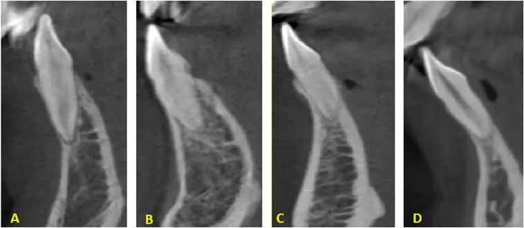
Buccal sagittal root position and higher alveolar bone concavity angle may predict outcomes in immediate implant placement: Study

Buccal sagittal root position, higher alveolar bone concavity angle, and maxillary sinus proximity may predict outcomes in immediate implant placement suggests a study published in the Journal of Prosthetic Dentistry.

Immediate implant placement in the maxillary premolar area is challenged by anatomic variations imposing risks such as perforation of the buccal bone and the maxillary sinus. Previous studies have addressed the potentially relevant factors individually; a study assessing all relevant variables comprehensively and with a large sample size is lacking. The purpose of this observational study was to analyze multiple anatomic considerations, including sagittal root position, alveolar bone concavity angle, buccal bone perforation, maxillary sinus floor root proximity, and maxillary sinus perforation in the maxillary premolar area, using cone beam computed tomography (CBCT) to inform, perceive, and simplify the placement of immediate implants. The analysis involved 333 CBCT images (189 of men, 144 of women). A total of 1332 first and second maxillary premolars were assessed for sagittal root position, alveolar bone concavity angle, buccal bone perforation, maxillary sinus floor root proximity, and maxillary sinus perforation. Chi-squared and kappa tests were used to analyze the distributions and agreement, respectively, while dependent and independent t tests were used to assess sex and tooth-specific differences. The Spearman correlation test was used to explore the potential correlations (α=.05). Results: The majority of sagittal root position distribution was on the buccal side, ranging from 79.3% to 88.3%, while maxillary sinus floor root proximity showed a predominance of the T0 category (roots separated from the maxillary sinus floor), with noticeable sex disparities in the second right premolar (73% in men versus 50.7% in women; P<.001). The “perforation” category of buccal bone perforation was highest in the right first premolar (54.1%), being higher in women across all teeth. The “perforation” category of maxillary sinus perforation was highest in the left second premolar (21.9%). Associations were found between buccal bone perforation and sagittal root position in the second premolars. The Spearman correlation between root proximity and sinus perforation was high, ranging from 0.68 to 0.78. The alveolar bone concavity angle in first premolars, compared with second premolars, poses a higher risk of buccal bone perforation, especially in women. The buccal position is the most common sagittal root position. The risk of sinus perforation is higher in maxillary second premolars, with proximity to or protrusion into the maxillary sinus floor categories strongly correlating with this complication. These findings highlight the importance of thorough assessment and strategic planning to ensure successful immediate implant placement and minimize potential complications.

Reference:

Alqutaibi AY, Aloufi AM, Hamadallah HH, Khaleefah FA, Tarawah RA, Almuzaini AS, Almashraqi A, Halboub E. Multifactorial analysis of the maxillary premolar area for immediate implant placement using cone beam computed tomography: A study of 333 maxillary images. J Prosthet Dent. 2024 Aug 1:S0022-3913(24)00468-2. doi: 10.1016/j.prosdent.2024.07.010. Epub ahead of print. PMID: 39095216.

Keywords:

Buccal, sagittal, root, position, higher, alveolar, bone, concavity, angle, maxillary sinus, proximity, predict, outcomes, immediate, implant placement, study, Journal of Prosthetic Dentistry, Alqutaibi AY, Aloufi AM, Hamadallah HH, Khaleefah FA, Tarawah RA, Almuzaini AS, Almashraqi A, Halboub E

Powered by WPeMatico