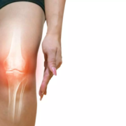
Adding oral methotrexate to care reduces pain in knee osteoarthritis

A randomized, placebo-controlled trial found that oral methotrexate added to usual analgesia care showed statistically significant reduction in knee osteoarthritis pain at 6 months, with improvements also noted in some secondary outcomes. This is important because knee osteoarthritis is associated with significant pain and disability, and treatments are limited. The findings are published in Annals of Internal Medicine.

Researchers from the University of Leeds randomly assigned 155 participants with symptomatic knee osteoarthritis to either oral methotrexate once weekly or matched placebo with continued usual analgesia over 12 months to assess symptomatic benefits of methotrexate. The participants had knee osteoarthritis diagnosed by radiography and knee pain (severity ≥4 out of 10) on most days in the past 3 months with inadequate response to current medication. The participants were assessed for knee pain at 6 months, with 12-month follow-up to assess longer term response. Secondarily, they were assessed for knee stiffness and function outcomes and adverse events.

The researchers found that methotrexate added to usual analgesia showed statistically significant reduction in pain, with improvements in some secondary outcomes. The two treatment groups had similar outcomes by 12 months, although loss to follow-up was higher and mean methotrexate dose lower by 12 months in the methotrexate group. According to the authors, further work is required to understand adequate methotrexate dosing, whether benefits are greater in those with elevated systemic inflammation levels, and to consider cost-effectiveness before introducing this therapy for a potentially large population.

Reference:

 Sarah R. Kingsbury, Puvan Tharmanathan, Ada Keding, Fiona E. Watt,  David L. Scott, PhD, Edward Roddy, Fraser Birrell, Pain Reduction With Oral Methotrexate in Knee Osteoarthritis: A Randomized, Placebo-Controlled Clinical Trial, Annals of Internal Medicine, https://doi.org/10.7326/M24-0303.

Powered by WPeMatico

Arthritis drugs may relieve long COVID lung symptoms, claims study

University of Virginia School of Medicine researchers have identified a potential treatment for the respiratory symptoms of long COVID after discovering an unknown cause of the condition inside the lungs.

The UVA researchers, led by Jie Sun, PhD, found that COVID-19 infection can cause sweeping changes in immune cells inside the lung tissues, promoting scarring and driving ongoing inflammation even after the initial infection has passed. This ongoing inflammation, they believe, drives the lasting respiratory symptoms, such as cough and difficulty breathing, associated with long COVID.

The new research from Sun and his colleagues indicates that doctors may be able to halt this chronic inflammation using a class of drugs, including baricitinib, that are already used to treat rheumatoid arthritis. The anti-inflammatory drugs previously received emergency authorization from the federal Food and Drug Administration to treat the uncontrolled inflammation seen in severe COVID-19 infections.

“Our study identified a root cause of the respiratory complication of long COVID by performing comparative analysis of both clinical samples and a relevant animal model,” said Sun, of UVA’s Carter Center for Immunology Research and UVA’s Division of Infectious Diseases and International Health. “We hope that the identification of the ‘driving’ mechanisms will help to rationally design clinical studies repurposing those FDA-approved drugs for respiratory long COVID soon.”

Millions struggle with long COVID

Long COVID is estimated to affect more than 60 million people around the world. For these patients, a COVID-19 infection turns into a seemingly endless ordeal, with symptoms lasting weeks, months or even years. Symptoms of long COVID can range from uncomfortable to debilitating; for example, respiratory symptoms can include shortness of breath, chest pain and even chronic lung scarring known as interstitial lung disease.

Prior research into long COVID has sought answers in patients’ blood, but Sun and his team wanted to see what changes were taking place in the lung tissues themselves. So the UVA researchers looked at cell samples collected from the lower airways of both lab mice and human patients. In both cases, they found that immune cells known as macrophages and T cells had gone haywire and were having faulty, harmful interactions. These cells normally help the body fight off the disease, but, in this case, they never stopped fighting, even after the initial COVID infection had passed.

The macrophages, the researchers found, had flooded into the lungs in abnormal numbers and were promoting tissue scarring. The T cells, meanwhile, were pumping out a substance called interferon that spurs continued inflammation.

Sun and his team believe that doctors may be able to break this cycle of inflammation using drugs that are already approved to treat the harmful inflammation seen in rheumatoid arthritis, a chronic autoimmune disease that affects joints. Additional research will be needed, but Sun hopes that UVA’s new discoveries will lead to much-needed new treatments for patients struggling with respiratory symptoms from long COVID.

“We hope our clinical colleagues around the globe could perform clinical trials soon to test the efficacty of baricitinib or other similar drugs targeting the same inflammatory pathway in long COVID,” Sun said. “Our new study has established a foundation for identification of new therapeutic interventions for long COVID by combining rigorous clinical testing and basic scientific research.”

Reference:

Chaofan Li et al, Comparative single-cell analysis reveals IFN-γ as a driver of respiratory sequelae after acute COVID-19, Science Translational Medicine (2024). DOI: 10.1126/scitranslmed.adn0136.

Powered by WPeMatico

Study assesses Effectiveness of Ondansetron for Prevention of Hypotension during Cesarean Section

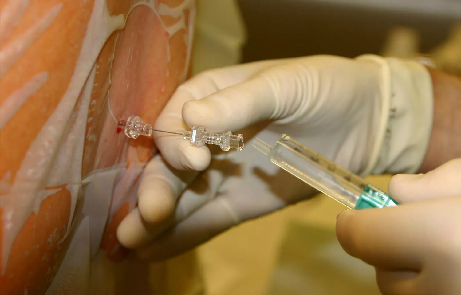
Spinal anesthesia is now the preferred technique for cesarean section because it has a rapid onset and poses considerably reduced anesthetic risks for both the mother and the fetus. Recent study aimed to investigate the effectiveness of ondansetron in preventing maternal hypotension during spinal anesthesia with ropivacaine for cesarean sections. A total of 138 primigravida parturients were randomly assigned to three groups: one received 4 mg of ondansetron, another received 8 mg of ondansetron, and the control group received normal saline. Noninvasive blood pressure, heart rate, doses of phenylephrine or ephedrine, and various other parameters were recorded.

The study concluded that there were no significant differences between the groups in terms of blood pressure, heart rate, doses of phenylephrine or ephedrine, time for the block, incidence of nausea/vomiting, umbilical artery pH, and neonatal Apgar scores at 1 and 5 minutes, showing that ondansetron did not have a significant effect in preventing maternal hypotension following spinal anesthesia with ropivacaine for cesarean section. The paper highlighted that spinal anesthesia is the preferred technique for cesarean sections due to its fast onset and lower anesthetic risks. However, the sudden decrease in afterload following lumbar sympathetic blockade may cause hypotension and fetal compromise. Ondansetron, a selective 5-hydroxytryptamine 3 (5-HT3) receptor antagonist, was suggested as a potential preventative measure for spinal-induced hypotension. Although previous studies have shown some beneficial effects of ondansetron, particularly when used with bupivacaine, the current study specifically focused on its interaction with ropivacaine and its impact on hemodynamic parameters.

The study followed ethical standards and included 138 parturients. Group allocation was concealed, and the solutions were prepared to ensure blinding. The results indicated no significant differences in blood pressure, heart rate, and doses of vasopressor agents among the different groups. Furthermore, there were no significant differences in the sensory and motor block characteristics, incidence of nausea/vomiting, shivering, and neonatal outcomes. The authors discussed potential reasons for the lack of significant differences, such as the slower onset of the spinal block after ropivacaine, the better hemodynamic profile of ropivacaine compared to bupivacaine, and the lower intrathecal volume of ropivacaine used in the study, all of which may have impacted the results. In conclusion, the study found that neither 4 mg nor 8 mg of ondansetron prevented hypotension in parturients undergoing spinal anesthesia with ropivacaine for cesarean section. The authors suggested the need for further studies to better understand the interaction between ropivacaine-induced neuraxial blockade and the preventive role of ondansetron for post-spinal hypotension, particularly with an emphasis on the pharmacokinetics of the administered drugs.

Key Points

– The study aimed to investigate the effectiveness of ondansetron in preventing maternal hypotension during spinal anesthesia with ropivacaine for cesarean sections. 138 primigravida parturients were randomly assigned to three groups: one received 4 mg of ondansetron, another received 8 mg of ondansetron, and the control group received normal saline.

– The study concluded that there were no significant differences between the groups in terms of blood pressure, heart rate, doses of phenylephrine or ephedrine, time for the block, incidence of nausea/vomiting, umbilical artery pH, and neonatal Apgar scores at 1 and 5 minutes, showing that ondansetron did not have a significant effect in preventing maternal hypotension following spinal anesthesia with ropivacaine for cesarean section.

– Spinal anesthesia is the preferred technique for cesarean sections due to its fast onset and lower anesthetic risks, but it may cause hypotension and fetal compromise. Ondansetron, a selective 5-hydroxytryptamine 3 (5-HT3) receptor antagonist, was suggested as a potential preventative measure for spinal-induced hypotension.

-The authors discussed potential reasons for the lack of significant differences, such as the slower onset of the spinal block after ropivacaine, the better hemodynamic profile of ropivacaine compared to bupivacaine, and the lower intrathecal volume of ropivacaine used in the study, all of which may have impacted the results.

– In conclusion, the study found that neither 4 mg nor 8 mg of ondansetron prevented hypotension in parturients undergoing spinal anesthesia with ropivacaine for cesarean section. The authors suggested the need for further studies to better understand the interaction between ropivacaine-induced neuraxial blockade and the preventive role of ondansetron for post-spinal hypotension, particularly with an emphasis on the pharmacokinetics of the administered drugs.

Reference –

Karachanidi S, Paraskeva A, Theodosopoulou P, et al. (July 22, 2024) Effect of Ondansetron on Maternal Hypotension During Spinal Anesthesia With Ropivacaine for Cesarean Sections: A Randomized, Double-Blind Trial. Cureus 16(7): e65073. DOI 10.7759/cureus.65073

Powered by WPeMatico
People in their 50s are at higher amputation risk than older people after leg surgery, reveals research

People in their 50s with severe peripheral artery disease or PAD may be more likely than people in their 80s to undergo leg amputation one to five years after emergency surgery to restore blood flow to the lower limbs, according to new research published today in the American Heart Association’s flagship journal Circulation.

Peripheral artery disease occurs when the arteries leading away from the heart narrow due to cholesterol deposits, preventing adequate blood flow throughout the body — commonly the lower extremities. The condition is estimated to affect 10 million to 12 million adults ages 40 and older in the U.S., according to 2024 guidelines from the American Heart Association and the American College of Cardiology.

Symptomatic PAD is characterized by painful muscle cramping in the hips, thighs, calves, or feet when walking, climbing stairs or exercising. The pain does not subside with rest. Modifiable risk factors for PAD include being a smoker, or former smoker, having diabetes, hypertension and/or abnormal cholesterol.

“People with severe forms of PAD requiring urgent surgery tend to have extremely poor disease progress. They are at high risk of limb loss and all-cause death following the initial surgery,” said study lead author Qiuju Li, Ph.D., a research fellow in medical statistics at the London School of Hygiene and Tropical Medicine in the United Kingdom. “Our primary finding is different from the traditional belief that older people were at an increased risk of major amputation. Our study, interestingly, shows the opposite relationship.”

This study analyzed previous data from nearly 95,000 adults older than age 50 with peripheral artery disease who had surgery to restore blood flow (known as revascularization) between 2013 and 2020 in England. The analysis projected the possibility of major limb amputation and death after revascularization. Major limb amputation was defined as leg amputation above the ankle.

Two-thirds of participants in the data review had undergone surgery to restore blood flow during an elective hospital admission, while the remainder had non-elective surgery during an emergency hospital admission. The analysis noted that:

  • The risk of major amputation among emergency admissions among patients between 50- to 54-years-old was 18% one year after revascularization and 28.8% five years after the surgery.
  • The risk of major amputation under the same circumstances for patients between ages 80 and 84-years-old was 11.9% at one year and 17% at five years.
  • Among patients who underwent a revascularization during an elective hospitalization, the risk of major amputation remained comparatively low regardless of the patient’s age: 10.8% for those 50- to 54-years-old, and 6.5% for those 80- to 84-years-old at five years.
  • However, the risk of death without a major amputation increased substantially among older patients after both elective and non-elective revascularizations: 48.7% and 58.9%, respectively, for patients between ages 80-84 at 5 years, vs. 12.9% and 16.9%, respectively, for those ages 50- 54 at 5 years.
  • After major amputation, the data showed participants were at an increased risk of death if they had undergone major amputation within six months of the revascularization.
  • More specifically, for participants between ages 80-84, the 1-year death rates after major amputation were 39.2% and 29.8% if they underwent major amputation at 3 months and 1 year, respectively, after revascularization, whereas the corresponding death rates were 20.3% and 14.9%, respectively, for those between ages 50 and 54.

“The findings also highlight how the association between the illness trajectories and patient characteristics is not straightforward,” Li said. “While being older at the time they had blood flow-restoring surgery was associated with a marked increase in the risk of death, the risk of major amputation after that surgery was lower among older patients rather than younger patients.”

In May 2024, joint guidelines from the American Heart Association, the American College of Cardiology and nine other medical societies highlighted the importance of early diagnosis and treatment to prevent amputation and other cardiovascular complications from lower extremity peripheral artery disease. The guidelines also called for coordinated care from a multispecialty team to treat the condition.

“This study shows that for patients with severe peripheral arterial disease, there is not one simple answer that can explain each patient’s condition,” said guidelines co-vice chair Philip Goodney, M.D., M.S., professor and section chief of Vascular Surgery at Dartmouth Health in New Hampshire. “For example, patients with severe disease who present at young ages have poor outcomes, irrespective of how they might be treated. This may be the result of severe disease or difficult circumstances for treatment. Either way, this information can help guide clinicians and researchers in determining the best treatments for these high-risk individuals. Similarly, the study also shows that older patients with moderate to severe disease can have good results when trying to prevent an amputation. Overall, these findings make it clear – in treatment for PAD, it is not ‘one size fits all.’”

This analysis had several limitations that may have affected the results. The data review did not account for subsequent surgeries to restore blood flow after the initial surgery; it could not account for disease severity or whether amputation occurred on the leg that underwent the initial surgery or the other leg; and race or ethnicity was not considered due to unreliable information from the database.

Study details and background:

  • The data review included 94,690 people with peripheral artery disease who underwent surgery to restore blood flow to the lower limbs between April 2013 and March 2020 in England.
  • Participants were ages 50 and older, the median age was 72 years and 66% of participants were men.
  • Data was collected from the Hospital Episode Statistics Admitted Patient Care database in England.
  • A statistical model was created from the data to describe patterns of survival after an initial lower limb revascularization; if and when patients experienced major amputation; and survival after amputation.
  • In total, 10.0% patients underwent major amputation during the follow-up period, which ended in March 2021. Of these, two thirds were emergency patients at time they were admitted to the hospital, and more than half had died by the end of study.
  • The revascularization procedure was labelled elective if performed during an elective admission, and non-elective if performed during an emergency admission or after transfer from another hospital.

Reference:

Qiuju Li, Panagiota Birmpili, Eleanor Atkins, Amundeep S. Johal, Robin Williams, Illness Trajectories After Revascularization in Patients With Peripheral Artery Disease: A Unified Approach to Understanding the Risk of Major Amputation and Death, Circulation, https://doi.org/10.1161/CIRCULATIONAHA.123.067687.

Powered by WPeMatico
Vitamin D supplements increase BMD and hand grip in patients with sickle cell disease: Study

A new study by Diana Hanna and team unveiled that in sickle cell disease (SCD) patients monthly oral high-dose vitamin D administration was safe and helpful in increasing the bone mineral density (BMD) scores, health-related quality of life (HRQoL), hand grip strength (HGS) and vitamin D levels. The findings of this study were published in the European Journal of Pediatrics.

Vaso-occlusive crises (VOCs) and persistent hemolytic anemia are two inherited symptoms of sickle cell disease. There are reports of musculoskeletal impairment in SCD patients and poorer disease outcomes have been associated with a high frequency of vitamin D insufficiency and poor bone mineral density in SCD patients. Thus, this study assessed the effects of monthly high-dose vitamin D3 supplementation on BMD, HGS, and HRQL in patients with sickle cell disease (SCD) and healthy controls.

42 SCD youngsters and 42 healthy matched controls participated in the research. Monthly high-dose oral vitamin D3 supplements were given to the trial participants. The functional ability was evaluated using the Childhood Health Assessment Questionnaire (CHAQ) and the HRQoL questionnaire, and changes in the blood levels of 25(OH) vitamin D3, maximal HGS, and BMD were measured from baseline to six months.

The results found SCD individuals to exhibit lower growth status at baseline throught negative Z scores. Suboptimal BMD was identified with a substantially lower Z score, poorer HGS, and worse HRQL indicators when compared to the controls. SCD patients had substantially decreased median 25(OH) vitamin D3 levels than the controls. The vitamin D supplementation improved DEXA Z-score, physical health limitations, pain ratings and CHAQ grades in SCD patients. The SCD group showed substantial improvements in HGS and CHAQ score compared to the controls.

Throughout the trial, there were no documented clinical adverse events (AEs) or new concomitant medicines (CMs), and both groups had safe levels of Ca and 25 (OH) D3 at 3 and 6 months.There was a strong positive link between HGS and overall physical score, but a negative correlation with CHAQ score. The study also found a substantial positive link between 6-month vitamin D levels and pain score, HGS and a negative correlation with CHAQ score. Overall, monthly oral high-dose vitamin D therapy was safe and effective in increasing BMD, HRQoL, hand grip, HGS, and vitamin D levels not just in SCD patients but also in healthy individuals.

Reference:

Hanna, D., Kamal, D. E., Fawzy, H. M., & Abd Elkhalek, R. (2024). Safety and efficacy of monthly high-dose vitamin D3 supplementation in children and adolescents with sickle cell disease. In European Journal of Pediatrics. Springer Science and Business Media LLC. https://doi.org/10.1007/s00431-024-05572-w

Powered by WPeMatico

Study Sheds Light on Impact of Intramural Leiomyomas on Fertility: Size Matters, Removal Offers No Benefit

USA: A recent systematic review and meta-analysis have delved into the relationship between intramural leiomyomas—commonly known as fibroids—and fertility, shedding light on implications for women’s reproductive health. Published in Obstetrics & Gynecology, the study synthesized findings from numerous research studies to elucidate how these benign uterine growths impact fertility outcomes.

The study found lower fertility rates for women with intramural leiomyomas and women with more than one leiomyoma in any location. The removal of leiomyoma does not improve outcomes.

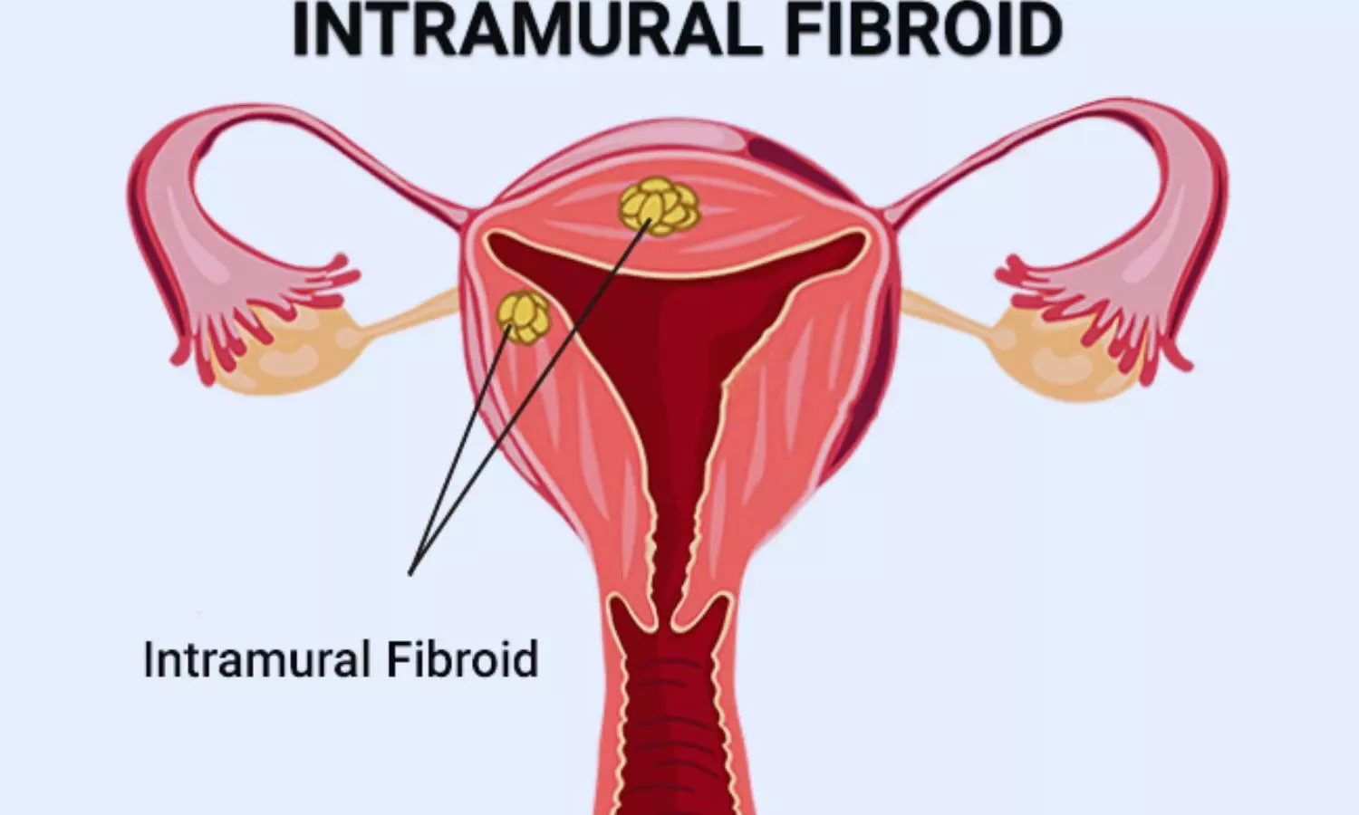
Intramural leiomyomas are non-cancerous tumors that develop within the muscular wall of the uterus. They are prevalent among women of reproductive age and can vary significantly in size and number. Concerns often arise regarding their potential interference with conception and pregnancy outcomes.

Against the above background, Elizabeth A. Pritts, Wisconsin Fertility Institute, Madison, WI, and colleagues aimed to evaluate fertility outcomes based on the size and number of intramural leiomyomas and outcomes after removal.

For this purpose, the researchers searched the online databases from 1994 to 2023. A total of 5,143 studies were identified, with the inclusion of 13 study groups. The study provided data on the size and number of leiomyomas, correlating these findings with clinical pregnancy rates and rates of ongoing pregnancies or live births.

The following were the key findings of the study:

  • In data sets with maximum leiomyoma diameters of less than 6 cm for study inclusion, women with leiomyomas smaller than 3 cm had lower clinical pregnancy rates than women without leiomyomas, with an odds ratio (OR) of 0.53 and, for ongoing pregnancy or live-birth rates, an OR of 0.59.
  • The ORs for clinical pregnancy rates in women with intermediately-sized leiomyomas (those between 3 cm and 6 cm) were lower than in women without leiomyomas, with an OR at 0.43 and, for ongoing pregnancy or live-birth rates, an OR at 0.38.
  • In data sets without exclusion for women with larger-sized leiomyomas, clinical pregnancy rates were lower for those with leiomyomas smaller than 5 cm compared with those without leiomyomas, with an OR of 0.75.
  • Women with leiomyomas larger than 5 cm showed no differences in clinical pregnancy rate compared with women without leiomyomas, with an OR of 0.71.
  • Although women with a single leiomyoma in any location had no differences in outcomes, those with more than one leiomyoma had lower clinical pregnancy rates and ongoing pregnancy or live birth rates, with an OR of 0.62 and 0.57, respectively.
  • The clinical pregnancy rate for women undergoing myomectomy for intramural leiomyomas was no different than those with intramural leiomyomas in situ, with an OR of 1.10.

“The findings imply that even small intramural leiomyomas are linked with lower fertility; removal does not confer benefit. Women with more than one leiomyoma in any location have lower fertility,” the researchers concluded.

Reference:

Pritts TL, Ogden M, Parker W, Ratcliffe J, Pritts EA. Intramural Leiomyomas and Fertility: A Systematic Review and Meta-Analysis. Obstet Gynecol. 2024 Jun 27. doi: 10.1097/AOG.0000000000005661. Epub ahead of print. PMID: 38935974.

Powered by WPeMatico

No Link Found Between COVID-19 Vaccination and Sudden Death in Young Adults, Study Reports

India: COVID-19 vaccination did not increase the risk of unexplained sudden death among young adults in India, a recent study published in the Indian Journal of Medical Research has concluded.

The study highlighted that a family history of sudden death, past COVID-19 hospitalization, and certain lifestyle behaviors increased the likelihood of unexplained sudden death.

In the wake of the COVID-19 era, there have been many instances of sudden unexplained death in the young Indian population, that is 18-45 years. This was attributed by many to the COVID-19 vaccination creating an atmosphere of worry.

With this background, more than 50 researchers across the country, including those from the ICMR – National Institute of Epidemiology, and those under the Sudden Adult Deaths Study Group got together and performed a multicentric matched case-control study to determine the factors associated with such deaths in individuals aged 18-45 years

The study was performed through the participation of 47 tertiary care hospitals across India and analysis was done on cases of healthy individuals aged 18-45 years without any known co-morbidity, who suddenly (<24 h of hospitalization or seen healthy 24 h before death) died of unexplained causes during 1st October 2021-31st March 2023.

Four controls were included per case matched for age, gender, and neighborhood. The researchers analyzed their data on COVID-19 vaccination/infection and post-COVID-19 conditions, family history of sudden death, smoking, recreational drug use, alcohol frequency and binge drinking, and vigorous-intensity physical activity two days before death/interviews and developed regression models considering COVID-19 vaccination ≤42 days before outcome, any vaccine received anytime and vaccine doses to compute an adjusted matched odds ratio (aOR). 

729 cases and 2916 controls were included in the analysis

They found the following results

  • Recipients of one dose of COVID-19 vaccine lowered the odds [aOR (95% CI)] for unexplained sudden death.

  • Past COVID-19 hospitalization, family history of sudden death, binge drinking 48 hours before death/interview, use of recreational drugs/substances, and performing vigorous-intensity physical activity 48 hours before death/interview were positively associated.

  • Two doses lowered the odds of unexplained sudden death, whereas a single dose did not.

“COVID-19 vaccination was not associated with an increased risk of unexplained sudden death among young adults. At the same time, a family history of sudden death, hospitalization for COVID-19, and lifestyle behaviors such as recent binge drinking and vigorous-intensity physical activity were risk factors for unexplained sudden death. Addressing these factors among young adults could potentially modify their risk of unexplained sudden death,” the researchers concluded.

Reference: 

Ponnaiah, M., Bhatnagar, T., Abdulkader, R. S., Elumalai, R., Surya, J., Jeyashree, K., Kumar, M. S., Govindaraju, R., Thangaraj, J. W. V., Aggarwal, H. K., Balan, S., Baruah, T. D., Basu, A., Bavaskar, Y., Bhadoria, A. S., Bhalla, A., Bhardwaj, P., Bhat, R., Chakravarty, J., Chandy, G. M., Gupta, B. K., Kakkar, R., Karnam, A. H. F., Kataria, S., Khambholja, J., Kumar, D., Kumar, N., Lyngdoh, M., Meena, M. S., Mehta, K., Sheethal, M. P., Mukherjee, S., Mundra, A., Murugan, A., Narayanan, S., Nathan, B., Ojah, J., Patil, P., Pawar, S., Ruban, A. C. P., Vadivelu, R., Rana, R. K., Boopathy, S. N., Priya, S., Sahoo, S. K., Shah, A., Shameem, M., Shanmugam, K., Shivnitwar, S. K., Singhai, A., Srivastava, S., Sulgante, S., Talukdar, A., Verma, A., Vohra, R., Wani, R. T., Bathula, B., Kumari, G., Kumar, D. S., Narasimhan, A., Krupa, N. C., Senguttuvan, T., Surendran, P., Tamilmani, D., Turuk, A., Kumar, G., Murkherjee, A., Aggarwal, R., & Murhekar, M. V. (2023). Factors associated with unexplained sudden deaths among adults aged 18-45 years in India: A multicentric matched case-control study. Indian Journal of Medical Research, 158(4), 351-362. https://doi.org/10.4103/ijmr.ijmr_2105_23

Powered by WPeMatico

High Diastolic Blood Pressure associated with increased risk of migraine among Females: Study

Having high blood pressure, specifically high diastolic blood pressure, was linked to a slightly higher odds of ever having migraine in female participants, according to a new study published in the July 31, 2024, online issue of Neurology®, the medical journal of the American Academy of Neurology. Diastolic pressure is when the heart is resting between beats. However, the study did not find an increased risk between other cardiovascular risk factors and migraine.

“Previous research shows that migraine is linked to a higher risk of cardiovascular events such as stroke, heart disease and heart attack, but less is known about how risk factors for cardiovascular events relate to having migraine,” said study author Antoinette Maassen van den Brink, PhD, of Erasmus MC University Medical Center in Rotterdam, the Netherlands. “Our study looked at well-known risk factors for cardiovascular disease, such as diabetes, smoking, obesity and high cholesterol and found an increased odds of having migraine only in female participants with higher diastolic blood pressure.”

The study involved 7,266 people, male and female, with a median age of 67 years, of whom 15% had previous or current migraine.

All participants had physical exams and provided blood samples. They were also asked questions about migraine, including if they had ever experienced a headache with severe pain that affected their daily activities.

After adjusting for multiple cardiovascular risk factors such as physical activity, as well as education level, researchers found female participants with higher diastolic blood pressure had 16% increased odds of having migraine per standard deviation increase in diastolic blood pressure. An increase per standard deviation is a measure to compare the diastolic blood pressure with other cardiovascular risk factors. No associations were found for systolic blood pressure. Maasen van den Brink said this contributes to the theory that migraine is associated with a slightly reduced function of the small blood vessels rather than a reduced function of the large blood vessels.

There were no associations for female participants with high cholesterol or obesity, and current smoking was associated with 28% lower odds of having migraine and diabetes with 26% lower odds of having migraine. Maassen van den Brink said, “These results should be interpreted with caution, as they do not prove that smoking causes a lower risk of migraine. Instead, smoking might trigger migraine attacks and therefore, people who choose to smoke are less likely to be people who have migraine.”

In male participants, researchers found no associations between cardiovascular risk factors and migraine.

“Our study suggests that overall, migraine is not directly related to traditional risk factors for cardiovascular disease,” said Maassen van den Brink. “Because we looked at people who were middle-age and older, future studies are needed in younger groups of people who are followed for longer periods of time.”

A limitation of the study was the small number of male participants with migraine. Maassen van den Brink said this could help explain why they found no associations for male participants between cardiovascular risk factors and migraine.

Reference:

Al-Hassany, L., et al. (2024) Sex-Specific Association of Cardiovascular Risk Factors With Migraine The Population-Based Rotterdam Study. Neurology. doi.org/10.1212/WNL.0000000000209700.

Powered by WPeMatico

Hydrogen-rich gas inhalation can alleviate exercise-induced fatigue, suggests study

A new study published in the International Journal of Sports Medicine, Inhalation of hydrogen-rich gas before acute exercise alleviates exercise fatigue , has found a possible novel intervention for exercise-induced fatigue. A cross-discipline international team of U.S. and Chinese researchers, including at Hebrew SeniorLife’s Hinda and Arthur Marcus Institute for Aging Research, found that inhaling hydrogen-rich gas (HRG) before engaging in acute exercise can significantly reduce fatigue and enhance performance.

The research involved 24 healthy adult men who were tested for their maximum cycling power (Wmax) and maximum cycling time (Tmax). In a double-blind, counterbalanced, randomized, and crossover design, participants inhaled either HRG or placebo gas (air) for 60 minutes before cycling at 80% Wmax until exhaustion.

Key findings from the study include:

  • Reduced Perceived Fatigue: Participants who inhaled HRG reported significantly lower scores on the Visual Analog Scale (VAS) for fatigue compared to those who inhaled placebo gas.
  • Improved Exercise Performance: HRG inhalation improved cycling frequency during the final 30 seconds of the exercise and reduced the rating of perceived exertion (RPE) at both the beginning and end of the ride.
  • Oxidative Stress Markers: HRG inhalation showed a significant improvement in the ability to inhibit hydroxyl radicals and lower serum lactate levels after exercise, indicating reduced oxidative stress.
  • Functional Performance: While HRG did not significantly impact counter-movement jump (CMJ) height or glutathione peroxidase activity, the overall benefits on fatigue and oxidative stress markers were notable.

“The study’s findings suggest that HRG inhalation prior to exercise could be a valuable strategy for athletes and fitness enthusiasts looking to enhance performance and recovery. By mitigating fatigue and improving markers of oxidative stress, HRG has the potential to support more effective training and better overall health outcomes,” said Junhong Zhou, PhD, assistant scientist II, Hinda and Arthur Marcus Institute for Aging Research, Hebrew SeniorLife.

The study opens new avenues for exploring the benefits of hydrogen-rich gas in sports and exercise science. As further research continues, HRG inhalation may become a widely adopted practice for enhancing athletic performance and combating exercise-induced fatigue.

Reference:

Gengxin Dong , Jianxin Wu , Yinglu Hong , Qian Li , Meng Liu , Guole Jiang , Dapeng Bao , Brad Manor , Junhong Zhou, Inhalation of hydrogen-rich gas before acute exercise alleviates exercise fatigue, Int J Sports Med, DOI: 10.1055/a-2318-1880

Powered by WPeMatico

AI model promising in accurately identifying dental implant systems on low-quality radiographs: Study

AI model promising in accurately identifying dental implant systems on low-quality radiographs suggests a study published in the Scientific Reports.

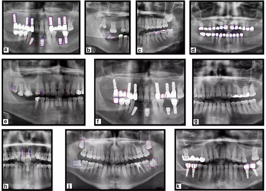
Most artificial intelligence (AI) studies have attempted to identify dental implant systems (DISs) while excluding low-quality and distorted dental radiographs, limiting their actual clinical use. This study aimed to evaluate the effectiveness of an AI model, trained on a large and multi-centre dataset, in identifying different types of DIS in low-quality and distorted dental radiographs. Based on the fine-tuned pre-trained ResNet-50 algorithm, 156,965 panoramic and periapical radiological images were used as training and validation datasets, and 530 low-quality and distorted images of four types (including those not perpendicular to the axis of the fixture, radiation overexposure, cut off the apex of the fixture, and containing foreign bodies) were used as test datasets. Moreover, the accuracy performance of low-quality and distorted DIS classification was compared using AI and five periodontists. Based on a test dataset, the performance evaluation of the AI model achieved accuracy, precision, recall, and F1 score metrics of 95.05%, 95.91%, 92.49%, and 94.17%, respectively. However, five periodontists classified nine types of DISs based on four different low-quality and distorted radiographs, achieving a mean overall accuracy of 37.2 ± 29.0%. Within the limitations of this study, AI demonstrated superior accuracy in identifying DIS from low-quality or distorted radiographs, outperforming dental professionals in classification tasks. However, extensive standardization research on low-quality and distorted radiographic images is essential for actual clinical application of AI.

Reference:

Lee, JH., Kim, YT. & Lee, JB. Identification of dental implant systems from low-quality and distorted dental radiographs using AI trained on a large multi-center dataset. Sci Rep 14, 12606 (2024). https://doi.org/10.1038/s41598-024-63422-z

Keywords:

AI model, promising, accurately, identifying, dental implant, systems, low-quality radiographs, study, Lee, JH., Kim, YT. & Lee, JB, Artificial intelligence, Diagnostic imaging, Deep learning, Scientific Reports

Powered by WPeMatico