Olpasiran effective therapeutic option for lowering lipoprotein(a) levels, suggests study
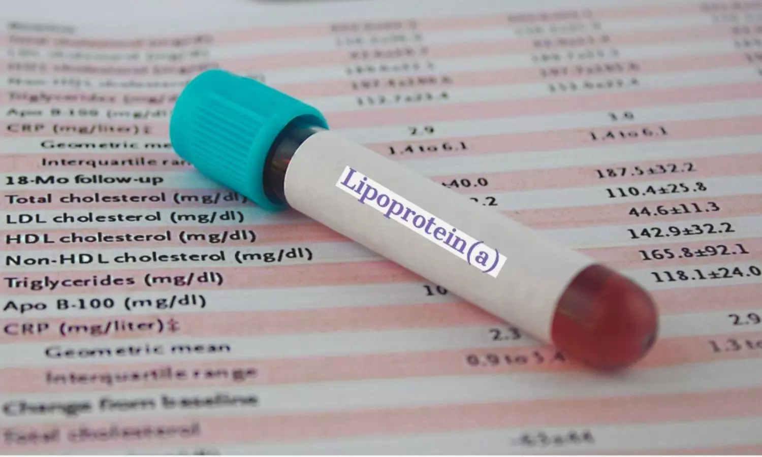
A new study published in the Journal of the American College of Cardiology found that strong siRNA that lowers lipoprotein(a) (Lp(a)) over an extended period of time is olpasiran. Nearly a year after the previous dosage, the individuals receiving doses over 75 mg Q12W saw a ∼40% to 50% drop in Lp(a) levels. Apolipoprotein(a) (apo(a)) is covalently bonded to apoB100 within a modified low-density lipoprotein (LDL) particle to form lipoprotein(a) (Lp(a)). A substantial amount of data points to Lp(a) as a causative factor in the processes that encourage calcific aortic valve disease and atherogenesis. Based on Mendelian randomization studies, a significant decrease in Lp(a) could be necessary in order to provide a significant therapeutic effect.

Olpasiran is a small interfering RNA (siRNA) molecule linked to N-acetylgalactosamine (GalNAc) that obstructs the expression of the LPA gene by causing the messenger RNA encoding apo(a) to break down, thereby stopping the hepatocyte from assembling the Lp(a) particle. Olpasiran is a small interfering RNA (siRNA) that inhibits the translation of apolipoprotein(a) mRNA, hence blocking the formation of lipoprotein(a) (Lp(a)). This research by Michelle O’Donoghue and colleagues evaluated both the longer-term safety and the timing of Lp(a) returning to baseline following olpasiran withdrawal.

In the phase 2 dose-finding trial OCEAN(a)-DOSE (Olpasiran Trials of Cardiovascular Events And LipoproteiN[a] Reduction–DOSE Finding Study), a total of 281 participants with atherosclerotic cardiovascular disease and Lp(a) >150 nmol/L were enrolled to receive one of four active doses of olpasiran versus placebo (10 mg, 75 mg, 225 mg Q12W, or an exploratory dose of 225 mg Q24W administered subcutaneously). Week 36 was the last dosage of olpasiran, and after week 48, there was a minimum 24-week prolonged off-treatment follow-up period.

276 individuals (98.2%) of the total study population started the post-treatment follow-up phase. The mean duration of the trial encompassed both treatment and non-treatment periods, was 86 weeks (Q1-Q3: 79-99 weeks). At 60, 72, 84, and 96 weeks, the off-treatment placebo-adjusted mean percent decrease from baseline in Lp(a) for the 75 mg Q12W dosage were −76.2%, −53.0%, −44.0%, and −27.9%, respectively (all P < 0.001).

For the 225 mg Q12W dosage, the corresponding off-treatment decreases in Lp(a) were −84.4%, −61.6%, −52.2%, and −36.4% (all P < 0.001). In the follow-up phase of the extension, no additional safety issues were found. Overall, the RNA interference of Olpasiran causes a significant decrease in Lp(a), with long-term pharmacodynamic effects that last for many months after therapy is stopped.

Source:

O’Donoghue, M. L., Rosenson, R. S., López, J. A. G., Lepor, N. E., Baum, S. J., Stout, E., Gaudet, D., Knusel, B., Kuder, J. F., Murphy, S. A., Wang, H., Wu, Y., Shah, T., Wang, J., Wilmanski, T., Sohn, W., Kassahun, H., & Sabatine, M. S. (2024). The Off-Treatment Effects of Olpasiran on Lipoprotein(a) Lowering. In Journal of the American College of Cardiology (Vol. 84, Issue 9, pp. 790–797). Elsevier BV. https://doi.org/10.1016/j.jacc.2024.05.058

Powered by WPeMatico

Researchers develop affordable, rapid blood test for brain cancer

Researchers at the University of Notre Dame have developed a novel, automated device capable of diagnosing glioblastoma, a fast-growing and incurable brain cancer, in less than an hour. The average glioblastoma patient survives 12-18 months after diagnosis.

The crux of the diagnostic is a biochip that uses electrokinetic technology to detect biomarkers, or active Epidermal Growth Factor Receptors (EGFRs), which are overexpressed in certain cancers such as glioblastoma and found in extracellular vesicles.

“Extracellular vesicles or exosomes are unique nanoparticles secreted by cells. They are big-10 to 50 times bigger than a molecule-and they have a weak charge. Our technology was specifically designed for these nanoparticles, using their features to our advantage,” said Hsueh-Chia Chang, the Bayer Professor of Chemical and Biomolecular Engineering at Notre Dame and lead author of the study about the diagnostic published in Communications Biology.

The challenge for researchers was two-fold: to develop a process that could distinguish between active and non-active EGFRs, and create a diagnostic technology that was sensitive yet selective in detecting active EGFRs on extracellular vesicles from blood samples.

To do this, researchers created a biochip that uses an inexpensive, electrokinetic sensor about the size of a ball in a ballpoint pen. Due to the size of the extracellular vesicles, antibodies on the sensor can form multiple bonds to the same extracellular vesicle. This method significantly enhances the sensitivity and selectivity of the diagnostic.

Then synthetic silica nanoparticles “report” the presence of active EGFRs on the captured extracellular vesicles, while bringing a high negative charge. When extracellular vesicles with active EGFRs are present, a voltage shift can be seen, indicating the presence of glioblastoma in the patient.

This charge-sensing strategy minimizes interference common in current sensor technologies that use electrochemical reactions or fluorescence.

“Our electrokinetic sensor allows us to do things other diagnostics cannot,” said Satyajyoti Senapati, a research associate professor of chemical and biomolecular engineering at Notre Dame and co-author of the study. “We can directly load blood without any pretreatment to isolate the extracellular vesicles because our sensor is not affected by other particles or molecules. It shows low noise and makes ours more sensitive for disease detection than other technologies.”

In total, the device includes three parts: an automation interface, a prototype of a portable machine that administers materials to run the test and the biochip. Each test requires a new biochip, but the automation interface and prototype are reusable.

Running one test takes under an hour, requiring only 100 microliters of blood. Each biochip costs less than $2 in materials to manufacture.

Although this diagnostic device was developed for glioblastoma, the researchers say it can be adapted for other types of biological nanoparticles. This opens up the possibility for the technology to detect a number of different biomarkers for other diseases. Chang said the team is exploring the technology for diagnosing pancreatic cancer and potentially other disorders such as cardiovascular disease, dementia and epilepsy.

“Our technique is not specific to glioblastoma, but it was particularly appropriate to start with it because of how deadly it is and the lack of early screening tests available,” Chang said. “Our hope is that if early detection is more feasible, then there is an increased chance of survival.”

Blood samples for testing the device were provided by the Centre for Research in Brain Cancer at the Olivia Newton-John Cancer Research Institute in Melbourne, Australia.

Reference:

Maniya, N.H., Kumar, S., Franklin, J.L. et al. An anion exchange membrane sensor detects EGFR and its activity state in plasma CD63 extracellular vesicles from patients with glioblastoma. Commun Biol 7, 677 (2024). https://doi.org/10.1038/s42003-024-06385-1.

Powered by WPeMatico

Maternal chronic hypertension increase risk of hypospadias in offspring, reveals study

A new meta-analysis published in the Journal of Pediatric Urology discovered a strong correlation between maternal chronic hypertension (CH) and the higher risk of hypospadias in the offspring. Hypospadias is a complicated congenital disorder which is typified by the abortive development of ventral urethral spongiosum and is sometimes accompanied by incomplete foreskin and ventral chordee. Hypertensive disorders of pregnancy (HDP) are among the maternal-placental variables that have been associated with a number of birth abnormalities, including the finding that HDP may be a stand-alone risk factor for hypospadias. Although earlier research has indicated that pregnancy-related hypertensive diseases raise the incidence of hypospadias, no studies have examined the impact of maternal chronic hypertension yet. Thus, this study evaluated the relationship with maternal CH and the risk of hypospadias by conducting a systematic review and meta-analysis of the observational data that are currently available.

EMBASE, Pubmed, SCOPUS, and manual techniques were searched in accordance with PRISMA 2020 standards and the MOOSE checklist. The Newcastle-Ottawa Scale (NOS) was used to rate the quality of the eligible papers that were included in the research. DerSimonian and Laird model pooled analysis for unadjusted and adjusted effect sizes was used to calculate OR and 95% CI. The I2 test was used to test for heterogeneity, while funnel plots were used to look at publication bias and to deal with uncertainty, sensitivity assessments are carried out.

The results of the study included a total of 1,130 publications through searches and 6 relevant studies with a high NOS quality score (6–9) were chosen. Among the 6 studies that qualified for analysis, a total of 519 hypospadias patients had maternal CH. One research is eliminated following sensitivity analysis because its definition of hypospadias is different. According to pooled unadjusted and adjusted OR, there is an increased risk of hypospadias in the setting of maternal CH among the five remaining trials. In the unadjusted pooled analysis, heterogeneity was considerable (I2= 73% P=0.005), but in the adjusted analysis, it was minimal (I2= 0% P=0.40). In all studies, the funnel plots were symmetrical which suggested no publication bias. Overall, results from both unadjusted and adjusted effect size models support the relationship between maternal CH and an increased incidence of hypospadias in children that is highlighted by this meta-analysis.

Source:

Situmorang, G. R., Hasan, Wahyudi, I., Abbas, T., Rodjani, A., & Raharja, P. A. R. (2024). Hypospadias Risk Associated with Chronic Hypertension During Pregnancy: A Systematic Review and Meta-analysis. In Journal of Pediatric Urology. Elsevier BV. https://doi.org/10.1016/j.jpurol.2024.07.029

Powered by WPeMatico

Survey Study Highlights Significant Financial Burden for Head and Neck Cancer Patients Facing Dental Complications

USA: A survey study published in JAMA Otolaryngology Head & Neck Surgery revealed that the majority of patients undergoing treatment for head and neck cancer required extensive dental care during their cancer treatment. This dental care imposed a financial burden on 39% of the patients, creating a significant barrier to receiving necessary care.

Patients receiving treatment for head and neck cancer (HNC) often face oral complications that necessitate significant dental care. However, this dental treatment is frequently not covered by medical insurance, resulting in a potential financial burden for these patients.

Betty Ben Dor, Department of Oral Medicine, Infection and Immunity at Harvard School of Dental Medicine Massachusetts et. al. discovered the financial burden of dental care among patients with head and neck cancer. The research aimed to characterize the dental care needs and the associated cost burden for these patients.

This survey study included members of the Head and Neck Cancer Alliance who were surveyed from March 23 to October 27, 2023, using the Qualtrics platform. The survey was promoted through the HNCA’s social media channels and email list. Data analysis took place between October 2023 and May 2024.

The following were key findings of the research:

  • Out of 100 individuals surveyed, 85 completed all required questions and were included in the analysis.
  • Of the 84 participants with age and sex data, 61% were aged 65 years or older, and 54% were female.
  • 70% of the 85 respondents reported that their current oral health was worse than before cancer treatment.
  • 86% of respondents experienced oral complications from cancer treatment, such as xerostomia (90%), caries (48%), and oral mucositis (40%).
  • 88% of respondents required follow-up dental treatment.
  • 14% of individuals before HNC treatment and 32% after treatment cited financial reasons for not receiving all recommended dental care.
  • 39% of respondents reported that their post-cancer dental care caused them financial hardship.
  • Individuals less likely to report financial hardship tended to have higher educational attainment, higher income, more frequent dental visits before HNC, same or better oral health after HNC, and lower out-of-pocket dental expenses after HNC.

The survey study concluded that most patients undergoing treatment for head and neck cancer (HNC) required extensive dental care during their cancer treatment, which posed a financial burden for 39% of these patients, acting as a significant barrier to receiving necessary care. Given that most private medical insurers do not cover dental treatment, there is a need for more comprehensive insurance coverage, warranting policy attention.

Reference 

Ben Dor B, Villa A, Hayes C, Alpert E, Shepard DS, Sonis ST. Financial Burden of Dental Care Among Patients With Head and Neck Cancer. JAMA Otolaryngol Head Neck Surg. 2024 Aug 1:e242260. doi: 10.1001/jamaoto.2024.2260. Epub ahead of print. PMID: 39088224; PMCID: PMC11295059.

Powered by WPeMatico

Tracheal Extubation in Children in lateral and supine positions may reduce respiratory complications: Study

Tracheal extubation marks a crucial phase in transitioning the child to the post-anesthetic phase and is frequently accompanied by an elevated perioperative risk. Recent research paper titled “A randomised controlled trial to compare tracheal extubation quality in lateral and supine positions after general anaesthesia in children” published in J Anaesthesiol Clin Pharmacology aimed to compare the quality of tracheal extubation in lateral and supine positions in children undergoing elective surgeries under general anaesthesia. The study found that positioning children in the lateral position during extubation resulted in improved extubation quality, as evidenced by lower cough scores with fewer incidents of complications compared to the supine position. The primary objective of the study was to compare tracheal extubation quality as judged by the Modified Minogue cough score, while the secondary objectives included assessment of the incidence of oxygen desaturation, laryngospasm, bronchospasm, and upper airway obstruction during emergence from general anaesthesia. The study involved a single-blinded randomised trial with 110 children in the age group of 2–12 years enrolled to be positioned in either the lateral (group L) or supine (group S) position during extubation at the end of surgery. The patients received a standardised anaesthesia regimen, and vital parameters, extubation quality, sedation score, and the incidence of adverse respiratory events were recorded every 5 minutes till 30 minutes post extubation. The findings revealed that children in the lateral position had a significantly lower cough score at the 15th minute post extubation compared to those in the supine position. Additionally, children in the lateral position had a lower incidence of adverse respiratory events (18%) compared to those in the supine position (30%) with a relative risk of 1.67. The study concluded that positioning children in the lateral position during extubation resulted in improved extubation quality.

Study Findings

The paper discussed various factors influencing tracheal extubation, such as the risks of respiratory complications, the lack of clear clinical protocols, and the decision-making process based on clinician experience. The study highlighted the importance of airway patency and the potential advantages of lateral positioning during extubation. It also recognized the need for further research on the impact of lateral positioning in different patient populations and identified limitations in blinding and the subjective nature of defining adverse events.

Discussion of Factors and Implications

In conclusion, the study provides valuable insights into the impact of lateral positioning on tracheal extubation quality in children, emphasizing the potential benefits of the lateral position in reducing respiratory complications and improving extubation outcomes. This detailed summary accurately conveys the main points and findings of the original research paper in 400 words.

Key Points

1. The research aimed to compare the quality of tracheal extubation in lateral and supine positions in children undergoing elective surgeries under general anesthesia. It found that positioning children in the lateral position during extubation resulted in improved extubation quality, as evidenced by lower cough scores with fewer incidents of complications compared to the supine position.

2. The primary objective of the study was to compare tracheal extubation quality as judged by the Modified Minogue cough score. Secondary objectives included assessment of the incidence of oxygen desaturation, laryngospasm, bronchospasm, and upper airway obstruction during emergence from general anesthesia.

3. The study involved a single-blinded randomized trial with 110 children in the age group of 2–12 years. The patients received a standardized anesthesia regimen, and vital parameters, extubation quality, sedation score, and the incidence of adverse respiratory events were recorded every 5 minutes till 30 minutes post extubation.

4. Findings revealed that children in the lateral position had a significantly lower cough score at the 15th minute post-extubation compared to those in the supine position. Additionally, children in the lateral position had a lower incidence of adverse respiratory events (18%) compared to those in the supine position (30%) with a relative risk of 1.67.

5. The study discussed factors influencing tracheal extubation, such as the risks of respiratory complications, the lack of clear clinical protocols, and the decision-making process based on clinician experience. It highlighted the importance of airway patency and the potential advantages of lateral positioning during extubation, while also recognizing the need for further research on the impact of lateral positioning in different patient populations and identifying limitations in blinding and the subjective nature of defining adverse events.

6. In conclusion, the study provides valuable insights into the impact of lateral positioning on tracheal extubation quality in children, emphasizing the potential benefits of the lateral position in reducing respiratory complications and improving extubation outcomes.

Reference –

Ganigara, Anuradha; Bhavana, D.A1; Chandrika, Y.R; Sharma, Trishi. A randomised controlled trial to compare tracheal extubation quality in lateral and supine positions after general anaesthesia in children. Journal of Anaesthesiology Clinical Pharmacology ():10.4103/joacp.joacp_506_23, June 27, 2024. | DOI: 10.4103/joacp.joacp_506_23

Powered by WPeMatico

Single Botulinum toxin-A Injection Outperforms Four FMR Sessions in Managing Primary Axillary Hyperhidrosis: Study

Egypt: In patients with primary axillary hyperhidrosis (PAH), botulinum toxin A (BT-A) treatment demonstrates superior efficacy for up to 12 months, is less painful, and necessitates fewer sessions compared to fractional microneedling radiofrequency (FMR), a recent study has found.

The findings published online in Lasers in Medical Science suggest that fractional microneedle radiofrequency is not a better substitute for BT-A in primary axillary hyperhidrosis.

This study is the first to compare both procedures over a 12-month longitudinal follow-up. While both treatment methods were found to be equally safe, the FMR procedure was more painful than the BT-A injection, the researchers stated. They found that a single session of BT-A injection is more effective in PAH management and yields higher patient satisfaction than four FMR sessions with the microneedle type and parameters used in the present study. After three months, both interventions showed the best efficacy, and then a gradual decline was observed, although some efficacy level was maintained in a few patients for up to 12 months.

Primary axillary hyperhidrosis is an idiopathic condition characterized by excessive and uncontrollable sweating, leading to significant psycho-social challenges. While various therapeutic options are available, each comes with its limitations. Recently, fractional microneedling radiofrequency has emerged as a promising new treatment option.

Against the above background, Vanessa Hafez, Department of Dermatology, Kasr Al-Ainy Faculty of Medicine, Cairo University, Cairo, Egypt, and colleagues aimed to determine the safety and efficacy of FMR in comparison to Botulinum toxin-A in patients with primary axillary hyperhidrosis.

For this purpose, the researchers conducted a randomized controlled clinical trial comprising 20 patients (40 sides). Participants were randomly assigned to receive either fractional microneedle radiofrequency (administered in 4 sessions at 3-week intervals) or botulinum toxin A (administered in a single session), with each side of their body treated with one of these modalities.

Efficacy was assessed at 3, 6, and 12 months using Minor’s starch iodine test, HDSS score, HQoL questionnaire, and patient satisfaction ratings.

The following were the key findings of the study:

  • Fractional microneedle radiofrequency, although showed moderate efficacy, is inferior to BT-A regarding longitudinal efficacy at 12 months, as well as patient satisfaction.
  • Both treatment modalities were equally safe, but the fractional microneedle radiofrequency procedure was substantially more painful.

The researchers revealed that fractional microneedle radiofrequency, even with four sessions, does not surpass botulinum toxin A (BT-A) in treating primary axillary hyperhidrosis. BT-A offers superior efficacy for up to 12 months, is less painful, more cost-effective, and requires fewer sessions.

“The single-pass technique with non-insulated needles for fractional microneedle radiofrequency (FMR) in this study yields results similar to those of previous studies using a multiple-pass method with insulated needles, though with shorter-lasting efficacy,” they concluded.

Reference:

Eid, R.O., Shaarawi, E., Hegazy, R.A. et al. Long-term efficacy of fractional microneedle radiofrequency versus botulinum toxin-A in primary axillary hyperhidrosis: a randomized controlled trial. Lasers Med Sci 39, 177 (2024). https://doi.org/10.1007/s10103-024-04115-x

Powered by WPeMatico

Nutritional insulin after meals may significantly reduce hypoglycemia risk in hospitalized patients: Study

A new study published in the journal of Diabetes Care and Clinical Practice showed that nutritional insulin administered after meals instead of before decreased the risk of hypoglycemia in hospitalized patients without appreciably lengthening their stay or causing severe hyperglycemia.

Hypoglycemia is a frequent occurrence on the patient days in intensive care units (10%) and non-intensive care units (3.5%), at least one hypoglycemic incident was seen. Hypoglycemia is associated with a considerable morbidity and mortality risk, particularly in the older population, with some studies suggesting a 3.4-fold greater risk of death. Insulin dosage errors are the most frequent cause of hypoglycemia in hospitalized patients when such patients have reduced calorie intake. While postprandial administration of most rapid-acting insulin analogs is allowed, they are often provided before meal consumption to enable enough time for absorption and an increase in plasma insulin levels to counteract postprandial blood sugar spikes. Thereby, Merit George and colleagues thus set out to investigate whether alterations in glycemic control or length of stay (LOS) might be brought about by a hospital-wide strategy that switched nutritional insulin delivery from pre-meal to post-meal.

This retrospective cohort research investigated the association between glycemia measurements and when nutritional insulin was administered to patients admitted to the Johns Hopkins Howard County Medical Center in Columbia, Maryland. This retrospective research, conducted over three time periods in a community hospital, assessed adult inpatients receiving nutritional insulin as Pre-intervention, post-intervention right away, and post-intervention later. These results encompassed the daily mean glucose level, LOS, severe hypoglycemia (≤ 40 mg/dL), moderate hypoglycemia (< 54 mg/dL) and severe hyperglycemia (≥ 300 mg/dL).

Between the three cohorts, the total number of patient-days studied were 1948, 1751, and 3244, respectively. The likelihood of experiencing severe hypoglycemia and any hypoglycemia reduced with time after multivariate correction. Over time, the daily mean glucose increased (194.6 ± 62.5 vs 196.8 ± 65.5 vs 199.3 ± 61.5 mg/dL; p = 0.003), despite the incidence of severe hyperglycemia (p = 0.10) and LOS (p = 0.74) did not change significantly.

By adopting a postprandial nutritional insulin delivery strategy throughout the whole hospital lowered the incidence of hypoglycemia while keeping the rate of severe hyperglycemia at bay. This implies a viable approach to enhance patient safety and more prospective randomized controlled studies are imperative to validate these results.

Source:

George, M., Zilbermint, M., Sokolinsky, S., Batty, K., Motevalli, M., Stanback, C., Gonzales, E., Miller, C., Sequeira, L., & Demidowich, A. P. (2024). Effects of preprandial versus postprandial nutritional insulin administration in the inpatient setting. In Diabetes Research and Clinical Practice (Vol. 214, p. 111785). Elsevier BV. https://doi.org/10.1016/j.diabres.2024.111785

Powered by WPeMatico

Combined vitamin C and D deficiency lowered BMD and elevated osteoporosis risk: Study

A new study published in the BMC Journal of Orthopaedic Surgery and Research observed that combined vitamin C and D deficiency dramatically reduced bone mineral density and osteoporosis risk were linked to combination deficits in vitamins C and D.

Osteoporosis has exponentially increased due to an increase in bone fragility and fracture susceptibility. Due to their effectiveness, antiresorptive medications such bisphosphonates have been the first-line of treatment. It has frequently been proposed that vitamin C mediates osteoclast differentiation in animal studies. A reduction in the quantity of osteoblasts and inhibition of osteoblast development were noted in mice lacking in vitamin C. Vitamin D insufficiency has been linked positively to reduced bone mineral density (BMD) and an increased risk of fracture because of its function in calcium homeostasis. In clinical practice, vitamin D and C deficits are very frequent, particularly in the older population.

Also, there is a lack of awareness regarding the contribution of vitamin C insufficiency to the consequences of osteoporosis. Thus, Lei He and colleagues investigated if a combined vitamin D and C insufficiency would be related to osteoporotic vertebral fracture (OVF) and bone mineral density.

The study included 99 postmenopausal female patients who were hospitalized to the spine surgery department of Sun Yat-sen University’s third affiliated hospital. The subjects were separated into four groups namely, the comparison group, the group with just vitamin D insufficiency, the group with only vitamin C deficiency, and the experimental group with both vitamin C and D deficiency combined. Analysis was done on the OVF condition, BMD, calcium, phosphorus, vitamin C, and vitamin D levels.

The results found that vitamin D and C levels differed significantly amongst the groups. There were substantial changes in lumbar BMD between vitamin D deficit alone and combination vitamin D and C deficiency. Only combined vitamin D and C insufficiency showed a substantial deleterious impact on lumbar BMD and T-scores.

Similarly, vitamin C and D insufficiency expressed a substantial positive correlation with lumbar osteoporosis. None of the categories showed a significant connection with OVF. Vitamin C and D insufficiency were shown to be substantially linked with reduced lumbar bone mineral density and osteoporosis. Overall, a combined vitamin D and C deficiency raises the risk of osteoporosis and lowers bone mineral density.

Source:

He, L., chhantyal, K., Chen, Z., Zhu, R., & Zhang, L. (2024). The association of combined vitamin C and D deficiency with bone mineral density and vertebral fracture. In Journal of Orthopaedic Surgery and Research (Vol. 19, Issue 1). Springer Science and Business Media LLC. https://doi.org/10.1186/s13018-024-04953-z

Powered by WPeMatico

Maternal Depression during pregnancy may negatively impact Executive Functioning of child: Study

Maternal depression affects approximately 1 in 7 pregnancies and is associated with pregnancy complications and adverse fetal outcomes. However, there are significant gaps in identifying and treating this condition. Recent study focused on examining the impact of maternal depression and anxiety during pregnancy on child executive functioning at 4.5 years of age. The research involved 323 mother-child dyads from the Ontario Birth Study, an open pregnancy cohort in Toronto, Canada. The study aimed to determine whether the timing and severity of maternal depression and/or anxiety during pregnancy affect child executive functioning, which is crucial for school readiness and long-term quality of life.

Impact of Maternal Depression and Anxiety

The results indicated that greater maternal depressive symptoms at 12 to 16 weeks of gestation were associated with impaired executive functioning in children at age 4.5 years. The study found that children of mothers meeting screening criteria for major depression in early pregnancy scored 11.3% lower on the Flanker test (a measure of attention) and 9.8% lower on the Dimensional Change Card Sort (a measure of cognitive flexibility) compared to children of mothers without maternal depressive symptoms in early pregnancy. The study also demonstrated that mild depressive symptoms had no significant effect on executive function scores. However, there was no significant effect of anxiety symptoms or maternal antidepressant use in early pregnancy or at later pregnancy stages.

Urgent Need for Maternal Depression Treatment

The study emphasized the urgent need to improve the recognition and treatment of maternal major depression, particularly in early pregnancy, to minimize its negative impact on child cognitive development. The findings suggested that fetal exposure to maternal major depression, but not milder forms of depression, at 12 to 16 weeks of gestation is associated with impaired executive functioning in the preschool years. The study provided detailed insights and nuanced findings related to the specific timing and severity of maternal depressive symptoms during pregnancy and their impact on child executive functioning. It analyzed the association between maternal depression and child executive functioning using standardized computerized assessments of attention and cognitive flexibility, highlighting the significance of early recognition and treatment of maternal major depression to minimize its negative effects on child cognitive development. The study results have implications for guiding clinical interventions and emphasizing the importance of addressing maternal mental health during pregnancy to optimize child neurodevelopment.

Key Points

– The study examined the impact of maternal depression and anxiety during pregnancy on child executive functioning at 4.5 years of age using a sample of 323 mother-child dyads from the Ontario Birth Study in Toronto, Canada.

– Greater maternal depressive symptoms at 12 to 16 weeks of gestation were found to be associated with impaired executive functioning in children at age 4.5 years. Children of mothers meeting screening criteria for major depression in early pregnancy scored significantly lower on measures of attention and cognitive flexibility compared to children of mothers without maternal depressive symptoms in early pregnancy.

– The study found no significant effect of anxiety symptoms or maternal antidepressant use in early pregnancy or at later pregnancy stages on child executive functioning. – The findings emphasized the urgent need to improve the recognition and treatment of maternal major depression, particularly in early pregnancy, to minimize its negative impact on child cognitive development. It was suggested that fetal exposure to maternal major depression at 12 to 16 weeks of gestation is associated with impaired executive functioning in the preschool years.

– The study provided detailed insights into the specific timing and severity of maternal depressive symptoms during pregnancy and their impact on child executive functioning through standardized computerized assessments of attention and cognitive flexibility.

– The results have implications for guiding clinical interventions and emphasize the importance of addressing maternal mental health during pregnancy to optimize child neurodevelopment. The study’s findings underscore the critical need for early recognition and treatment of maternal major depression in pregnancy to mitigate its adverse effects on child cognitive development, particularly in relation to executive functioning. These findings are crucial for informing clinical interventions and supporting the prioritization of maternal mental health during pregnancy to enhance child neurodevelopment.

Reference –

Levitan RD, Atkinson L, Knight JA, et al. Maternal major depression during early pregnancy is associated with impaired child executive functioning at 4.5 years of age. Am J Obstet Gynecol 2024;231:246.e1-10

Powered by WPeMatico

Lifestyle modifications can bring down the incidence of GDM and gestational hypertension: Study

The body mass index (BMI) before pregnancy and gestational
weight gain (GWG) may have an association with the outcome of pregnancies. Preeclampsia,
gestational diabetes, macrosomia show an association with the BMI. Small-for-gestational
age (SGA) infants and preterm births are more seen with mothers with low BMI. Obese
women are likely to benefit from low GWG. Both BMI and GWG are closely related
to lifestyle, and genetic traits and other medical conditions. Physiologic
weight gain in pregnancy is contributed to by the foetus (3.2-3.6 kg), fat
deposition (2.7-3.6 kg), increased blood volume (1.4-1.8 kg), increased
extravascular fluid volume (0.9-1.4 kg), amniotic fluid volume (0.9 kg), breast
enlargement (0.45-1.4 kg), uterine hypertrophy (0..9kg), and placenta (0.7kg).

Glucose intolerance of first onset in pregnancy or first
recognition during pregnancy is considered as gestational diabetes mellitus
(GDM). GDM is a known risk factor for perinatal complications, and later
development of type2 diabetes mellitus. The pregnancy related weight gain can
contribute to fat deposition and insulin resistance and it is usually in the
second trimester. Insulin resistance is more if there is a rapid or disproportionate
increase of weight.

A retrospective analysis of the data collected from 720
pregnant mothers during the period from January 2017 to January 2019 in a
tertiary health care centre.

Gestational hypertension was significant in overweight women
and those who gained weight above recommended range. (22.4% Vs. 0%; p
<0.001).

GDM was noted in a significant percentage of pregnant women
within the recommended weight gain group. (12.4% Vs. 0%; p<0.001). Obesity
in pregnancy ranges from 1.8% to 25.3%.

In this study, authors had 75 (10.4%) overweight women and
22 (3.1%) obese women. The results show that both obesity and overweight are
high-risk factors for gestational hypertension. Study results showed that 78.7%
of pre- pregnancy underweight, 4.6% normal weight, and 16.7% overweight had
inadequate GWG. All women with normal pre- pregnancy BMI had adequate GWG.
Further excessive GWG was seen most in pre-pregnancy overweight women (66.4%)
than those with normal weight (30.8%) or underweight (2.8%). GWG was higher in
higher BMI groups, showing that overweight and obese women are more likely to
have more than recommended GWG. Excessive and inadequate GWG both can lead to
adverse pregnancy outcomes. This is echoed by several studies which show that
women with weight gain outside the recommended range have a higher incidence of
pregnancy complications. The study also
showed that excessive weight gain was associated with hypertensive disorders in
pregnancy.

About three fourth of the antenatal patients had normal
weight gain. All patients with normal BMI had recommended weight gain. Most
women with low pre-pregnancy BMI, had low GWG and most women with high BMI had
more GWG. Gestational hypertension was associated with high prepregnancy BMI and
more than recommended GWG. No mothers with recommended weight gain
developedgestational hypertension. Both Women with recommended and less than
recommended GWG developed GDM, while no women in more than recommended GWG
group had GDM. Pre- pregnancy dietary counselling, physical and lifestyle
modifications have a role in bringing down the incidence of gestational
diabetes mellitus and gestational hypertension. Efforts should be taken to
reduce weight before pregnancy and control excessive GWG during pregnancy to
reduce such complications.

Source: Sreelakshmy K and Shahnas M / Indian Journal of
Obstetrics and Gynecology Research 2024;11(1):66–69; https://doi.org/10.18231/j.ijogr.2024.012

Powered by WPeMatico