Eating Mediterranean diet may offer some protection against COVID-19, unravels study

According to the researchers, good adherence to the Mediterranean Diet in research might be related to a reduced possibility of getting infected with COVID-19; however, in respect to symptoms and severity of COVID-19 itself, this impact remains controversial. A recent systematic review by Ceria Halim and colleagues was published in PloS One.

The growing interest in the Mediterranean diet has noted its anti-inflammatory and immunomodulatory properties, directed towards immune responses. Due to the potential importance of immune modulation in COVID-19 immunopathogenesis, the Mediterranean diet has been proposed to influence COVID-19 risks and severity. The objective of this systematic review was to synthesize all available evidence concerning the relationship between adherence to the Mediterranean Diet and COVID-19 outcomes in terms of the risk of infection, the manifestation of symptoms, and the severity of disease.

The protocol of the current systematic review had been registered in the International Prospective Register of Systematic Reviews (PROSPERO), under the identification number CRD42023451794. A literature search in PubMed, ProQuest, and Google Scholar was conducted in August 2023. Six observational studies comprising a total of 55,489 patients, was conducted to evaluate the relationship between Mediterranean Diet consumption and COVID-19 outcomes.Studies included in this review were those on human populations assessing the relationship of adherence to the Mediterranean Diet with the risk of COVID-19 infection, symptom severity, or disease outcomes. On the contrary, studies excluded were those that had full text in other languages apart from English. Reviews, editorials, letters, animal studies, and duplicates were also removed from the search results.

The risk of bias was evaluated using Newcastle-Ottawa Scale. The review presented a narrative synthesis, making comparisons across studies to provide a structured summary.

Key Findings

  • Of the articles reviewed, six studies met the inclusion criteria, covering a combined sample of 55,489 patients. All the studies were observational and used food frequency questionnaires for assessing adherence to the Mediterranean Diet. Adherence scoring systems differed across studies.

  • Of the six studies, four showed a statistically significant association of higher adherence to the Mediterranean Diet with a lower COVID-19 infection risk. The measure of association, across studies, for reduced COVID-19 risk in high-adherence groups ranged from an (OR:0.06-0.992). However, one study showed that the association was not significant.

  • One of the studies found a significant association between better adherence to the Mediterranean Diet and lower COVID-19 severity; three other studies did not show a significant association.

  • Among the studies done assessing COVID-19 severity, one of the researches indicated that higher adherence to the Mediterranean Diet was associated with low chances of severe illness development; another two studies were inconclusive, and non-significant results indicated the effect of the diet in the severity of COVID-19.

The majority of the studies included in this review Used self-administered food frequency questionnaires to measure adherence to the MD. These are then liable to bias in recall and estimation of dietary intake. Moreover, there is always potential inconsistency related to heterogeneity in scoring tools and dietary patterns across studies.

In summary, a high degree of adherence to the Mediterranean Diet seems to act as a protective factor against COVID-19 infection. Its benefits with respect to COVID-19 symptoms and the severity of disease are less certain. The review reinforces that dietary habits play an important role in disease prevention and that the use of the Mediterranean Diet could be an important addition to reducing the burden of COVID-19.

Reference:

Halim, C., Howen, M., Fitrisubroto, A. A. N. B., Pratama, T., Harahap, I. R., Ganesh, L. J., & Siahaan, A. M. P. (2024). Relevance of Mediterranean diet as a nutritional strategy in diminishing COVID-19 risk: A systematic review. PloS One, 19(8), e0301564. https://doi.org/10.1371/journal.pone.0301564

Powered by WPeMatico

Prenatal smoking may risk academic achievement of unborn babies, finds study

Smoking harms almost every part of your body. But if you smoke when pregnant, the toxic chemicals in tobacco will also harm your unborn baby, with new research showing that it could lead to reduced academic outcomes at school.

In a systematic review of 19 studies and 1.25 million participants, researchers at the University of South Australia along with a team at Curtin University, SAHMRI, Harvard University and others* found that 79% of studies reported reduced academic achievement in children exposed to maternal prenatal smoking.

An additional meta-analysis of eight primary studies with 723,877 participants showed that children exposed to maternal prenatal tobacco smoking were 49% more likely to struggle with poor academic achievement in comparison to those who had not been exposed to smoking in utero.

In Australia, 8.7% (or 26,433) of all mothers who gave birth in 2021 smoked at some time during their pregnancy.

Lead researcher, UniSA’s Dr Bereket Duko, says that despite what is already known about smoking, research is still uncovering additional negative effects.

“For decades, agencies across the globe have pushed anti-smoking campaigns about the dangers of smoking. But despite these efforts, tobacco smoking remains a pervasive global public health issue,” Dr Bereket says.

“Prenatal smoking is known to cause multiple pregnancy complications, including a higher risk of miscarriage, stillbirth, restricted growth and development, and serious birth defects. It is also linked with adverse mental health outcomes and behavioural issues.

“Our new research adds to this, by showing that maternal prenatal smoking has a significant risk of limiting a child’s academic performance, putting them well behind their peers at school.

“We all want children to have the best start in life. But clearly, we must do better to educate mothers and families about the noxious effects of smoking while pregnant on mother and baby.

“Remember, the fight against smoking is not one we have already won. Yes, we have made big steps to reduce the number of people smoking, and we have made many aware of the health risks. But this is an ongoing battle, and we must continue to educate people about the dangers of tobacco so that the next generations do not unnecessarily suffer.”

Reference:

Bereket Duko, Asres Bedaso, Berihun Assefa Dachew, Elizabeth Newnham, Amanuel Tesfay Gebremedhin, Gizachew Tessema, Kristjana Einarsdottir, Rosa Alati, Gavin Pereira, The effect of maternal prenatal tobacco smoking on offspring academic achievement: A systematic review and meta-analysis, Addictive Behaviors, https://doi.org/10.1016/j.addbeh.2024.107985.

Powered by WPeMatico

MRI may help doctors predict more aggressive prostate cancer in patients, suggests study

New Corewell Health research suggests an MRI scan can help predict whether patients with intermediate-risk prostate cancer (cancer confined to the entire prostate) may have more aggressive cancer in five years.

Knowing this could potentially help doctors determine if treatment is needed up front vs. using a method called active surveillance where the disease is closely monitored over time. The study, recently published in the Journal of Urology, is the first to evaluate this risk group.

Currently, there has been a growing trend to manage low-risk prostate cancer patients with active surveillance to help patients avoid side effects associated with surgery or radiation. Patients are considered low risk when less than half of one lobe of the prostate is affected, and the cancer cells haven’t mutated or changed much. However, for intermediate-risk patients, choosing what treatment path is best can be a bit more challenging for doctors.

“While active surveillance is the standard of care for low-risk patients, it’s been unclear whether patients with a slower-growing form of intermediate-risk prostate cancer should be carefully watched or undergo immediate treatment,” said Kiran Nandalur, M.D., principal investigator of the study and a radiologist at Corewell Health William Beaumont University Hospital. “Our data suggests that an MRI can show suspicious lesions based on size and markers of tumor aggression, which may help doctors differentiate a treatment path for these patients.”

In the study, about 1,500 low- and intermediate-risk patients across Michigan were examined to determine if individuals with suspicious findings on an MRI test returned with a more advanced stage of the disease within five years. Here’s what the study found:

Overall, 36% of the study participants who were watching their prostate cancer demonstrated more aggressive disease within five years.

Considering traditional risk factors and using an MRI classification system that rates lesion suspicion, patients with high-risk imaging features were approximately 130% more likely to have more aggressive disease on follow-up than those with low-risk imaging results.

Suspicious lesions on an MRI indicated more than twice the risk of progressive disease in both low-risk and intermediate-risk prostate cancer patients, which has not been previously shown in the intermediate-risk patients.

“The implication for patients and doctors is that an upfront MRI is important before undertaking any active surveillance for prostate cancer since it may help predict if an individual might come back with worse disease later,” Dr. Nandalur said. “This type of imaging helps pave the way for treatment planning so patients can live their lives to the fullest whether they decide to watch their cancer or seek treatment.”

Additional institutions contributing to the study include the Department of Urology at Wayne State University in Detroit, Michigan; Michigan Institute of Urology in West Bloomfield, Michigan; Brady Urologic Institute at Johns Hopkins University in Baltimore, Maryland; and the Michigan Urological Surgery Improvement Collaborative.

References: Kiran R. Nandalur, Chen Shen, Lil Zhao, Sayf Al-Katib, Joseph Lee, Brian Seifman, Hong Ye, Kevin Ginsburg, Thomas Quinn, Sirisha Nandalur, Arvin George, David Gangwish, Abhay Dhaliwal, Connor Erwin, Amanda Young, Akram Albeer, and Jason Hafron https://doi.org/10.1097/JU.0000000000004117

Powered by WPeMatico

Ohvira syndrome with rare presentations: Case report

OHVIRA syndrome, also known as Herlyn-Werner Wunderlich
syndrome (HWW syndrome), is a Mullerian duct anomaly which is associated with
uterus didelphys, unilateral obstructed hemivagina, and ipsilateral renal
agenesis. OHVIRA syndrome belongs to the group of ORTAs (Obstructive
reproductive tract abnormalities) with incidence varying between 0.1% and 3.8%
in the general female population and 7% in all mullerian anomalies. The patient
presents with varying symptoms with most common symptoms being pelvic pain,
vaginal mass and rarely primary infertility ; and usually presenting after
menarche. The average age of diagnosis ranged from 10- 29 years with 14 years
as the median age and pain being the most common symptom.

A 20-year old unmarried patient who was apparently normal
one month ago when she developed spotting per vagina for one week, changed around
1 pad per day and was associated with white discharge per vagina. It was not
associated with pain or passage of clots. She had no history of similar
complaints in the past. Menarche attained at 11 years of age with past cycles
of regular length and no menstrual complaints. Patient had consulted on OPD
basis for the same for which USG abdomen and pelvis was done and it showed a
bicornuate uterus with early PCOS changes and right lateral wall vaginal cyst.
Patient was a known case of unilateral renal agensis since birth. To rule out
anomalies, MRI pelvis was done which showed OHVIRA syndrome with pyometra.
Other haematological parameters were within normal limits.

Purslow, in 1922, first reported this syndrome of obstructed
hemivagina and ipsilateral renal anomaly. Subsequently, in 1983, Herlyn and
Werner recognised similar cases analogous to the anomaly and since then the
anomaly has been termed as “Herlyn – Werner – Wunderlich” syndrome. To aid in
easy communication of the syndrome, in 2007, Smith and Laufer proposed the
acronym of OHVIRA.

High level of clinical acumen is required for the early
diagnosis of this syndrome and prompt correction of the abnormality to preserve
the fertility of female. The modalities available for diagnosis and surgical planning
include Ultrasound and MRI. Even though USG can help in diagnosis, MRI is
superior to USG in that it aids in better characterization of uterine shape and
relationship of adjacent organs with the uterus due to wider field of view and
multiplanar images. It also indicates the presence of pyometra / hematosalphinx
which are uncommon presentations associated with the syndrome.

The preferred method of treating obstructed hemivagina is
resection of the vaginal septum. A limited resection marsupialization and the
insertion of a Foley’s catheter may be carried out during an initial surgical
procedure in situations when the obstructed hemivagina reaches the hymeneal
ring, enabling the remaining vaginal septum to be removed later. In addition,
particularly in young girls, hysteroscopic excision of the septum under
transabdominal ultrasound guidance may be performed to preserve hymenal
integrity. Unless tubal disease is suspected, diagnostic laparoscopy is not
often advised.

Early diagnosis and surgical correction of the hemivagina is
essential for preserving the reproductive potential of the patients. With the
development of high tech diagnostic tools, such rare syndromes can be diagnosed
early and defects can be promptly corrected for the patient to have a normal
reproductive life.

Source: Sujatha M. S et al. / Indian Journal of Obstetrics
and Gynecology Research 2024;11(1):100–104; https://doi.org/10.18231/j.ijogr.2024.019

Powered by WPeMatico

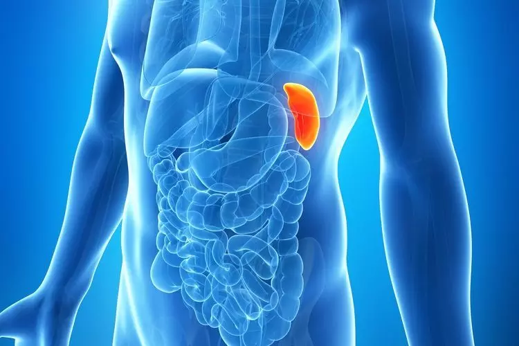
Splenic repair bests splenectomy for lowering mortality rate in traumatic injury cases: JAMA

A new study published in the Journal of American Medical Association showed that after severe splenic damage, splenic repair was independently linked to a reduced death rate after laparotomy than splenectomy. One of the organs most often hurt in abdominal trauma instances is the spleen. After suffering a severe abdominal injury, the spleen is frequently removed via a laparotomy with minimal attempt to preserve it. Dominik Jakob and colleagues conducted this study to investigate whether splenic repair is associated with lower mortality when compared with splenectomy, as well as the relationship between surgical management (splenic repair vs. splenectomy) and outcomes in patients with traumatic splenic injuries undergoing laparotomy.

From January 2013 to December 2019, a cohort research based on the trauma registry was conducted utilizing data from the American College of Surgeons Trauma Quality Improvement Program. Adult patients receiving laparotomy following traumatic injury within 6 hours of arrival and with significant splenic damage were among the participants. The period of data analysis in 2023 was April through August. The exposure criterion was splenic repair vs splenectomy in individuals with severe traumatic splenic damage. In-hospital mortality was the main outcome. Several statistical techniques were used to compare the results including multivariable logistic regression, propensity score matching, inverse-probability weighting, and 1:1 exact matching with sequential conditional logistic regression analysis as the major analysis.

11,244 individuals having laparotomy with significant traumatic splenic damage were identified by this study. Of them, 427 patients (3.8%) had splenic repair while 10,820 patients (96.2%) had splenectomy. 23 (5.3%) of the patients who had their spleen salvaged initially needed to have one during their follow-up hospital stay. 400 patients who had their spleen preserved were matched with 400 patients who had their spleen removed. When comparing to the splenectomy group, the mortality rate was considerably reduced in the splenic repair group. The conditional regression analysis was used to confirm the link between splenic repair and decreased mortality. Also, this link was verified using inverse-probability weighting, propensity score matching, and multivariable logistic regression.

Overall, the current study found that among patients who had laparotomies for severe traumatic splenic damage, there was a negative correlation between mortality with splenic repair as opposed to splenectomy. After the first splenic repair, around 5% of patients needed a splenectomy. The results imply that in certain instances of severe splenic damage, attempts to preserve the spleen may be warranted.

Source:

Jakob, D. A., Müller, M., Kolitsas, A., Exadaktylos, A. K., & Demetriades, D. (2024). Surgical Repair vs Splenectomy in Patients With Severe Traumatic Spleen Injuries. In JAMA Network Open (Vol. 7, Issue 8, p. e2425300). American Medical Association (AMA). https://doi.org/10.1001/jamanetworkopen.2024.25300

Powered by WPeMatico

Unique Case of Accelerated Bone Growth Precipitating a One-Stage Masquelet: A Report

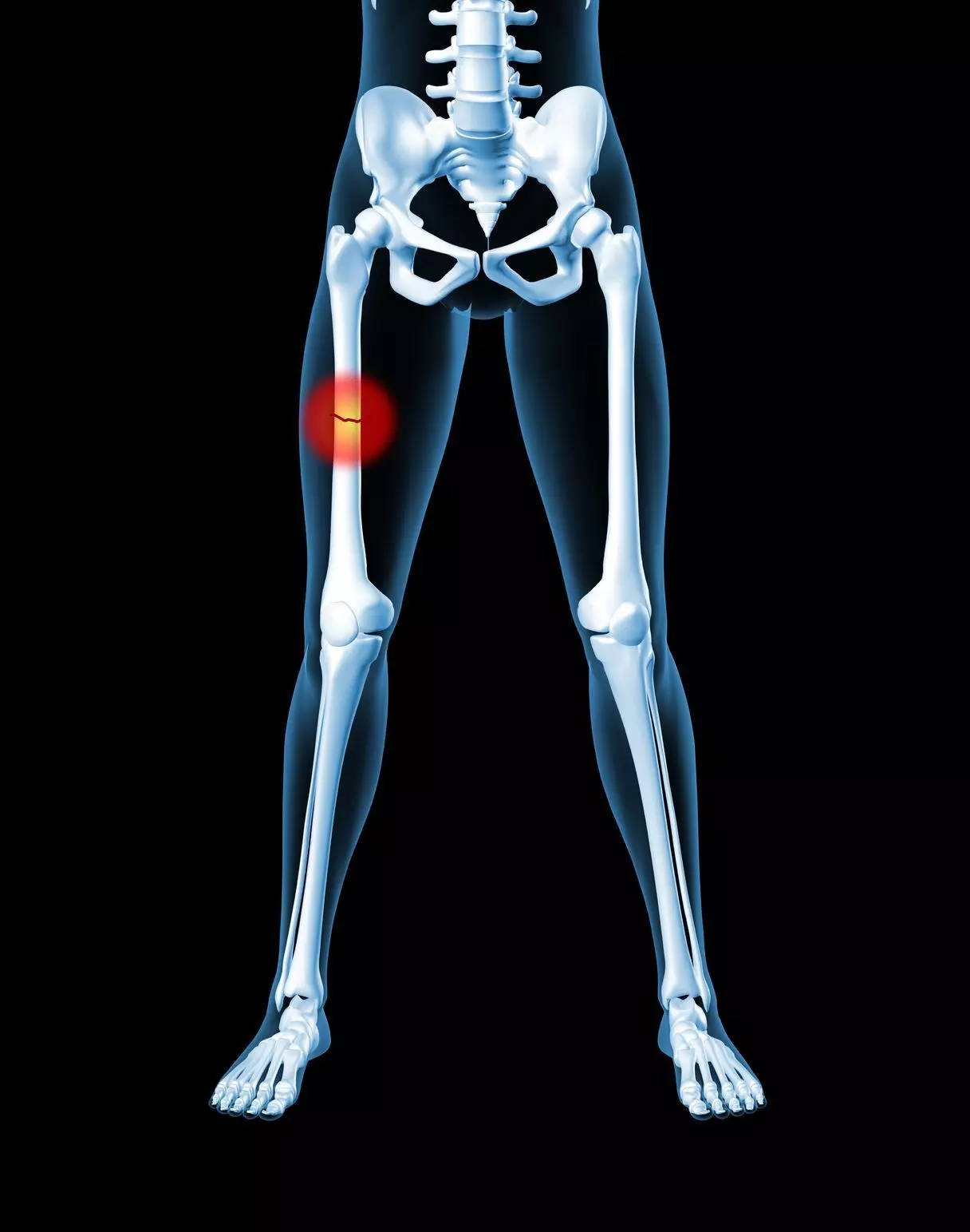
The masquelet technique is a two-stage procedure used by orthopedic surgeons to treat large segmental bone defects secondary to infection, trauma, and tumor resection. This technique characteristically requires the placement of a temporary cement spacer and subsequent bone grafting for complete reconstruction. Megan Tersteeg et al describe a unique case of segmental bone loss reconstruction in which a patient successfully achieved fracture union after the first step of the masquelet technique without bone grafting.

A 21-year-old male with a history of mild scoliosis, no previous fractures, and daily smoking for 2 years sustained multiple musculoskeletal injuries after a motorcycle collision. The injuries included a closed left femoral shaft injury and an open right Gutstilo and Anderson Type III A femur fracture with 10 cm of segmental bone loss. On the night of the injury, the patient underwent emergent irrigation, debridement, and temporary stabilization of his fracture with external fixation. Tobramycin antibiotic cement with vancomycin powder was placed within the thigh wound with a negative-pressure wound vacuum to cover the wound. 2 days later, once resuscitated, the right femur underwent another irrigation and debridement with an antibiotic bead and wound vacuum exchange. After multiple irrigation and debridement procedures of the lower extremity, the wound bed was amenable to definitive fixation of the femur. On day 5, the femur underwent intramedullary retrograde nailing with the placement of two proximal and two distal interlocking screws, revealing a 10-cm segmental bone defect. The defect was filled with tobramycin antibiotic cement mixed with vancomycin powder and underwent delayed primary wound closure. This was the first stage of a proposed masquelet technique treatment plan for this injury. He was made non-weight-bearing in his right lower extremity due to additional ipsilateral musculoskeletal injuries, but the patient was encouraged to range the knee from 0° extension to 140° flexion as often as possible. The remainder of his musculoskeletal injuries eventually underwent definitive fixation, with subsequent discharge to a rehabilitation center on hospital day 13.

The patient was seen in clinic 1 month after his initial injury, at which time he was non-weight-bearing in his right lower extremity and receiving outpatient physical therapy. His X-rays demonstrated unchanged osseous alignment with early callus formation. He was made to be a weight bearing as tolerated and continued his home exercise program to improve his knee and ankle range of motion (ROM) until his subsequent follow-up. At the 8-week follow-up visit, the patient’s pain was well controlled, and he was weight-bearing as tolerated on the right lower extremity up to his full bodyweight of 65.8 kg. There were no signs of infection, and his X-rays at the time showed abundant callus formation bridging the fracture site around the cement spacer. At this time, the second stage of the masquelet technique with bone grafting was considered. However, due to significant callus formation and minimal pain with ambulation, the decision was made to continue to monitor his progress and reevaluate at his next visit. Six-month post-injury, he was weight-bearing without assistive devices, without any gait abnormalities, and with no pain. Radiographs revealed the interval progression of callus formation. At the last follow-up, 1 year after definitive fixation, the patient reported no complaints regarding his right lower extremity. He had full ROM of knees, a normal, non antalgic gait, and had fully returned to work in construction. Radiographs demonstrated bridging callus formation in three out of four cortices and unchanged alignment.

The patient concluded that – “this case identifies a unique instance of successful fracture union of a 10-cm segmental bone defect despite the completion of only the first step in the masquelet procedure. This warrants further research on the mechanisms behind masquelet-induced regeneration without bone grafting and consideration of circumstances in which fracture union can be obtained following stage one of the masquelet procedures.”

Further reading:

Unique Case of Accelerated Bone Growth Precipitating a One-Stage Masquelet: A Case Report

Megan Tersteeg et al

Journal of Orthopaedic Case Reports 2024 May:14(5):Page 94-98

Powered by WPeMatico

Serum leptin can be a predictable marker for gestational diabetes mellitus: IJOGR

Gestational Diabetes Mellitus (GDM) is one of the most
prevalent conditions associated with pregnancy, and if untreated it can have a
major negative impact on both the mother’s and the child’s health. It affects
3-8% of pregnancies and is defined by glucose intolerance identified during
pregnancy due to obesity risk. The etiology of GDM remains poorly understood.
The most widely accepted explanation states that human placental lactogen,
prolactin, glucocorticoids, and progesterone all of which are released by the
placenta during pregnancy cause GDM by lowering insulin sensitivity and boosting
anti-insulin hormones. Adipocytes produce adipocytokines likes leptin that
contribute to energy balance, metabolism of glucose and insulin. According to
certain research, leptin may also play a major role in controlling the body’s
overall glucose homeostasis. Leptin may therefore play a significant role in the
onset and prognosis of GDM. There is a favorable relationship between plasma
leptin concentrations and both direct and indirect markers of obesity. Leptin
levels rise in expectant mothers who have altered maternal fat reserves and
glucose metabolism. The concentration of leptin in mothers rises two to three
times higher than in non-pregnant women, peaking at 28 weeks of gestation.

This study assessed the serum leptin concentrations for
development of gestational diabetes mellitus. It is a case-control study,
included 120 participants (60: Gestational Diabetes Mellitus Cases and 60:
Healthy Controls). Authors assed BMI, FBS, HbA1c, Lipid Profile and serum
leptin. Results: The serum leptin concentrations were significantly elevated in
patients with gestational diabetes mellitus when compared to controls (P=0.001).
There was a significant positive correlation between the serum leptin and BMI,
FBS, HbA1c, Total Cholesterol and LDL (P=0.001).

The current study illustrated how maternal circulating
leptin changes during gestational diabetes mellitus. Several studies have
reported varying patterns of leptin variation with increasing gestational age
in a typical pregnancy. The results of this investigation support the
hypothesis that GDM and the mother’s circulating leptin profile are related. An
adaptive reaction to insulin resistance and the inflammatory response linked to
GDM could account for the variation in circulating leptin concentration
observed in early gestation between women with and without GDM. By directly
regulating the insulin receptor or indirectly through central neuronal
pathways, leptin may enhance insulin sensitivity and it can also directly
promote the absorption of glucose and the synthesis of glycogen in skeletal
muscle.

It was also believed that the upregulation of the leptin
gene and protein production constituted a sort of defense mechanism against the
imbalance of proinflammatory cytokines that occurs in many abnormal
pregnancies, including GDM.

Based on the present study findings, authors concluded that
significant elevated levels of serum leptin might be served as a marker for
detection of gestational diabetes mellitus.

Source: Thilak, Harika and Narayanswamy / Indian Journal of
Obstetrics and Gynecology Research 2024;11(2):198–202

Powered by WPeMatico

Novel sugar test practical, user-friendly, and efficient test for hyposalivation among elderly, claims study

Novel sugar test was practical, user-friendly, and efficient test for hyposalivation among elderly, claims study published in the Journal of Oral Rehabilitation.

Hyposalivation is a highly prevalent condition in old persons. The aim of this study was to assess a novel tool for the diagnosis of hyposalivation using a sugar cube and to compare its reliability to other validated test methods. Five tests were performed: unstimulated salivary flow test by draining method (USFT), oral moisture device Mucus® (MCS), sugar cube test (SCT), stimulated salivary flow test by chewing gum (CG) and Saxon test (SX). The sugar cube test consisted in measuring the time needed to disintegrate a standard-sized sugar cube N°4 (5.6 g) placed beneath the tongue. Bivariate correlation analyses were performed. ROC curve analysis and the Youden Index were used to determine the cut-off value. Sensitivity and specificity were calculated according to the determined cut-off point. Results: A total of 121 participants were recruited, with an average age of 49.8 ± 18.2 years. The USFT was best correlated with the SCT. No correlation was found between SCT and the stimulated saliva tests (CG, SX) as well as the MCS test. According to the Youden index, 140 s is appropriate to diagnose hyposalivation. Sensitivity of .64 and specificity of .91 were found for SCT using USFT as gold standard. The mean subjective evaluation score of the SCT was 3.1 ± 1, significantly higher than USFT 2.6 ± 1.1 (p < .05). The sugar cube test proved to be a useful method for assessing unstimulated salivation, reducing test execution time and causing less participant discomfort than the gold standard unstimulated salivary flow test.


Reference: Spyraki F, Maniewicz S, Mojon P, Müller F. A novel test for hyposalivation using a sugar cube. J Oral Rehabil. 2024; 51: 1433-1439. doi:10.1111/joor.13715


Keywords: Novel, sugar, test practical, user-friendly, efficient, test, hyposalivation, elderly, claims, study, Spyraki F, Maniewicz S, Mojon P, Müller F, Journal of Oral Rehabilitation

Powered by WPeMatico

Knowing you have brain aneurysm may raise anxiety risk, other mental health conditions, suggests research

Among people who had aneurysms (weakened areas in brain blood vessels), the risk of developing a mental health condition was higher than among peers without an aneurysm. The highest risk was seen in patients younger than age 40, according to research published today in Stroke, the peer-reviewed scientific journal of the American Stroke Association, a division of the American Heart Association.

An unruptured intracranial aneurysm occurs when the wall of a blood vessel in the brain becomes weak and bulges out, posing a potential risk of future rupture and life-threatening bleeding in the brain.

“As a neurosurgeon who treats cerebral aneurysms, I often see people who do not undergo surgery yet feel fear and/or anxiety about their condition before each imaging or screening test to monitor their condition. Even when it is medically judged and explained that follow-up observation is the best course for their aneurysm rather than surgery, they still worry about the very slim chance of developing a fatal brain bleed,” said study co-author Na-Rae Yang, M.D., Ph.D., an assistant professor of neurosurgery in the department of neurosurgery at Ewha Womans University Mokdong Hospital, Ewha Womans University College of Medicine in Seoul, South Korea.

”This study did not specifically target brain aneurysms that were small enough not to require treatment and had a low probability of rupture. We included all diagnosed, untreated brain aneurysms that were being followed,” Yang said. “The reason for not treating and only following up may be that the aneurysm is small and unlikely to rupture, making treatment unnecessary. However, the decision is likely based on various circumstances, including the patient’s overall condition, and such specific reasons were not captured in this study group.”

While other studies have observed patterns of stress and anxiety in patients living with an aneurysm diagnosis, the researchers said this is one of the first large studies to estimate the risk of a mental health condition after aneurysm diagnosis.

In this study, researchers examined data from the National Health Information Database in South Korea, which includes over 20 years (2004 to 2024) of health data for more than 85,000 people who received care from hospitals, clinics and pharmacies throughout the country. It is one of the world’s largest and most comprehensive health information databases. More than six months after diagnosis with an aneurysm, researchers compared how many people developed a mental health condition, such as anxiety, stress, depression, bipolar and eating disorders, insomnia and alcohol or drug misuse. They then compared them to the rate of mental health condition diagnoses among similar adults without aneurysm who received care for an upper respiratory infection.

The analysis took place over a 10-year period and found, when compared to people without an aneurysm:

  • people with an aneurysm were 10% more likely to be diagnosed with a mental health condition;
  • the risk of a mental health condition was particularly pronounced in people with an aneurysm under the age of 40; and
  • in cases where a mental health condition was diagnosed by a psychiatrist, the difference was even greater, showing a three-fold increased risk.

“Our finding of a higher rate of a severe mental health condition in younger adults with aneurysm underscores the significant psychological burden for this group of patients who may already be dealing with other life stressors, such as building their career and/or raising a family,” Yang said. “This elevated rate of mental health conditions suggests that younger people might be particularly vulnerable, highlighting the need for targeted mental health support and interventions for this age group. This includes providing clear explanations and tailoring treatment decisions for each person,” Yang said.

An aneurysm may be diagnosed when a person has imaging (such as a CT scan or MRI) to evaluate neurological symptoms (such as a headache or vision changes), or it can be discovered by chance if imaging is done for an unrelated issue.

Study limitations include that the analysis was based on diagnostic and prescription codes in a national database, which may be imprecise and may not reflect the nuances of individual patient care and mental health status. In addition, the specific size and location of aneurysms were not considered. This study design highlights a possible association but cannot establish a cause-and-effect relationship between an aneurysm diagnosis and later mental health status. This study population was from South Korea, so the findings may not be consistent with populations in other countries.

Study details, background or design:

  • The study compared the occurrence of several mental health conditions over a 10-year period in 85,438 adults (51% male; average age of 56 years) with untreated aneurysms diagnosed between 2011 and 2019, to more than 331,000 (49% male; average age of 57) without aneurysms treated for acute upper respiratory infections during the same period. The adults without aneurysms were matched to the diagnosis group for age, sex, economic status and the presence of other medical conditions.
  • Mental health conditions were defined as a diagnosis of anxiety, stress, depressive disorder, bipolar disorder, eating disorder, insomnia, or alcohol or substance misuse according to the International Classification of Diseases coding database. Cases defined as having been diagnosed with a mental health condition plus receiving treatment from a psychiatrist included psychotherapy with or without medication.
  • Differences in the risk of mental health conditions were calculated after adjusting for several factors, including age, sex, health insurance status, disability, other medical conditions, smoking, alcohol consumption or obesity.

Reference:

Young Goo Kim, Hyungmi An, Ga Eun Kim, Hyang Woon Lee,and Na Rae Yang, Higher Risk of Mental Illness in Patients With Diagnosed and Untreated Unruptured Intracranial Aneurysm: Findings From a Nationwide Cohort Study, Stroke, https://doi.org/10.1161/STROKEAHA.123.045393.

Powered by WPeMatico

Robotic arm can be used to perform remote echocardiograms, shows study

New research presented at this year’s ESC Congress 2024 in London, UK (30 Aug-2 Sept) shows that performing echocardiograms remotely using a 5G cellular network has similar accuracy to those performed in person by cardiologists.

“Comprehensive echocardiographic exam with a 5G cellular network and robotic arm-based remote system is feasible with relatively good diagnostic accuracy,” said study author Dr Yu Liu, Zhongshan Hospital, Shanghai, China.

Echocardiography is the test-of-choice for the initial evaluation of many cardiac diseases and requires the expertise of a cardiologist for interpretation. However, this expertise is often limited or unavailable in rural or indeed smaller urban areas. Robotic arm-assisted remote echocardiograms have been attempted for teleconsultation in previous studies but analysis was limited to heart failure patients, primarily due to the network delay in telecommunications and the subsequent inadequate control of the robotic arm equipEuropean Society of Cardiologyment.

In this study, the authors assessed the feasibility and diagnostic accuracy of a 5G cellular network and robotic arm-based remote echocardiographic system in an outpatient clinic based 20 kilometres away from Zhongshan Hospital. A total of 51 patients were enrolled from the outpatient cardiology clinic. All underwent standard comprehensive echocardiography on a 5G cellular network robotic arm-based remote echocardiographic system, as well as a conventional echocardiographic platform (at Zhongshan Hospital) successively.

The order in which patients were examined on the remote and conventional instruments was randomly determined. There was no interval between the two examinations, and examinations of the same patient were performed by experienced but different cardiologists, who were blinded to each other’s diagnosis. The doctor who used the remote system was also randomly allocated and had not been previously specifically trained. The examinations were real time and diagnoses were made immediately after the examinations.

From the 51 patients, the image quality was sufficient for diagnosis in 50 patients (24 (48%) female). A single patient was excluded because some key views could not be obtained using the remote system, meaning 98% of the examinations had been technically successful.

Around one third (17 patients) had a heart problem identified using conventional in person echocardiography, including 10 with a primary diagnosis of valvulopathy (1 Barlow’s syndrome, 1 bicuspid aortic valve and 8 less-than-moderate regurgitation), 2 cardiac surgery follow-ups (1 case of aortic valve replacement and septal myectomy, and 1 case of mitral valve replacement and tricuspid annuloplasty), and 2 hypertrophy cardiomyopathy (including 1 case of obstruction at papillary muscle level), 2 with abnormal left ventricular wall motion (including 1 case of apical mural thrombus), and 1 with congenital heart disease (secumdum atrial septal defect).

Echocardiograms using the robotic arm resulted in the same diagnosis as conventional in-person echocardiography in 98% of cases (papillary muscle level obstruction was missed in one case).

Time for image acquisition using remote echocardiography was significantly longer (around 50% longer) than conventional (24 mins 36 secs vs. 16 mins 15 secs).

A previous version of the robotic arm has been cleared for clinical use in scanning the abdomen (China, Europe, Australia and Singapore), which requires less complex scanning manoeuvres. However, the authors say a multi-centre study at a larger scale with both other local hospital and referral centres involved should be carried out before this new technology should be used.

Although 5G technology is not available everywhere, lead-author Xianhong Shu, also of Zhongshan Hospital, said: “This system would increase the accessibility of better medical resources as patients may travel less to get diagnosis and medical advice from cardiologists based in referral centres.”

She adds there are further potential advantages: “A remote robotic echo system may help protect more health professionals from the risk of exposure during pandemics like the COVID-19 as the cardiologist may not need to be in close contact with the patient if only echocardiogram consultation is required.”

Powered by WPeMatico