High Initial fractional inspired oxygen May Reduce Mortality in Preterm Infants, reveals study

Researchers have found that resuscitation with higher fractional inspired oxygen (FiO2) levels significantly reduces mortality in preterm infants born at less than 32 weeks’ gestation. This conclusion emerges from a comprehensive network meta-analysis (NMA) of individual participant data (IPD) from multiple randomized clinical trials. This study was recently published in JAMA Pediatrics by James X. and colleagues.

While the benefits of lower FiO2 levels in term and near-term infants are well-documented, their impact on very preterm infants has been unclear. This study aims to evaluate the relative effectiveness of different initial FiO2 levels in reducing mortality, severe morbidities, and achieving optimal oxygen saturations (SpO2) in infants born before 32 weeks’ gestation.

The analysis included data from MEDLINE, Embase, CENTRAL, CINAHL, ClinicalTrials.gov, and WHO ICTRP, spanning from 1980 to October 10, 2023. Eligible studies were randomized clinical trials involving infants born at less than 32 weeks’ gestation. These trials compared at least two different initial oxygen concentrations for delivery room resuscitation, categorized as low (≤0.3), intermediate (0.5-0.65), or high (≥0.90) FiO2. Investigators from eligible studies were invited to contribute their IPD. The data were meticulously processed and verified for quality and integrity. A one-stage contrast-based Bayesian IPD-NMA was conducted using noninformative priors and random effects, adjusted for key covariates.

The primary outcome was all-cause mortality at hospital discharge. Secondary outcomes included morbidities associated with prematurity and SpO2 levels at 5 minutes post-resuscitation.

  • Data were obtained for 1055 infants from 12 of the 13 eligible studies conducted between 2005 and 2019.

  • The findings revealed that resuscitation with high initial FiO2 (≥0.90) was associated with significantly reduced mortality compared to both low (≤0.3) FiO2 (odds ratio [OR], 0.45; 95% credible interval [CrI], 0.23-0.86; low certainty) and intermediate (0.5-0.65) FiO2 (OR, 0.34; 95% CrI, 0.11-0.99; very low certainty).

  • High initial FiO2 had a 97% probability of ranking first in reducing mortality. However, the effects on other morbidities were inconclusive.

These results suggest that a high initial FiO2 (≥0.90) may be more effective in reducing mortality in preterm infants born at less than 32 weeks’ gestation compared to lower FiO2 levels. While the certainty of these findings varies, the data supports the consideration of higher FiO2 levels during resuscitation in this vulnerable population.

High initial FiO2 (≥0.90) may significantly reduce mortality in preterm infants born before 32 weeks’ gestation compared to low initial FiO2, albeit with low certainty. The potential advantage of high FiO2 over intermediate FiO2 is suggested but requires further evidence. These findings could influence resuscitation protocols to improve outcomes for preterm infants.

Reference:

Sotiropoulos, J. X., Oei, J. L., Schmölzer, G. M., Libesman, S., Hunter, K. E., Williams, J. G., Webster, A. C., Vento, M., Kapadia, V., Rabi, Y., Dekker, J., Vermeulen, M. J., Sundaram, V., Kumar, P., Kaban, R. K., Rohsiswatmo, R., Saugstad, O. D., & Seidler, A. L. (2024). Initial oxygen concentration for the resuscitation of infants born at less than 32 weeks’ gestation: A systematic review and individual participant data network meta-analysis. JAMA Pediatrics. https://doi.org/10.1001/jamapediatrics.2024.1848

Powered by WPeMatico

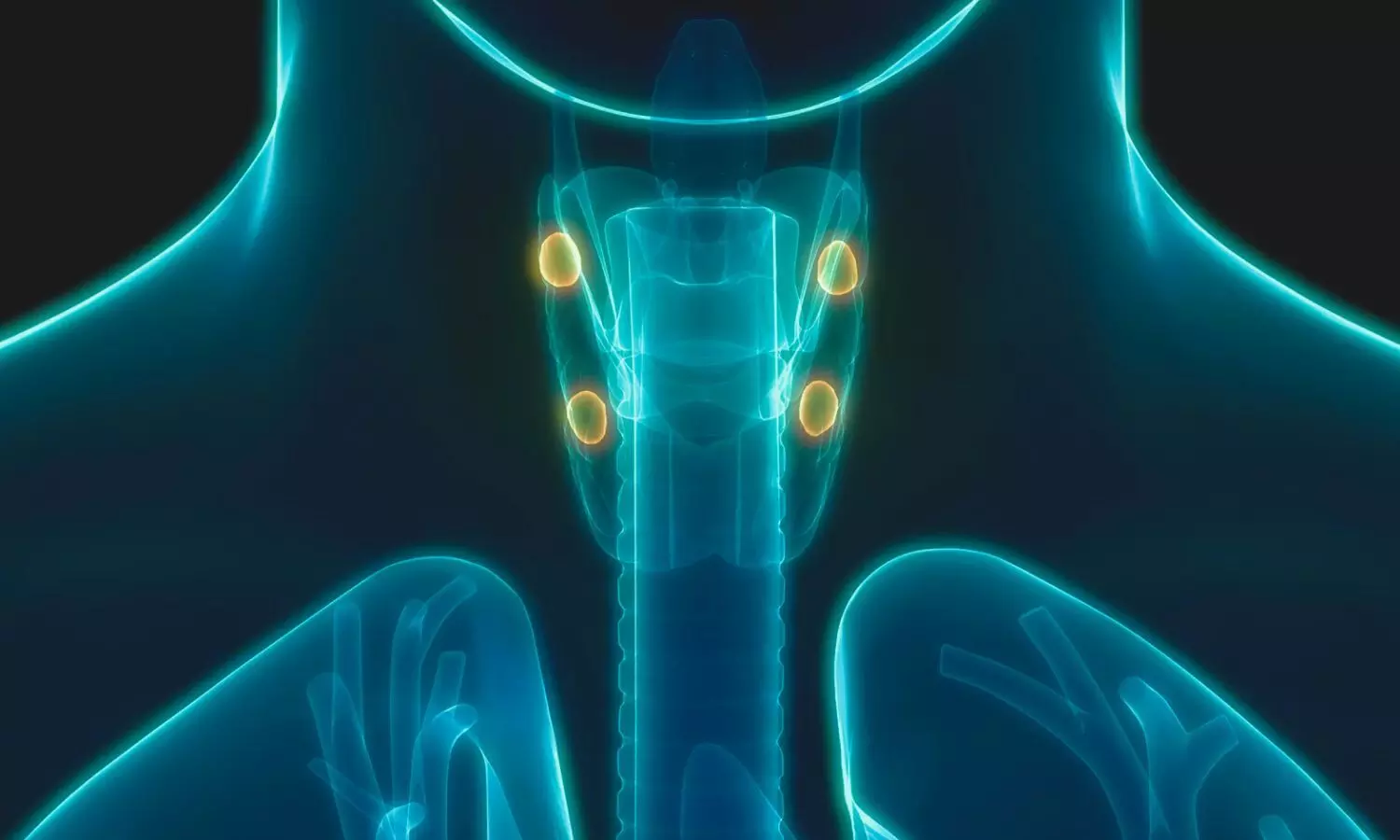
Precision Imaging Triumphs: FCH PET/CT Outshines MIBI SPECT/CT in Parathyroidectomy Outcomes in new study

France: Advanced imaging technologies such as F18-Choline PET/CT and MIBI SPECT/CT are revolutionizing the surgical management of primary hyperparathyroidism, according to findings from a recent diagnostic randomized clinical trial. The study investigates how these imaging modalities enhance precision in localizing abnormal parathyroid glands, optimizing patients’ surgical outcomes.

The study, published in JAMA Otolaryngology – Head & Neck Surgery, revealed that F-18 fluorocholine (FCH) PET/CT could be a first-line technique over technetium-99m sestamibi (Tc-99m MIBI) SPECT/CT for imaging parathyroid tumors before surgery.

“In the diagnostic randomized clinical trial involving 57 evaluable patients, achieving normocalcemia one month after initial positive imaging-guided minimally invasive parathyroidectomy (MIP) was more common following FCH PET/CT imaging as the first-line approach compared to MIBI SPECT/CT imaging,” the researchers reported.

Primary hyperparathyroidism, characterized by excessive production of parathyroid hormone, can lead to elevated calcium levels and various symptoms such as bone pain, kidney stones, and fatigue. Traditionally, surgeons relied on ultrasound and sestamibi scans for preoperative localization of parathyroid adenomas or hyperplasia. However, these methods are not always effective, leading to challenges during surgery.

Against the above background, Elske Quak, Department of Nuclear Medicine and Thyroid Unit, Centre François Baclesse, Caen, France, and colleagues aimed to compare first-line FCH PET/CT vs MIBI SPECT/CT for optimal care in PHPT patients requiring parathyroidectomy. They also compared the proportions of patients in whom the first-line imaging method resulted in successful MIP and normalization of calcemia one month after surgery.

For this purpose, the researchers conducted a French multicenter randomized open diagnostic intervention phase 3 trial; enrolling patients from 2019 to 2022 and participating up to 6 months after surgery. The study enrolled adult patients diagnosed with primary hyperparathyroidism (PHPT) who required surgical intervention. Individuals with a history of prior parathyroid surgery or those diagnosed with multiple endocrine neoplasia type 1 (MEN1) were not eligible to participate.

Patients were randomly assigned in equal numbers to undergo either FCH PET/CT (FCH1) or MIBI SPECT/CT (MIBI1) as their initial imaging modality. If the first-line imaging was negative or inconclusive, they subsequently underwent FCH PET/CT (FCH2) after MIBI1 or MIBI SPECT/CT (MIBI2) after FCH1.

All patients underwent surgery under general anesthesia within 12 weeks after their last imaging session. Clinical and biochemical assessments, including serum calcium and parathyroid hormone levels, were conducted at 1 and 6 months post-surgery.

The primary endpoint was defined as achieving a true-positive result in the first-line imaging-guided minimally invasive parathyroidectomy (MIP), alongside serum calcium levels of 2.55 mmol/l or lower one-month post-surgery, aligned with the local upper limit of normal.

The study led to the following findings:

  • 57 patients received FCH1 (n = 29) or MIBI1 (n = 28). The mean age of patients was 62.8 years, with 74% female patients. Baseline patient characteristics were similar between groups.
  • Normocalcemia was found one month after positive first-line imaging-guided MIP in 85% of patients in the FCH1 group and 56% of patients in the MIBI1 group.
  • Sensitivity was 82% and 63% for FCH1 and MIBI1, respectively.
  • Follow-up at six months with biochemical measures was available in 43 patients, confirming that all patients with normocalcemia one month after surgery still had it at six months.
  • There were no adverse events related to imaging and four adverse events related to surgery.

“First-line FCH PET/CT is a safe and suitable replacement for MIBI SPECT/CT. FCH PET/CT leads more patients with PHPT to correct imaging-guided MIP and normocalcemia compared to MIBI SPECT/CT thanks to its superior sensitivity,” the researchers concluded.

Reference:

Quak E, Lasne-Cardon A, Cavarec M, et al. F18-Choline PET/CT or MIBI SPECT/CT in the Surgical Management of Primary Hyperparathyroidism: A Diagnostic Randomized Clinical Trial. JAMA Otolaryngol Head Neck Surg. Published online June 20, 2024. doi:10.1001/jamaoto.2024.1421

Powered by WPeMatico

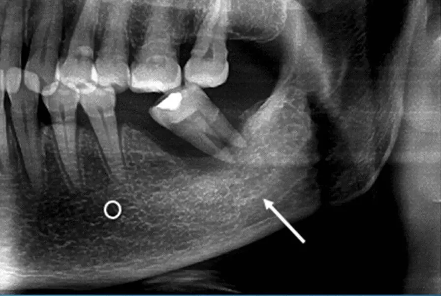
Tooth extraction significant risk factor for osteoradionecrosis, particularly when post-extraction sockets receive high RT dosage: Study

Tooth extraction is a significant risk factor for osteoradionecrosis, particularly when post-extraction sockets receive a high RT dosage suggests a study published in the Oral Diseases.

A study was done to evaluate osteoradionecrosis (ORN) incidence in a cohort of patients undergoing tooth extraction (TE) before radiotherapy (RT) for head and neck cancers. The Ethics Committee of Università Cattolica del Sacro Cuore (ID-2132) approved the study protocol and registered it at clinicaltrials.gov (ID: NCT04009161). TE was performed in case of signs of pericoronitis, periapical lesions, restorative impossibility, and severe periodontitis. ORN was defined as an exposed bone at an unhealed post-extraction socket in the absence of oncological recurrence. The RT plans were reviewed, and each post-extractive socket was contoured to calculate the received radiation dose. RESULTS

In total, 156 patients with 610 TE were enrolled. The mean follow-up was 567 days. ORN was diagnosed in four patients (2.6% of patients and 0.7% of TE). Need for osteotomy and radiation dose at the extraction site were associated with ORN (OR for osteotomy: 21.9, 95% CI: 2.17-222.2, p = 0.009; OR for RT dose: 1.1, 95% CI: 1-1.15, p = 0.05). TE appears to be a significant risk factor for ORN, particularly when osteotomy is required, and post-extraction sockets receive a high RT dosage. This study proposes a decision-making algorithm for TE and outlines a straightforward surgical protocol.

Reference:

Oral Dis 2024 Apr 09;[EPub Ahead of Print], C Rupe, G Gioco, M Massaccesi, L Tagliaferri, F Pastore, F Micciché, J Galli, D Mele, ML Specchia, A Cassano, M Cordaro, C Lajolo

Keywords:

Tooth extraction, significant, risk, factor, osteoradionecrosis, particularly, post-extraction, sockets, receive, high RT dosage, study, oral diseases

Powered by WPeMatico

Open bypass for infrapopliteal revascularization lowers major adverse limb events, suggests study

A recent study published in the Journal of Vascular Surgery found that among the patients having suitable single segment great saphenous vein who undergo infrapopliteal revascularization for chronic limb threatening ischemia (CLTI), open bypass surgery was associated with a lower incidence of major adverse limb event (MALE) or death and less significant amputation than endovascular treatments. This study by Kristina Giles and colleagues compared the BEST-CLI patients with severe infrapopliteal disease who received open tibial bypass to endovascular tibial procedures.

This comprehensive research focused on a subset of BEST-CLI trial participants who had a suitable single-segment great saphenous vein conduit and significant infrapopliteal disease. These patients were randomized into two groups, where one group had patients who were undergoing open bypass surgery and the other group had patients who were receiving endovascular tibial interventions. The primary outcome measured was the incidence of MALE or all-cause death. MALE included major limb amputation or major re-interventions. The outcomes were assessed using Cox proportional regression models.

The key findings of this study were;

  • The analyzed subgroup consisted of 665 patients, where 339 patients were in the tibial endovascular and 326 patients were in the open tibial bypass group procedure group. The main endpoint of MALE or all-cause mortality at 3 years was considerably lower in the surgical group (48.5%) than in the endovascular group (56.7%).
  • Mortality rates were 35.5% in the surgical group and 35.8% in the endovascular group. However, the surgical group had a notably lower rate of MALE events at 23.3% when compared to 35.0% in the endovascular group. This included a reduced rate of major re-interventions in the surgical group (10.9%) when compared to the endovascular group (20.2%).
  • While the surgical group had fewer above-ankle amputations (13.5%) than the endovascular group, the freedom from above-ankle amputation or all-cause mortality was equal between treatment groups, with 43.6% in the surgical group when compared to 45.3% in the endovascular group. Also, perioperative (30-day) mortality was comparable across treatment groups (2.5% open vs 2.4% endovascular; p=0.93), as it was observed to be 30-day MACE (5.3% open vs 2.7% endovascular; p=0.12).

Overall, when compared to endovascular treatments, open bypass surgery was linked to a reduced risk of major amputation and a lower incidence of MALE or death in patients with acceptable single segment great saphenous veins who had infrapopliteal revascularization for CLTI. Further research is imperative to examine variations in anatomic extent, comorbidities and lesion complexity.

Reference:

Giles, K. A., Farber, A., Menard, M. T., Conte, M. S., Nolan, B. W., Siracuse, J. J., Strong, M., Doros, G., Venermo, M., Azene, E., Rosenfield, K., & Powell, R. J. (2024). Surgery or Endovascular Therapy for Patients with Chronic Limb-threatening Ischemia Requiring Infrapopliteal Interventions. In Journal of Vascular Surgery. Elsevier BV. https://doi.org/10.1016/j.jvs.2024.05.049

Powered by WPeMatico

Dyslipidemia in pregnancy may increase risk of preterm birth: Study

A recent study found a significant causal relationship between blood lipid levels and preterm birth which is the leading cause of neonatal mortality throughout the globe. The findings published in the recent issue of European Journal of Obstetrics & Gynecology and Reproductive Biology highlighted how dyslipidemia contributes to the risk of preterm delivery.

This research extracted data from genome-wide association studies and included participants from the FinnGen database and the UK Biobank. The study included data from a total of 115,082 participants who had their lipid levels measured, along with 8,507 patients who underwent preterm birth. The study focused at single-nucleotide polymorphisms (SNPs) that strongly associated with blood lipid levels. These SNPs were uncorrelated (R2 < 0.001) and had a significant association (p < 5 × 10–8) with lipid measurements. The research also conducted various sensitivity analyses to account for genetic pleiotropy, heterogeneity, and the directionality of causality to ensure the robustness of the findings.

The results revealed that increased levels of several blood lipids were associated with an elevated risk of preterm birth. Also, increased levels of apolipoprotein B was linked to a 12% higher risk (odds ratio [OR], 1.12; 95% confidence interval [CI], 1.02–1.23; p = 0.019) and higher levels of low-density lipoprotein cholesterol (LDL-C) were associated with an 11% higher risk (OR, 1.11; 95% CI, 1.00–1.22; p = 0.040).

Other lipids that showed a significant association included non-high-density lipoprotein cholesterol (non-HDL-C), remnant cholesterol and total free cholesterol, where each of these contributed to about an 11% increase in the risk of preterm delivery. The study identified that triglycerides in low-density lipoprotein (LDL) also had a causal relationship with the risk of preterm birth. The results of the  sensitivity analyses confirmed the robustness of the main findings, with no evidence of horizontal pleiotropy or reverse causal relationships detected.

This study provides strong proof of the detrimental impact of dyslipidemia on preterm birth by emphasizing the role of elevated LDL cholesterol. These findings highlight the importance of monitoring and managing blood lipid levels in pregnant women to potentially reduce the risk of preterm delivery. Overall, the research illuminates the need for further studies to explore potential interventions and treatments that could reduce this risk to improve neonatal outcomes and reduce the global burden of preterm births.

Source:

Zhang, W., Liu, L., Yang, X., Wang, K., Yao, H., & Wang, F. (2024). Dyslipidemia characterized by low density lipoprotein cholesterol and risk of preterm Birth: A Mendelian randomization study. In European Journal of Obstetrics & Gynecology and Reproductive Biology. Elsevier BV. https://doi.org/10.1016/j.ejogrb.2024.06.035

Powered by WPeMatico

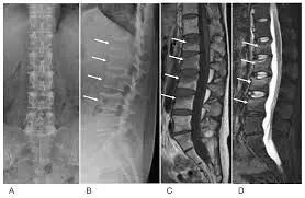
Estimated incidence of pregnancy and lactation-associated premenopausal osteoporosis higher with risk of Vertebral fracture: Study

The estimated incidence of pregnancy and lactation-associated premenopausal osteoporosis is higher with the risk of Vertebral fracture suggests a study published in the BMC Pregnancy and Childbirth.

Pregnancy and lactation-associated osteoporosis (PLO), as well as premenopausal osteoporosis, might be a predictor of future fracture. This study aimed to describe the clinical features of PLO as a subtype of premenopausal osteoporosis and to evaluate medical interventions for it. From an administrative claims database including 4,224,246 people in Japan, we classified women for whom the date of childbirth had been defined and who had suffered low-trauma fracture between the ages of 18–47 years as the premenopausal osteoporosis group. A fracture site for which the odds ratio for fractures occurring between 5 months before and 12 months after childbirth (around childbirth) was greater than 1 was considered the PLO site. We classified patients with a fracture at the PLO site around childbirth as the PLO group. The control group consisted of 500 women without fragility fractures. We investigated some drugs and diseases to explore fracture-causing factors, as well as medical interventions such as osteoporosis diagnosis, bone densitometry, anti-osteoporosis pharmacotherapy, and lactation inhibitors. Results: In total, 231 parous women were classified into the premenopausal osteoporosis group. The most common fracture was vertebral fracture and was likely to occur around childbirth, followed by distal radius and sacral fractures, which were rare around childbirth. Considering vertebral, pelvic, and proximal femoral fractures as PLO sites, 56 women with 57 PLO fractures were classified into the PLO group. The incidence of PLO was estimated at 460 per million deliveries. Ovulation disorder and high maternal age were associated with the development of PLO. Vertebral fracture was the most common PLO fracture. It was mainly diagnosed a few months, and possibly up to 1 year, postpartum. PLO patients with vertebral fractures underwent more medical interventions than did those with other fractures, but they were still inadequate. PLO with vertebral fracture was one of the major types of premenopausal osteoporosis. The prevalence of PLO is considered to be higher than previously thought, indicating the presence of potentially overlooked patients. More timely interventions for PLO might lead to the improved management of latent patients with premenopausal osteoporosis and reduce future fracture risk.

Reference:

Kasahara, K., Tanaka-Mizuno, S., Tsuji, S. et al. Pregnancy and lactation-associated osteoporosis as a major type of premenopausal osteoporosis: a retrospective cohort study based on real-world data. BMC Pregnancy Childbirth 24, 301 (2024). https://doi.org/10.1186/s12884-024-06520-0

Keywords:

BMC Pregnancy and Childbirth, Kasahara, K., Tanaka-Mizuno, S., Tsuji, S, Fracture, Lactation, Osteoporosis, Pregnancy, Premenopausal

Powered by WPeMatico

Vaccination may reduce risk of cognitive impairment in patients with COVID 19 infections: Study  

Since the outset of the COVID 19 pandemic, 10 to 30 per cent of the general population has experienced some form of virus-induced cognitive impairment, including trouble concentrating, brain fog or memory loss.

This led a team of researchers to explore the mechanism behind this phenomenon and pinpoint a specific protein that appears to be driving these cognitive changes. 

A new study published in Nature Immunology, led by researchers at Western and Washington University School of Medicine in St. Louis, Missouri, also looked at how vaccination may help reduce the impacts of memory loss following COVID-19 infections.  

The research team, including Schulich School of Medicine & Dentistry professor Dr. Robyn Klein, who joined Western from Washington University used rodent models to better understand how COVID-19 impacts cognitive impairment.   

“We looked carefully at their brains during acute infection and then later after recovery to discover what was abnormal in terms of the different immune cells trafficking into the brain and their effects on neural cells,” said Klein, who holds the Canada Excellence Research Chair in Neurovirology and Neuroimmunology.  

Klein said she was concerned by reports of cognitive impairment in the early days of the pandemic, which led researchers to question whether the virus was invading the central nervous system.  

Klein’s previous work studied viruses that invade the brain.  

“We had previously shown that the virus could not be detected in human or hamster brains, and this study also showed that the virus was not invading the central nervous system,” said Klein. The finding means some other mechanism is leading to cognitive impairment.      

The team identified SARS-CoV-2 infection increased levels of brain Interleukin-1 beta (IL-1β), a cytokine protein that impacts the immune system. The team observed that the models with increased levels of IL-1β experienced loss of neurogenesis, the process by which new neurons are formed in the brain, and also displayed memory loss.  

Vaccination reduces cognitive symptoms

The team concluded IL-1β was one potential mechanism driving SARS-CoV-2-induced cognitive impairment, and wondered whether this may be prevented by vaccination.

Researchers then investigated how vaccinated models were impacted. They found a promising correlation between vaccination and reduced cognitive impairments like memory loss.

The researchers showed that prior vaccination reduced inflammation of the brain and lowered levels of IL-1β. As a result, the vaccinated models experienced less of an impact on memory and brain function. 

Klein says there is still more work to be done to fully understand how vaccinations are achieving this result, and whether it will translate to humans.  

“We know there’s anecdotal evidence that humans who’ve been vaccinated have a much lower risk of developing this long COVID brain fog,” said Klein.  

The vaccine used in the study is not the same as the vaccines available to people, Klein stressed, meaning more studies will need to be conducted to further investigate the connection between vaccination and reduced long COVID impacts.  

“What we do know is that if you’re vaccinated you have much less inflammation,” said Klein.  

Vaccination is about lowering the risk of the impacts of infection, not completely preventing infection, she added. For example, a vaccine can protect individuals from developing severe pneumonia, but that doesn’t mean it completely protects against pneumonia. 

The same is likely true for cognitive impacts. 

“People need to understand that about vaccines,” Klein said. “They need to know what vaccines can do and what they can’t do.” 

References: Abigail Vanderheiden, Jeremy D. Hill, Xiaoping Jiang, Ben Deppen, Gayan Bamunuarachchi, Nadia Soudani, Astha Joshi, Matthew D. Cain, Adrianus C. M. Boon & Robyn S. Klein Nature Immunology (2024)

Powered by WPeMatico

Common BP drug may make leukemia more responsive to chemotherapy while protecting heart: Study

Researchers from the University of Missouri School of Medicine found that a targeted gene therapy may make acute myeloid leukemia (AML) more sensitive to chemotherapy, while also protecting the heart against toxicity often caused by cancer treatments.

Acute myeloid leukemia is the most common type of leukemia in adults and the resulting chemotherapy treatment can put patients at an increased risk for cardiac damage. Associate Professor of Medicine Dr. Xunlei Kang and PhD students Yi Pan and Chen Wang led a study looking at similarities between leukemia and cardiovascular disease. They found a shared target — AGTR1, a receptor responsible for cell reproduction, was overabundant in the blood cells of patients with leukemia.

The researchers used losartan, a common medicine for treating high blood pressure, to inhibit the AGTR1 receptor in mice. This disrupted cancer growth, slowing the development of leukemia and led to longer survival. The next step is to further investigate losartan’s effectiveness in treating human leukemia patients.

“Mouse models of leukemia differ from human disease in several ways, including differences in the immune system, the bone marrow microenvironment and responses to treatments,” Pan said. “We will now carefully interpret and validate these findings in human studies to ensure translational relevance,” Pan said.

If these findings are confirmed in human clinical trials, the approval process to use losartan would be shorter compared to other medications, since it’s already FDA-approved and will not require comprehensive data about the drug.

“When we treated mice with the AGTR1 inhibitor losartan, we observed that this commercially available drug shows great promise in reducing AML development while protecting against chemotherapy-induced cardiotoxicity,” Kang said. “This finding shows great potential to both enhance the success of chemotherapy while protecting the heart.”

Dr. Xunlei Kang, MD, PhD is an associate professor of medicine at the MU School of Medicine and focuses his research on blood conditions and stem cell study. He received his medical degree and doctorate from Shanghai Jiao Tong University in China.

“Inhibiting AGTR1 reduces AML burden and protects the heart from cardiotoxicity in mouse models” was recently published in the journal of Science Translational Medicine. In addition to Kang, Pan and Chen, authors include research specialists Wenxuan Zhou and Yao Shi; PhD student XiaDuo Meng, Hematology and Medical Oncology fellow Yasir Muhammad, MD; Richard D. Hammer, MD, professor of clinical pathology and anatomical sciences; De-Pei Li, MD, professor of medicine and associate director of the Center for Precision Medicine; Zhenguo Liu, MD, professor of medicine and chief of cardiology; and Gerhard Hildebrandt, MD, Chief of the Division of Hematology and Medical Oncology. Bei Jia and Hong Zheng from Penn State University College of Medicine also contributed to the paper.

Powered by WPeMatico

Tranexamic Acid Injection may Reduce Postoperative Bruising in Eyelid Surgery, reveals research

Researchers have found that administering subcutaneous tranexamic acid injections without a waiting period before eyelid surgery significantly reduces postoperative bruising. A recent study was published in the journal Ophthalmic Plastic and Reconstructive Surgery by Paramo and colleagues. This prospective, randomized, multicenter study investigated the effects of immediate surgery post-injection on intraoperative bleeding and postoperative ecchymosis.

Previous pilot studies indicated that waiting 15 minutes after a subcutaneous tranexamic acid injection could reduce intraoperative bleeding and postoperative bruising in eyelid surgery. However, the effects of initiating surgery immediately after the injection were previously unexplored. This study aimed to determine the outcomes of such an approach.

The study was designed as a prospective, randomized, multicenter, double-masked, controlled trial. It included patients undergoing bilateral symmetric upper and/or lower lid blepharoplasty or ptosis repair. Participants received tranexamic acid in one eyelid and a control in the other. Surgeons noted the side with more intraoperative bleeding, and two masked graders evaluated periocular ecchymosis at postoperative day 0 and postoperative week 1 (POW 1) using a 5-point scale. At POW 1, patients reported their subjective grading of bruising, indicating whether it was more pronounced on one side or similar on both sides. Data were analyzed using Wilcoxon signed-rank and sign tests.

  • A total of 130 patients participated in the study. Findings revealed significantly less eyelid ecchymosis on the tranexamic side at both postoperative day 0 (p = 0.001) and POW 1 (p < 0.001).

  • Specifically, the 69 levator advancement surgeries showed markedly less ecchymosis at postoperative day 0 (p < 0.001) and POW 1 (p = 0.001).

  • Upper eyelid blepharoplasty, combined upper and lower lid blepharoplasty, and conjunctivomullerectomy showed trends towards significance.

  • Among 68 patients who reported their grading at POW 1, 69% noted less bruising on the tranexamic side (p < 0.001).

  • No significant difference in intraoperative bleeding was observed between the sides (p = 0.930).

The study demonstrated that administering subcutaneous tranexamic acid without a waiting period before eyelid surgery significantly reduces postoperative bruising. This effect was evident on both postoperative day 0 and POW 1, particularly in levator advancement surgeries. The lack of significant difference in intraoperative bleeding suggests that the immediate initiation of surgery post-injection does not increase intraoperative risks.

Administering subcutaneous tranexamic acid without a waiting period before eyelid surgery effectively reduces postoperative bruising without affecting intraoperative bleeding. These findings suggest that a higher dose of tranexamic acid can be a promising intervention to improve surgical outcomes and patient satisfaction in eyelid surgery.

Reference:

Paramo, R., Cheng, T., Malik, A., Fan, J., & Barmettler, A. (2024). Effect of tranexamic acid on intra- and postoperative bleeding in eyelid surgery: A prospective, randomized, multicenter, double-masked, control trial. Ophthalmic Plastic and Reconstructive Surgery, 40(3), 331–335. https://doi.org/10.1097/iop.0000000000002583

Powered by WPeMatico

FDA Permits Marketing of First Point-of-Care Hepatitis C RNA Test

Today, the US Food and Drug Administration granted marketing authorization to Cepheid for the Xpert HCV test and GeneXpert Xpress System, the first hepatitis C virus (HCV) test that can be used to bring diagnosis to appropriately certified point-of-care settings for individuals at risk for hepatitis C.

The test may be performed in settings operating under a CLIA (Clinical Laboratory Improvement Amendments) Certificate of Waiver, such as certain substance use disorder treatment facilities, correctional facilities, syringe service programs, doctor’s offices, emergency departments and urgent care clinics. Rather than requiring a sample to be sent to a central lab for testing, the test detects HCV RNA and delivers results in about an hour using a blood sample from the fingertip.

The authorization of this test enables a test-and-treat approach where a person can be tested for HCV, and if positive for HCV RNA, be linked to care and potentially receive treatment during the same health care visit. Prior to the availability of a rapid, point-of-care test, HCV testing has been a multi-step process which often results in patients needing follow-up appointments for test results and additional testing, which can lead to patients not receiving a diagnosis and not receiving necessary treatment.

“Despite the existence of a safe and highly effective oral cure for hepatitis C, many people do not know they have the disease due partly to the lack of availability of convenient, widespread testing options,” said Jeff Shuren, M.D., J.D., director of the FDA’s Center for Devices and Radiological Health. “Equipping health care providers with tools to diagnose and treat patients in the same visit can result in hundreds of thousands more hepatitis C patients being diagnosed and treated, preventing individual disease progression and additional spread of the virus.”

According to the U.S. Centers for Disease Control and Prevention, hepatitis C is a liver infection caused by the hepatitis C virus. Hepatitis C is spread through contact with blood from a person with hepatitis C. For some people, hepatitis C is a short-term illness, but for more than half of people with HCV infection, it becomes a long-term, chronic infection.

It is estimated more than 2.4 million people – and as many as 4 million people – in the United States have hepatitis C, which if left untreated, often leads to serious and sometimes deadly outcomes such as liver cancer and liver failure. The infection contributed to more than 12,000 deaths in 2022 alone.

The proposed fiscal year 2025 budget for the Department of Health and Human Services includes a proposed five-year program to eliminate hepatitis C in the U.S. The program aims to significantly expand testing, treatment, prevention and monitoring of hepatitis C infections in the U.S. With hepatitis C being the most common cause of liver cancer today, this initiative is an important contribution to President Biden’s Cancer Moonshot.

“A third of people with hepatitis C in the United States don’t even know they have a deadly, yet curable, infection,” said Jonathan Mermin, M.D., M.P.H., director of CDC’s National Center for HIV, Viral Hepatitis, STD and TB Prevention. “This new test provides hope that more people will be cured, but it will only succeed if it is affordable and available.”

Validation data for the Xpert HCV test and GeneXpert Xpress System was gathered through the Independent Test Assessment Program (ITAP), a National Institutes of Health (NIH) Rapid Acceleration of Diagnostics (RADx®) Tech program, in collaboration with the FDA. ITAP was launched in 2021 to accelerate test evaluation to support the FDA’s regulatory review and the availability of high-quality, accurate and reliable diagnostic tests to the public.

“Today’s announcement by the FDA of marketing authorization for a rapid diagnostic to detect hepatitis C RNA is an example of the power of the RADx Tech model to deliver a much-needed test to millions of people in record time,” said National Institute of Biomedical Imaging and Bioengineering (NIBIB) director Bruce J. Tromberg, Ph.D. “Although our ITAP partnership with the FDA was originally designed to accelerate regulatory authorization of reliable home and point-of-care tests for COVID-19, we’ve successfully expanded the program across HHS to include tests for hepatitis C and several other innovative diagnostics.”

The test is indicated for adults with signs or symptoms of, or at risk for hepatitis C and is not intended for use in monitoring patients undergoing treatment or for use in screening blood, plasma or tissue donors.

The risks associated with the test include the possibility of false positive and false negative test results. False negative test results can delay effective treatment and potentially increase spread of infection to other persons throughout the community. False positive results could lead to an inappropriate diagnosis of, and unnecessary treatment for hepatitis C. This could cause psychological distress and delay receiving a correct diagnosis, in addition to the expense and risk of side effects from unnecessary treatment.

The FDA reviewed the Xpert HCV test and GeneXpert Xpress System under the FDA’s De Novo premarket review pathway, a regulatory pathway for low- to moderate-risk devices of a new type. Along with this De Novo authorization, the FDA is establishing special controls that define the requirements related to labeling and performance testing. When met, the special controls, in combination with general controls, provide a reasonable assurance of safety and effectiveness for tests of this type.

This action creates a new regulatory classification, which means that subsequent devices of the same type with the same intended use may go through FDA’s 510(k) premarket process, whereby devices can obtain marketing authorization by demonstrating substantial equivalence to a predicate device, which may save a developer time and expense compared to other review pathways.

Powered by WPeMatico