Two sessions of low-level light therapy may decrease ocular pain and dry eye disease symptoms after cataract: BMJ

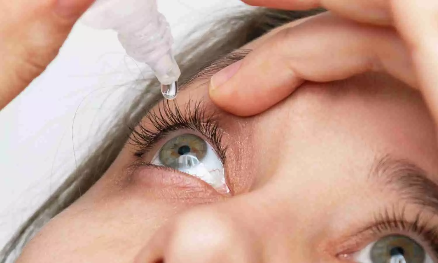
A new study published in the British Journal of Ophthalmology found that low-level light therapy (LLLT) can dramatically lessen symptoms of dry eyes after cataract surgery. Despite the growing evidence that patients with pre-existing dry eye disease (DED) must optimize their ocular surface prior to cataract surgery in order to achieve the intended postoperative outcomes, it is still worth to investigate the prophylactic treatment of healthy patients who were undergoing surgery in order to prevent iatrogenic DED. Low-level laser therapy is a rapidly emerging technique used to treat a wide range of diseases requiring healing stimulation, pain and inflammation reduction, and function restoration. Therefore, Giuseppe Giannaccare and team carried out this study in order to assess the effectiveness of low-level light treatment in iatrogenic dry eye disease.

This prospective, interventional, randomized, controlled, and double-masked randomized patients 1:1 to receive either sham treatment (LLLT with a power output less than 30%) or real light therapy. Two therapy sessions were performed on the patients where one was 7±2 day prior to cataract surgery (T0) and one was 7±2 day following (T1). The Ocular Surface Disease Index (OSDI) questionnaire, tear meniscus height, non-invasive break-up time (NIBUT), meibomian gland loss (MGL) and redness score were among the outcome markers assessed 30±4 days following surgery (T2).

The study found that 131 patients (70 men and 61 women, mean age 73.53±7.29 years) were randomly assigned to undergo LLLT (n = 73) or sham therapy (n = 80) and successfully completed the trial on a regular basis. At T1 and T2, the OSDI scores of patients treated with LLLT were significantly lower than those of controls. Additionally, there were higher NIBUT values at T2 and lower MGL Meiboscore values at T1. The patients treated with LLLT showed substantially higher NIBUT values at T2 when compared with T0 and considerably lower OSDI scores at T2 compared with controls.

This study demonstrates that after cataract surgery, low-level light treatment, or LLLT, can greatly reduce the symptoms of dry eyes. When comparing LLLT recipients before and after surgery to controls, it was evident that they had improved non-invasive break-up time values and significantly lower Ocular Surface Disease Index scores. Following surgery, the patients who received LLLT in particular reported decreased ocular pain and better tear film stability.

Source:

Giannaccare, G., Rossi, C., Borselli, M., Carnovale Scalzo, G., Scalia, G., Pietropaolo, R., Fratto, B., Pellegrini, M., Yu, A. C., & Scorcia, V. (2023). Outcomes of low-level light therapy before and after cataract surgery for the prophylaxis of postoperative dry eye: a prospective randomised double-masked controlled clinical trial. In British Journal of Ophthalmology (Vol. 108, Issue 8, pp. 1172–1176). BMJ. https://doi.org/10.1136/bjo-2023-323920

Powered by WPeMatico

FDA approves oral deuruxolitinib for severe alopecia areata

The US Food and Drug Administration (FDA) has approved oral selective inhibitor of Janus Kinases (JAK) JAK1 and JAK2 deuruxolitinib (Lesqselvi) 8 mg for adult patients with severe alopecia areata.

Deuruxolitinib in twice daily dose functions by disrupting these pathways which are suggested to contribute to severe alopecia areata patients’ loss of hair.

Alopecia areata affects around 700,000 people in the United States, and 300,000 have severe alopecia areata. Alopecia often leads patients to self-treat before seeking professional help, driven by dissatisfaction with the slow progress of existing treatments. 

“LEQSELVI offers a new and effective solution that will significantly enhance options for long-suffering patients battling severe alopecia areata and their physicians,” said Abhay Gandhi, CEO, North America Business, Sun Pharma. “Our fast-growing dermatology business is excited to add this novel treatment to its portfolio.”

Alopecia areata is a common autoimmune disease in which hair loss is thought to occur due to the collapse of immune privilege, leading to the immune system targeting the hair follicles and causing sudden hair loss on the scalp, face and sometimes other areas of the body. LEQSELVI is a new, twice-daily oral selective inhibitor of Janus Kinases (JAK) JAK1 and JAK2. As a JAK inhibitor, LEQSELVI interrupts the pathways thought to contribute to hair loss in severe alopecia areata.

“We welcome the approval of LEQSELVI as a significant step for the alopecia areata community,” said Nicole Friedland, President and CEO, National Alopecia Areata Foundation (NAAF). “Alopecia areata is an autoimmune disease, with significant physical, emotional and financial impacts that go beyond hair loss. Today’s announcement empowers the alopecia community with even more choices, to which NAAF is committed, and provides another important option for those living with severe alopecia areata.”

The approval is based on data from two multicenter, randomized, double-blind, placebo-controlled Phase 3 clinical trials THRIVE-AA1 and THRIVE-AA2, which enrolled a total of 1,220 patients with alopecia areata who had at least 50% scalp hair loss as measured by Severity of Alopecia Tool (SALT) for more than six months. Data were also collected from two open-label, long-term extension trials in which patients were eligible to enroll upon completion of the 24-week trials.

At study baseline, the average patient had only 13% of their scalp hair coverage. In the trials, at 24 weeks, the primary endpoint was met, with more than 30% of patients taking LEQSELVI experiencing 80% or more scalp hair coverage (SALT ≤20).1 The number of patients taking LEQSELVI and achieving a SALT score of ≤20 showed a consistent upward trend with no plateau through 24 weeks.1 Additionally, up to 25% of patients had almost all of their scalp hair back at 24 weeks (≥90% coverage).

“For many people with severe alopecia areata, early intervention with effective treatment is critical,” said Natasha Mesinkovska, MD, PhD, Associate Professor and Vice Chair for Clinical Research of Dermatology, University of California, Irvine, and investigator in the LEQSELVI clinical development program. “An oral JAK that delivers proven results will be impactful for the alopecia areata community.”

Across the Phase 2 dose-ranging study and Phase 3 randomized, placebo-controlled trials, few patients (3.1%) receiving LEQSELVI 8 mg twice daily were discontinued from the trials due to adverse reactions.1 In clinical trials, more than 100 people continued taking deuruxolitinib for more than three years.

LEQSELVI may cause serious side effects including serious infections, malignancies, thrombosis, gastrointestinal perforations, and certain laboratory abnormalities. There also may be an increased risk of mortality and major cardiovascular events. LEQSELVI should not be used in patients who are CYP2C9 poor metabolizers or who are taking moderate or strong CYP2C9 inhibitors. In placebo-controlled trials, the three most common adverse events were headache (12.4% as compared to 9.4% with placebo), acne (10% as compared to 4.3% with placebo), and nasopharyngitis (8.1% as compared to 6.7% with placebo). Please see full Prescribing Information Including BOXED WARNING and Medication Guide and see below for Important Safety Information.

“We are pleased with the timely approval of LEQSELVI by the U.S. FDA,” said Marek Honczarenko, MD, PhD, Senior Vice President, Head of Development, Sun Pharma. “This validates our team’s capability to effectively bring treatments from research and development to approval in a way that is meaningful for physicians and patients. I want to thank all the investigators and patients for their participation in the clinical trials.”

Sun Pharma is committed to delivering support to the alopecia areata community and will introduce an access program to help eligible patients get started and stay on track with their treatment. 

About LEQSELVI™ and alopecia areata

LEQSELVI (deuruxolitinib) 8 mg tablets is an oral selective inhibitor of Janus kinases JAK1 and JAK2 approved for the treatment of adult patients with severe alopecia areata. Alopecia areata is an autoimmune disease in which the immune system attacks hair follicles, resulting in partial or complete loss of hair on the scalp and body. Alopecia areata may affect up to 2.5% of the United States and global population during their lifetime.3,6,7 The scalp is the most commonly affected area, but any hair-bearing site can be affected alone or together with the scalp. Onset of the disease can occur throughout life and affects both women and men. Alopecia areata can be associated with serious psychological consequences, including anxiety and depression. There are currently limited approved treatment options available for alopecia areata.

About THRIVE-AA1 and THRIVE-AA2 trial design

THRIVE-AA1 and THRIVE-AA2 (NCT04518995 and NCT04797650) were randomized, double-blind, placebo-controlled clinical trials in 1223 adult patients ages 18-65 with severe alopecia areata at sites in the U.S., Canada and Europe evaluating the regrowth of scalp hair after 24 weeks of dosing using the SALT score. Patients were randomized to receive either 8 mg twice daily or 12 mg twice daily of deuruxolitinib or placebo for 24 weeks. The primary endpoint was the percentage of patients achieving a SALT score of 20 or less at 24 weeks. Patients enrolled in THRIVE-AA1 and THRIVE-AA2 were required to have at least 50 percent scalp hair loss due to alopecia areata, as measured by SALT. A SALT score of 100 represents total scalp hair loss, whereas a score of 0 represents no scalp hair loss. The average baseline SALT score across all patients in THRIVE-AA1 and THRIVE-AA2 was approximately 85.9 and 87.9 respectively.
Powered by WPeMatico

Nonoperative Treatment Equally Effective as Operative Approach for Proximal Hamstring Avulsions: NEJM

A new study conducted by researchers revealed that nonoperative treatment for proximal hamstring avulsions is as effective and noninferior to operative treatment. Drawing on a stringent noninferiority test across 10 centers in Sweden and Norway, it compared these two modalities of treatment between patients aged 30 to 70 years. These results were published in the The New England Journal of Medicine by Elsa Pihl and colleagues.

This was a randomized controlled trial with a parallel non-randomized observational cohort. Of the patients, 119 were enrolled in the randomized trial and 97 participated in the observational cohort. Patients were randomized either to operative reinsertion of tendons or to non-operative management, with a primary end-point of Perth Hamstring Assessment Tool (PHAT) score at the 2-year follow-up period. Secondary outcome measures included scores on the Lower Extremity Functional Scale (LEFS).

The key findings of the study were:

  • In the randomized trial, the mean PHAT scores were 79.9 ± 19.5 for the operative group and 78.5 ± 19.4 for the nonoperative group.

  • The noninferiority limit of 10 points was not crossed, with a mean difference of −1.2 (95% CI, −8.6 to 6.2; P=0.009 for noninferiority), indicating that nonoperative treatment is comparable to operative treatment in terms of functional outcomes.

  • The LEFS scores showed a mean difference of −1.6 (95% CI, −5.2 to 2.0) between the groups, aligning with the primary outcome.

  • The operative group experienced 9 adverse events, compared to 3 in the nonoperative group, resulting in an odds ratio of 0.3 (95% CI, 0.1 to 1.2), suggesting a lower incidence of complications with nonoperative treatment.

  • In the observational cohort, the mean PHAT score difference between nonoperative and operative treatment groups was −2.6 (95% CI, −9.9 to 4.6), further supporting the equivalence of the nonoperative approach.

These findings of the study present that nonoperative treatment for avulsions of the proximal hamstring is not worse than operative treatment, and it is a feasible alternative to surgical intervention without harming the functional outcomes. Now, there is evidence for revisiting these traditional management strategies for such injuries and possibly even favoring less invasive methods.

Reference:

Pihl, E., Laszlo, S., Rosenlund, A.-M., Kristoffersen, M. H., Schilcher, J., Hedbeck, C. J., Skorpil, M., Micoli, C., Eklund, M., Sköldenberg, O., Frihagen, F., & Jonsson, K. B. (2024). Operative versus nonoperative treatment of proximal hamstring avulsions. NEJM Evidence, 3(8). https://doi.org/10.1056/evidoa2400056

Powered by WPeMatico

Microconvex probes provide more reliable outcomes in newborns undergoing lung USG: Study

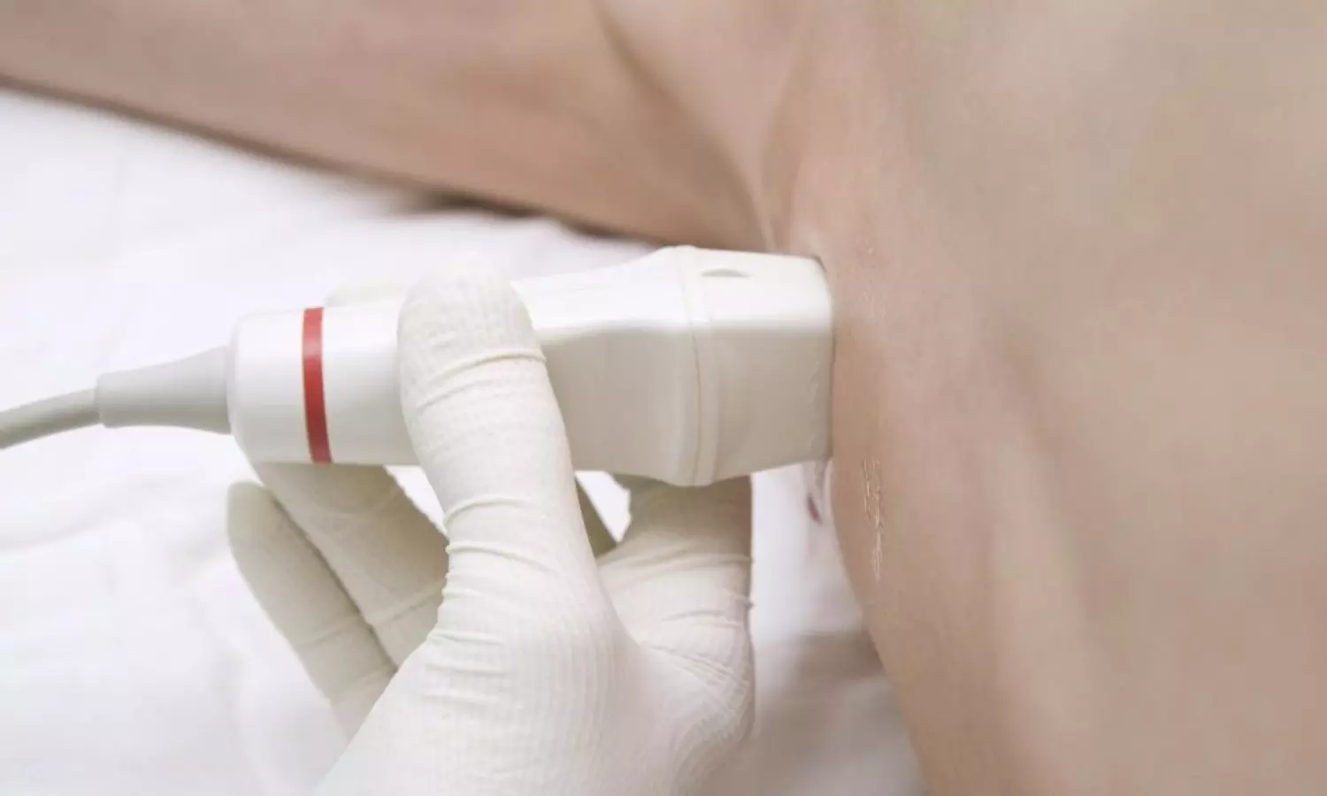
A new study by Lukas Aichhorn and team unveiled that the linear probe performs better with the microconvex probe than echo probes for lung ultrasonography in newborns. The findings of this study were published in the recent issue of journal of PLOS ONE.

In recent years, lung ultrasonography (LUS) has gained popularity in pediatric and neonatal critical care units. Many neonatologist performed lung ultrasound (NPLUS) protocols have been used in the regular environment to help in the identification of respiratory diseases, to provide guidance for operations like the insertion of chest tubes and for the administration of surfactants. The impact of probes used in ultrasound on lung ultrasound scores and picture quality is still unclear. As a result, this study compared the performance of three types of ultrasound probes in hemodynamically stable neonates, namely linear, microconvex and echo.

This prospective, randomized, blinded, comparative research was conducted between 2020 and 2022. The participants included were the hemodynamically stable newborns with nasal continuous positive airway pressure, a high flow nasal cannula, or no respiratory assistance. Lung ultrasonography with carried out with either an echo or a microconvex probe and the linear probe was the control. The main outcome measured was the NPLUS score. The additional outcome indicators were the number of B-lines, the thickness of the pleural line and subjective image quality. Furthermore, the association between NPLUS findings and clinical data was investigated.

An comprehensive analysis was conducted on 1584 video loops from a total of 66 newborns, whose mean corrected gestational age was 33.8 weeks and their weight was 1950g. 

The NPLUS score was observed to be lower when using the echo- and microconvex probes in comparison to the linear probe with the corresponding coefficients of -1.09 and -2.95, respectively.

A reasonably good correlation was found between the NPLUS score and the pulse oximetric saturation/fraction of inspired oxygen ratio, with the greatest results obtained when utilizing the microconvex probe.

The findings help validate the current guidelines and also highlight the extent to which outcomes vary when alternative probes are utilized. The discrepancies revealed need careful interpretation of results, particularly when guiding therapy and presenting prognoses. Overall, the association between the NPLUS score and clinical characteristics will help to validate the usage of this diagnostic tool.

Source:

Aichhorn, L., Habrina, L., Werther, T., Berger, A., & Küng, E. (2024). Comparison of different types of ultrasound probes for lung ultrasound in neonates-A prospective randomized comparison study. In S. Yadav (Ed.), PLOS ONE (Vol. 19, Issue 7, p. e0306472). Public Library of Science (PLoS). https://doi.org/10.1371/journal.pone.0306472

Powered by WPeMatico

Extended periods of daytime sleep increase risk of non-alcoholic fatty liver disease: Study

A new study by Qiao X and team suggests that sleeping for extended periods of time during the day may have a role in the development of non-alcoholic fatty liver disease (NAFLD). The findings of this study were published in the journal of Nature and Science of Sleep. A major public health concern is the growing prevalence of NAFLD and its myriad of consequences, which are mostly caused by the changes in food and lifestyle habits. Taking naps throughout the day is a common practice in many countries, especially China. It has not yet been conclusively determined if napping and the prevalence of NAFLD are causally related.

And so, conventional observational studies, including carefully designed prospective studies with sizable sample sizes, continue to be susceptible to residual confounding variables and the potential for reverse causation. Therefore, this study was conducted to examine how taking naps during the day might affect the likelihood of developing NAFLD by offering further insight into the causal relationship and helping to identify risk factors for the condition.

Mendelian randomization (MR) and meta-analysis were employed to combine observational and genetic data. Using 105 single-nucleotide polymorphisms (SNPs) linked to daytime napping habits, a two-sample MR analysis was carried out. Also, summary-level information about the outcomes of NAFLD was obtained from the extensive UK Biobank project. The association between extensive daytime napping and NAFLD was examined using network meta-analyses, and a subgroup analysis was also conducted.

This comprehensive study found significant links between sleeping throughout the day and non-alcoholic fatty liver disease. This systematic review and meta-analysis also showed that the individuals who slept for more than 30 minutes during the day had a higher chance of developing non-alcoholic fatty liver disease when compared to people who did not sleep. Additionally, MR analysis showed a significant correlation between a genetic predisposition for longer naps during the day and an elevated risk of NAFLD.

This MR study showed a link between midday sleeping and an increased chance of incident non-alcoholic fatty liver disease. Also, the individuals with NMA who slept for more than thirty minutes on average were shown to be at a higher risk of developing NAFLD than the individuals who napped for shorter periods of time or not at all.

Reference:

Qiao, X., Wang, X., Guo, L., & Pan, Q. (2024). Excessive Daytime Napping Increases the Risk of Non-Alcoholic Fatty Liver Disease: A Meta-Analysis and a Mendelian Randomization Study. In Nature and Science of Sleep: Vol. Volume 16 (pp. 1067–1074). Informa UK Limited. https://doi.org/10.2147/nss.s468444

Powered by WPeMatico

Two narrow-diameter titanium-zirconium implants with stud-attachments tied to highly satisfactory outcomes in mandible : Study

Two narrow-diameter titanium-zirconium implants with stud attachments tied to highly satisfactory outcomes in mandible suggest a study published in the Clinical Oral Implant Research.

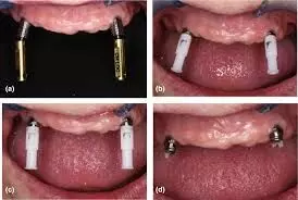
A study evaluated the survival rates and marginal bone loss of narrow-diameter titanium-zirconium implants supporting complete maxillary and mandibular overdentures up to 3 years after loading. Ten completely edentulous patients who were dissatisfied with their complete dentures were enrolled. Two narrow-diameter implants were placed in the canine region of the maxilla and mandible. After second-stage surgery, implant-supported overdentures (palatal-free) attached by parallel alignable stud-attachments were placed. Patients were followed periodically for up to 36 months. Standardised radiographs were taken at baseline, 12 and 36 months to analyse mean marginal bone level changes around the implants. Results: The Kaplan–Meier survival rates were 100% for mandibular and 68.0% (SE ± 10.9%) for maxillary implants at 36 months (p = .008). Six maxillary implants failed after loading; no mandibular implants were lost. Five implants failed due to loss of osseointegration. One implant fractured. The mean marginal bone level changes around the analyzed implants (n = 28, 9 patients) were −0.71 ± 0.82 mm in the mandible and −2.08 ± 1.52 mm in the maxilla at the 36-month follow-up. The difference in marginal bone level changes between the maxilla and mandible was significant (p = .019) at the 12- and 36-month follow-ups. Two narrow-diameter titanium-zirconium implants with stud attachments showed a highly satisfactory outcome in the mandible. The maxillary implants showed a high failure rate and significantly more bone loss over time than the mandibular implants. The minimal concept of two implants and an overdenture should be limited to the edentulous mandible.

Reference:

Kern, J.-S., Salin, E., Elvers, D., Rittich, A., Tuna, T., Hölzle, F., & Wolfart, S. (2024). Maxillary and mandibular overdentures retained by two unsplinted narrow-diameter titanium-zirconium implants – A clinical pilot study. Clinical Oral Implants Research, 00, 1–13. https://doi.org/10.1111/clr.14303

Keywords:

Two, narrow-diameter, titanium-zirconium, implants, stud-attachments, tied, highly, satisfactory, outcome, mandible, study, Kern, J.-S., Salin, E., Elvers, D., Rittich, A., Tuna, T., Hölzle, F., & Wolfart, S

Powered by WPeMatico

Endometrial thickness in abnormal uterine bleeding may predict endometrial cancer among peri and post menopausal women: Study

The endometrium constitutes a dynamic organ in female
reproductive years subjected to cyclical hormonal variations. Perimenopause is
a transition phase of women’s life marked by erratic cycle and fluctuating
hormonal milieu. Abnormal uterine bleeding constitutes majority of
gynecological consultations and responsible for all two – third hysterectomies.
The investigations in them should be targeted to exclude serious pathology such
as endometrial cancer and its precursor lesion endometrial hyperplasia.

Trans vaginal sonography (TVS), which is a simple,
non-invasive technique for endometrial evaluation plays a major role in this
regard. Use of high resolution transvaginal probes and colour Doppler further
enhanced the ability to study the endometrium beyond its thickness. Findings
such as focal thickening, heterogenecity in the endometrium, irregular
endometrial thickening, polypoidal mass lesion, intrauterine fluid collection
and disruption of sub endometrial halo prompt further evaluation to rule out
serious pathology even if the endometrial thickness is normal.

In postmenopausal lady the cut-off for endometrial thickness
for further evaluation is almost defined. However, there is a lack of clear
cut-off criteria for abnormal endometrial thickness for peri-menopausal women.
The present study tries to correlate the endometrial thickness in TVS
evaluation with the histolpathological abnormality in the peri menopausal and
post-menopausal women.

Present study by Mishra et al. was conducted with the
following objectives: 1. To assess the endometrial thickness by trans vaginal
ultrasonography. 2. To see correlation of histopathology with endometrial
thickness in abnormal uterine bleeding (AUB) in perimenopausal and
post-menopausal women. 3. The current study also throws some light about
various causes of AUB as per PALM-COIEN prevalent in our locality. 4. To find
the cutoff for endometrial thickness that delineates the abnormal from normal
histology.

This was an observational study conducted on 242
peri-menopausal and postmenopausal women ≥40 years of age presenting with
abnormal uterine bleeding (AUB). Based on the ROC curve, the cutoff point that
delineates the abnormal histology and normal histology for endometrial
thickness was 14.20 mm. Area under the curve (AUC) was 0.783 with 95% CI as
0.686 – 0.880 having sensitivity 71% and specificity 90% (p < 0.001)

In the present study peri menopausal AUB may present to
gynecological outpatient department with varied complaints. Out of them heavy
menstrual bleed and frequent cycle were found to be commonly associated with
gynaecological pathology in these age groups. In all peri menopausal AUB Trans
vaginal sonography for endometrial thickness is the initial best modality of
investigation. Combining colour Doppler is complementary and it enhances
further diagnostic accuracy of endometrial evaluation. Endometrial biopsy for
histopathological examination should be offered to all perimenopausal AUB with
cut off value equal to or more than 13mm. However, with positive doppler
findings, focal abnormality, coexisting risk factors such as obesity, diabetes,
age greater than or equal to 45 years or presence of post-menopausal bleeding,
endometrial biopsy should be considered irrespective of the thickness as there
is increased chance of having significant endometrial pathology.

Source: Mishra et al. / Indian Journal of Obstetrics and
Gynecology Research 2024;11(2):185–190

https://doi.org/10.18231/j.ijogr.2024.038

Powered by WPeMatico

Hypertensive disorders of pregnancy on the rise, claims study

In Canada, rates of hypertensive disorders of pregnancy (HDP) have increased, but the good news is there has been a decline in some related health conditions, according to new research published in CMAJ.

Hypertensive disorders of pregnancy include chronic hypertension (high blood pressure), gestational hypertension, and preeclampsia or eclampsia. These disorders affect 5%–10% of pregnancies worldwide, and cause more than 50 000 maternal deaths and 500 000 deaths in fetuses and infants every year.

A large study of more than 2.8 million births in hospital in Canada (excluding Quebec) between 2012 and 2021 dentified females with HDP, with the aim of understanding trends in HDP and related health outcomes. Over the study period, the rate of any HDP increased from 6.1% to 8.5% in absolute numbers, with a relative increase of 40%. Pre-existing hypertension temporally increased from 0.6% to 0.9%, gestational hypertension rose from 3.9% to 5.1%, and preeclampsia from 1.2% to 2.6%

The researchers noted several trends. Rates of HDP were higher in females under age 20 and in those older than 34 years. There was also variability in rates across provinces and territories, with the Northwest Territories (6.5%) and Ontario (6.9%) having the lowest rates, and the highest rate (10.7%) in Newfoundland and Labrador.

The authors adjusted for risk factors for HDP, including maternal age, number of previous livebirths, pre-existing diabetes, and rural residence, but doing so did not significantly affect the risk of HDP. “[T]he rise in HDP may be explained by factors that we did not account for, including body mass index (BMI),” writes lead author Dr. Susie Dzakpasu, senior epidemiologist at the Maternal and Infant Health Section of the Public Health Agency of Canada, with coauthors. “High BMI is a known risk factor for hypertension, including during pregnancy.”

Between 2015 and 2021 in Canada, rates of overweight or obese BMI status increased from 41% to 48% among females aged 18–34 years, and from 56% to 64% in those aged 35–49 years. The authors also found that provinces with higher rates of overweight and obesity had higher rates of HDP.

“[D]ownward trends in other adverse outcomes suggest that the clinical management of HDP may have improved over time,” write the authors. “This underscores the importance of standard measurement of blood pressure at each prenatal visit and the institution of evidence-based antihypertensive therapy.”

Dr. Catherine Varner, deputy editor, CMAJ, writes that the “growing population of high-risk obstetrical patients should alert health policy-makers that more people need easily accessed, specialized obstetrical care.”

Females with hypertension and other disorders should be monitored in the early postpartum period, yet there is a shortage of qualified health care providers.

The findings of Dzakpasu and colleagues “highlight the increasing demand for obstetrical care providers with the expertise to care for high-risk patients, supported by appropriate models of care both before and after delivery. As the number of obstetricians will be unable to conceivably meet the needs of the growing population entering their reproductive lives with more comorbidities than the preceding generation, scaling team-based maternity care may help people receive the right care, at the right time, in the right place, by the right provider,” Dr. Varner concludes.

Reference:

Susie Dzakpasu, Chantal Nelson, Elizabeth K. Darling, Wesley Edwards, Phil A. Murphy, Heather Scott, Michiel Van den Hof and Joel G. Ray, Trends in rate of hypertensive disorders of pregnancy and associated morbidities in Canada: a population-based study (2012–2021), Canadian Medical Association Journal, DOI: https://doi.org/10.1503/cmaj.231547.

Powered by WPeMatico

Ultrasound-Guided Suprazygomatic Maxillary Nerve Block effective for Postoperative Pain in Functional Endoscopic Sinus Surgery: Study

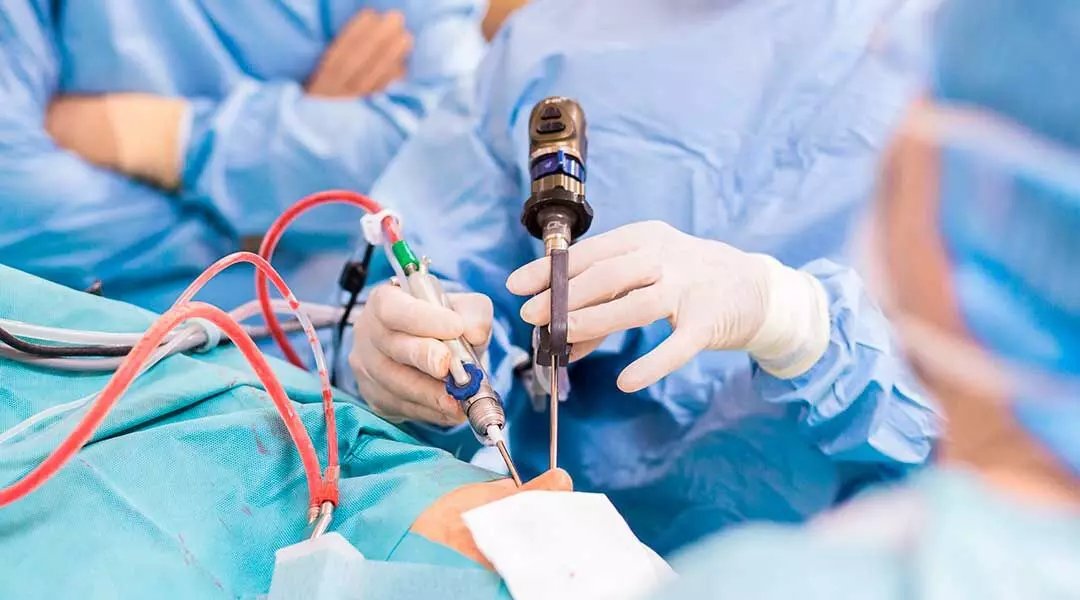
FESS is presently the conventional surgical approach for managing sinonasal pathologies. Recent randomized controlled trial aimed to assess the efficacy of ultrasound-guided suprazygomatic maxillary nerve block (SZMNB) for postoperative pain management in patients undergoing functional endoscopic sinus surgery (FESS). The study enrolled 48 adult patients aged 18-65 years with ASA physical status I-II who were scheduled for FESS. Patients were randomly allocated to receive either SZMNB with general anesthesia (GA) (n=24) or GA alone (n=24).

Study Outcomes

The primary outcome was the numerical rating scale (NRS) pain score in the immediate postoperative period. Secondary outcomes included 24-hour postoperative rescue analgesic consumption, surgeon satisfaction score, postoperative hemodynamics, and postoperative complications.

Postoperative Pain Scores

The median (IQR) NRS pain score in the immediate postoperative period was significantly lower in the SZMNB group compared to the control group [0 (0-0.25) vs 2 (1.75-3), p<0.001]. Pain scores were also significantly reduced at all time points up to 24 hours after surgery in the SZMNB group compared to control (p<0.001).

Rescue Analgesia

None of the patients in the SZMNB group required rescue analgesia, whereas 8 patients in the control group required intravenous diclofenac within 1 hour of surgery and 10 patients within 1-6 hours. The time taken to perform the SZMNB block was 485 (SD 26.0) seconds. Successful spread of the local anesthetic was confirmed in 37 out of 40 blocks, and the maxillary artery was visualized in 35 out of 40 blocks.

Postoperative Parameters

Postoperative heart rate was significantly lower in the SZMNB group compared to control up to 2 hours after surgery (p<0.05), but mean arterial pressure was similar between groups. There was no significant difference in surgeon satisfaction scores or incidence of postoperative complications between groups.

Conclusion

In conclusion, the ultrasound-guided SZMNB provided excellent postoperative analgesia for patients undergoing FESS without significant side effects. This regional nerve block technique can be effectively utilized to improve postoperative pain management in patients undergoing FESS.

Key Points

Here are the 6 key points from the research paper: 1. The study was a randomized controlled trial that aimed to assess the efficacy of ultrasound-guided suprazygomatic maxillary nerve block (SZMNB) for postoperative pain management in patients undergoing functional endoscopic sinus surgery (FESS). 2. The primary outcome was the numerical rating scale (NRS) pain score in the immediate postoperative period, while secondary outcomes included 24-hour postoperative rescue analgesic consumption, surgeon satisfaction score, postoperative hemodynamics, and postoperative complications. 3. The median NRS pain score in the immediate postoperative period was significantly lower in the SZMNB group compared to the control group, and pain scores were also significantly reduced at all time points up to 24 hours after surgery in the SZMNB group. 4. None of the patients in the SZMNB group required rescue analgesia, whereas 8 patients in the control group required intravenous diclofenac within 1 hour of surgery and 10 patients within 1-6 hours. 5. Postoperative heart rate was significantly lower in the SZMNB group compared to the control group up to 2 hours after surgery, but mean arterial pressure was similar between groups. There was no significant difference in surgeon satisfaction scores or incidence of postoperative complications between groups. 6. The ultrasound-guided SZMNB provided excellent postoperative analgesia for patients undergoing FESS without significant side effects, and this regional nerve block technique can be effectively utilized to improve postoperative pain management in patients undergoing FESS.

Reference –

Neupane A, Jain D, Arora S, Gandhi K, Singla V, Goel N, et al. Evaluation of ultrasound‑guided suprazygomatic maxillary nerve block in functional endoscopic sinus surgery for postoperative pain relief: A randomised controlled trial. Indian J Anaesth 2024;68:706‑11

Powered by WPeMatico

Wearable devices can increase health anxiety. Could they adversely affect health?

Using a wearable device, such as a smart watch, to track health data and symptoms, is supposed to help people monitor their health and address symptoms as quickly as possible to spur positive health outcomes. But for people with atrial fibrillation, also known as Afib, using a wearable device to monitor the heart rate and to alert wearers of an irregular heartbeat might not be as helpful as wearers think.

A new study in the Journal of the American Heart Association, led by Lindsay Rosman, PhD, assistant professor of medicine in the division of cardiology at the University of North Carolina School of Medicine, is the first to show that wearable devices, such as smart watches, can significantly amplify anxiety and increase healthcare use in patients with Afib.

The study included 172 patients from UNC Health with a prior diagnosis of Afib who completed a survey and had their information linked to electronic health records. About half of the study sample had a wearable device and their data was compared to individuals without a wearable device. Rosman and her team found that patients with Afib who use wearables are more likely to be preoccupied with their heart symptoms, report concerns about their AFib treatment, and use healthcare resources compared to Afib patients without these devices. Providers and healthcare clinics were also impacted, as wearable users were more likely to call the clinic and send messages to their healthcare providers than individuals who did not have a device.

Also, 1 in 5 AFib patients who used wearables in this study experienced intense fear and anxiety in response to irregular rhythm notifications from their device. And a similar proportion (20%) routinely contacted their doctors when ECG results were abnormal or indicative of possible AFib. But it’s unclear if they actually needed to see a doctor, due to the alerts from their devices. It’s also unclear if the reported anxiety contributed to the worsening of symptoms, although anxiety has been a well-documented contributing factor to various conditions, including AFib.

“Given the significant increase in use of wearable devices in this patient group (and the population in general),” said Rosman, “we believe prospective studies and randomized trials are needed to understand the net effects of wearables – including their alerts – on patients’ healthcare use and psychological well‐being, as well as the downstream effects on providers, hospitals, and health systems.”

Reference:

Lindsey Rosman, Rachel Lampert, Songcheng Zhuo, Tiffany Armbruster and Anil Gehi, Wearable Devices, Health Care Use, and Psychological Well‐Being in Patients With Atrial Fibrillation, Journal of the American Heart Association, https://doi.org/10.1161/JAHA.123.033750.

Powered by WPeMatico