Some companies are overinflating value of umbilical cord blood banking to expectant parents, experts warn
Powered by WPeMatico
Powered by WPeMatico

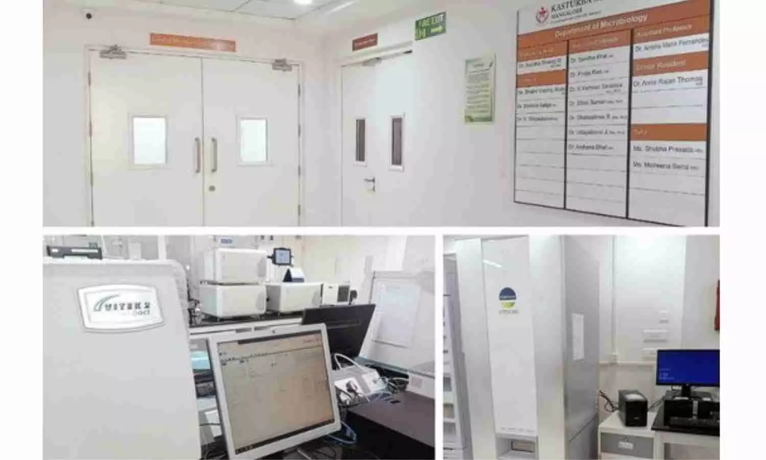
Mangaluru: The National Medical Council (NMC) has recognised Kasturba Medical College (KMC), Mangalore as a Regional Centre for the National Action Plan on Antimicrobial Resistance (NAP-AMR) Cell.
In a press note issued in this regard, KMC stated, “We are proud to announce that Kasturba Medical College, Mangalore, has been recognized as a Regional Center and Coordinator for the NMC-NAP-AMR (National Medical Commission – National Action Plan on Antimicrobial Resistance) activities.”
Following this, the medical college authorities appointed Dr Pooja Rao, Associate Professor of Microbiology as the Regional Coordinator.
Also read- 70th Anniversary: Kasturba Medical College Mangalore Commemorates College Day And Awards Ceremony
“The recognition is a significant milestone in the ongoing efforts to combat antimicrobial resistance and to enhance medical education and training in India. The NMC is developing resource faculties to train prescribers across medical colleges and institutions in India. As part of this initiative, KMC-Mangaluru has been nominated as one of the regional centres,” mentioned a press release as per The Hindu reports.
Speaking about this, MAHE Vice-Chancellor Lt Gen M.D. Venkatesh told TH, “Being recognised as a Regional Centre for NAP-AMR is a testament to our commitment to excellence in medical education and public health. This accolade underscores our dedication to nurturing healthcare professionals who are not only skilled but also acutely aware of the global challenges posed by antimicrobial resistance.”
KMC-Mangaluru Dean Unnikrishnan said, “This initiative will empower our faculty to lead the way in antimicrobial stewardship and to educate prescribers across the region. Our faculty members, as State-level master trainers, will be pivotal in disseminating critical knowledge and best practices to prescribers, thereby enhancing the overall quality of healthcare.”
As per the Daily, regional centres are required to form a “NAP-AMR Cell” with at least four faculty members, which should include a nodal officer at the rank of Professor, Additional Professor, or Associate Professor, from the departments of Microbiology, Pharmacology, Medicine, or Community Medicine.
Also read- Kasturba Medical College To Hold EREVNA 2024- National UG Medical Research Conference
Powered by WPeMatico

Switzerland: A recent study has found favorable long-term survival and reduced risk of valve dysfunction in patients undergoing TAVR with elevated pre-procedural CRP levels. However, they are at higher risk for periprocedural infections and subsequent hospital readmissions related to infections of various types during the follow-up period.
The findings were published online in Cardiovascular Revascularization Medicine on July 6, 2024.
Transcatheter aortic valve replacement (TAVR) has revolutionized the treatment of aortic valve stenosis, offering a minimally invasive alternative to open-heart surgery for many patients. Recent research has delved into the role of elevated C-reactive protein (CRP) levels before TAVR. However, there is no clarity on its impact on the long-term results of TAVR. Considering this, Stefan Toggweiler, From the Heart Center Lucerne, Cardiology Division, Luzerner Kantonsspital, Lucerne, Switzerland, and colleagues aimed to investigate the long-term (up to six years) clinical outcomes of TAVR patients with normal compared to elevated CRP levels before TAVR.
For this purpose, the researchers included consecutive patients undergoing TAVR between 2012 and 2023 at a tertiary cardiology facility. They were categorized into two cohorts based on the baseline CRP levels: normal CRP (≤ 5 mg/l) and elevated CRP (>5 mg/l). Patients were clinically monitored for up to six years following TAVR.
According to the authors, the study is the first to comprehensively examine outcomes after TAVR in patients with pre-procedural CRP elevation during a prolonged follow-up period of up to six years.
The study led to the following findings:
“Our study found that patients with elevated CRP levels undergoing TAVR experienced favorable long-term outcomes, with no significant differences in long-term survival or valve dysfunction compared to those with normal CRP levels. However, elevated baseline CRP was associated with increased rates of periprocedural infections and hospital readmissions due to infections,” the researchers concluded.
Reference:
Brunner, S., Moccetti, F., Loretz, L., Conrad, N., Bossard, M., Attinger-Toller, A., Kurmann, R., Cuculi, F., Wolfrum, M., & Toggweiler, S. (2024). The impact of elevated C-reactive protein levels on long-term outcomes of patients undergoing transcatheter aortic valve replacement. Cardiovascular Revascularization Medicine. https://doi.org/10.1016/j.carrev.2024.07.002
Powered by WPeMatico

Bhopal: Following a review by the Union Health Ministry and experts on cases of viral infection and Acute Encephalitis Syndrome (AES) in Gujarat, Rajasthan, and Madhya Pradesh, no cases of Chandipura virus have been reported in the state, Madhya Pradesh, state Health Minister Rajendra Shukla said on Sunday.
Shukla said the health department possesses all the necessary equipment and facilities to identify the virus, which is one of the causes of AES.
According to a PTI report, “The state health department is constantly monitoring the situation. No case of Chandipura virus has been found in Madhya Pradesh,” said Shukla, adding that the health department has been updating all details on the IDSP portal.
He said the state government will follow the guidelines of the Centre as per the situation.
“The Health Department is fully prepared to deal with this situation,” the Deputy Chief Minister said, adds news agency PTI.
Acute Encephalitis Syndrome (AES) is a group of clinically similar neurologic manifestations caused by several different viruses, bacteria, fungus, parasites, spirochetes, chemicals/ toxins, etc. The known viral causes of AES include JE, Dengue, HSV, CHPV and West Nile etc, according to the Union Health Ministry.
Chandipura Virus (CHPV) is a member of Rhabdoviridae family known to cause sporadic cases and outbreaks in western, central and southern parts of the country, especially during the monsoon season.
It is transmitted by vectors such as sand flies and ticks. It is to be noted that vector control, hygiene and awareness are the only measures available against the disease, according to a ministry statement.
Medical Dialogues team had earlier reported that Prof (Dr) Atul Goel, DGHS, Union Health Ministry, and Director of NCDC, along with experts from AIIMS, Kalawati Saran Children’s Hospital, and National Institute of Mental Health & Neurosciences (NIMHANS), as well as officials from Central and State surveillance units has reviewed the Chandipura virus and Acute Encephalitis Syndrome (AES) cases in Gujarat, Rajasthan, and Madhya Pradesh.
Powered by WPeMatico

China: A recent study revealed a potential association between the ratio of red blood cell distribution width (RDW) to albumin level and mortality risk. The study, published in JAMA Network Open, sheds light on a novel biomarker that could aid in predicting mortality outcomes in various clinical settings.
In the cohort study involving 469 572 participants at baseline and 44 383 deaths during follow-up periods, higher red blood cell distribution width to albumin concentration (RAR) levels ratio were associated with increased risks of all-cause and cause-specific mortality.
“These findings suggest that the RAR, assessed via routine laboratory tests, could be a promising indicator that is reliable, inexpensive, and simple for identifying individuals at high mortality risk in clinical practice,” the researchers wrote.
Red blood cell distribution width measures the variation in the size of red blood cells. At the same time, albumin is a protein produced by the liver that plays a crucial role in maintaining blood volume and regulating various physiological processes. RDW and albumin levels are routinely measured in clinical practice as part of routine blood tests, offering valuable insights into a patient’s health status.
The ratio of red blood cell distribution width (RDW) to albumin concentration (RAR) has emerged as a reliable prognostic marker for mortality in patients with several diseases. However, there is no information on whether RAR is associated with mortality in the general population. To fill this knowledge gap, Meng Hao, Fudan University Pudong Medical Center, Shanghai, China, and colleagues aimed to explore whether RAR is associated with all-cause and cause-specific mortality and to elucidate their dose-response association.
For this purpose, the researchers conducted a population-based prospective cohort study using data from participants in the 1998-2018 US National Health and Nutrition Examination Survey (NHANES) and from the UK Biobank with baseline information from 2006 to 2010. It included participants with complete data on serum albumin concentration, RDW, and cause of death. The NHANES data were linked to the National Death Index records through December 31, 2019. Causes and dates of death were obtained from the National Health Service Central Register Scotland (Scotland) and National Health Service Information Centre (England and Wales), for the UK Biobank.
Cox proportional hazards regression models evaluated the potential associations between RAR and all-cause and cause-specific mortality risk. Restricted cubic spline regressions were applied to estimate possible nonlinear associations.
The researchers reported the following findings:
The findings suggest that RAR may be a reliable, simple, and inexpensive indicator for identifying individuals at high mortality risk in clinical practice.
As the scientific community continues to explore the implications of these findings, the study represents a significant step forward in understanding the complex interplay between hematological parameters and mortality outcomes. Researchers aim to improve patient care and outcomes across diverse clinical settings by uncovering new insights into predictive biomarkers.
Reference:
Hao M, Jiang S, Tang J, et al. Ratio of Red Blood Cell Distribution Width to Albumin Level and Risk of Mortality. JAMA Netw Open. 2024;7(5):e2413213. doi:10.1001/jamanetworkopen.2024.13213
Powered by WPeMatico

New research presented at this year’s Annual Meeting of the European Association for Study of Diabetes (Madrid, Spain, 9-13 September) shows that the risk of developing type 1 diabetes (T1D) decreases markedly in girls after age 10 years, while the risk in boys stays the same.
Furthermore, risk of T1D is significantly higher boys with a single autoantibody than their female counterparts, suggesting the sex could be linked with autoantibody development, indicating the importance of incorporating sex in the assessment of risk. The study is by Erin L. Templeman and Dr Richard Oram, University of Exeter Medical School, Exeter, UK, and colleagues.
Autoantibodies (ABs) are proteins produced by the body’s immune system, which attack other proteins in autoimmune diseases including T1D.
In contrast to most autoimmune diseases, male sex is a risk factor for type 1 diabetes (T1D). This raises the hypothesis that either immune, metabolic, or other differences between sexes may impact risk or progression through stages of T1D. In this study, the authors aimed to assess the risk and rate of progression for individuals in the TrialNet Pathway to Prevention study that screens relatives of people with T1D for the presence of autoantibodies.
The authors studied 235,765 relatives of people with T1D. They used computer and statistical modelling to calculate risk of T1D, stated as estimated 5-year risk for females and males respectively, after adjusting for confounders.
The proportion of individuals who screened positive for ABs was higher in males (females: 5.0% males: 5.4%). Of these individuals, males were more likely to screen positive for multiple ABs (females: 1.8%, males: 2.6%). Absolute 5-year risk of progression to T1D in single AB positive individuals was significantly higher (in males (females: 14%, males: 21%).
However similar risk was displayed across sexes when presenting with stage 1 (at least 2 ABs) (females: 38%, males: 38%) or stage 2 (AB and abnormal blood sugar stability) (females: 57%, males: 59%). Risk remains significantly higher in single AB positive males when adjusting for the presence of a first-degree relative with T1D and age (27% higher risk in single AB positive males compared to single AB positive females).
A large decrease in 5-year T1D risk is displayed in females when screened, and autoantibody positive, after 10 years old as compared to before aged 10 years old (see fig. 1 of abstract). In contrast, a steady decline in 5-year T1D risk is displayed in males as age at screening increases.
The authors conclude: “The risk of T1D is significantly higher in males than females when presenting with a single autoantibody. Risk is similar between males and females in childhood, with the risk diverging at age 10. Risk in females then dramatically decreases, whereas risk is sustained in males. This suggests sex appears to be linked with autoantibody development, indicating the importance of incorporating sex in the assessment of risk.”
On the difference in risk between boys and girls, the authors add: “We don’t know and this is an interesting area where more research is needed. The change in risk at around the age of 10 raises the hypothesis that puberty related hormones may play a role.”
They add: “We found the biggest differences in risk of T1D in individuals who had not progressed to stage 1 T1D, with a similar rate and risk of progression in those who were stage 1 and stage 2 T1D. Therefore, it seems most likely that explaining the mechanism of sex differences may help us understand underlying pathogenesis of T1D and potential intervention targets, more than affecting how we screen, diagnose and intervene.”
Reference:
Trajectory of type 1 diabetes risk shifts after age 10 years between at-risk males and females, European Association for the Study of Obesity, Meeting: Annual Meeting of the European Association for the Study of Diabetes (EASD).
Powered by WPeMatico

China: A recent study utilizing cardiovascular magnetic resonance (CMR) imaging has provided significant insights into the subclinical left ventricular (LV) myocardial dysfunction among patients with Type 2 Diabetes Mellitus (T2DM) and diabetic peripheral neuropathy (DPN), highlighting the intersection between diabetes-related complications and cardiovascular health.
Powered by WPeMatico

Bangladesh: A recent cohort study published in Tropical Medicine and Health revealed an association between mid-trimester impaired femur growth and elevated prediabetic biomarkers in Bangladeshi adolescents.
Diabetes typically manifests more prominently in adulthood but can remain latent during childhood, often originating during early fetal development. Within fetal biometry, femur length (FL) measurement is critical in evaluating fetal growth and developmental progress. Urme Binte Sayeed, University of Tsukuba, Tsukuba, Ibaraki, Japan, and colleagues aimed to assess potential associations between fetal femur growth and prediabetic biomarkers in Bangladeshi children.
For this purpose, the researchers conducted a cohort study embedded in a population-based maternal food and micronutrient supplementation (MINIMat) trial in Matlab, Bangladesh. The cohort of children were monitored until they reached 15 years of age.
The original trial began by confirming pregnancy via ultrasound before reaching 13 gestational weeks (GWs). Subsequently, additional ultrasound assessments were conducted at 14, 19, and 30 GWs. Femur length (FL) was measured across its entirety to capture a comprehensive image of fetal development. Standardization of FL according to gestational week (GW) was performed, followed by calculation of z-scores. Plasma levels of fasting blood glucose (FBG) and glycated hemoglobin (HbA1c), as well as the triglyceride-glucose index—a marker of insulin resistance—were determined from whole blood.
The researchers conducted multivariable linear regression analysis using a generalized linear model to examine the impact of FL at 14, 19, and 30 GWs on prediabetic biomarkers at ages 9 and 15 years. Covariates in the analysis included maternal micronutrient and food supplementation group, parity, child sex, and BMI at ages 9 and 15 years.
The following were the key findings of the study:
· 1.2% of the participants had impaired fasting glucose during preadolescence, which increased to 3.5% during adolescence.
· At nine years, 6.3% of the participants had elevated HbA1c%, which increased to 28% at 15 years.
· There was an increase in the TyG index from 9.5% (during preadolescence) to 13% (during adolescence).
· A one standard deviation decrease in FL at 14 and 19 GWs was associated with increased FBG (β = − 0.44; β = − 0.59) and HbA1c (β = − 0.01; β = − 0.01) levels at 15 years. FL was not associated with diabetic biomarkers at nine years.
In conclusion, the researchers found that a shorter fetal femur length during mid-pregnancy may be associated with increased prediabetic biomarkers among Bangladeshi adolescents. The study provided evidence indicating that reduced fetal femur length may raise prediabetes risk in adolescents.
Adolescence marks a critical period for the early emergence of chronic diseases. Consequently, it is essential to conduct additional research to elucidate the potential link between fetal growth restriction and the development of metabolic disorders in adulthood, independent of lifestyle modifications. Furthermore, educating prospective parents on pre-pregnancy nutrition and adopting practices that support healthy fetal development are crucial steps to mitigate the occurrence of fetal growth restriction and low birth weight.
Reference:
Sayeed, U.B., Akhtar, E., Roy, A.K. et al. Fetal femur length and risk of diabetes in adolescence: a prospective cohort study. Trop Med Health 52, 44 (2024). https://doi.org/10.1186/s41182-024-00611-6
Powered by WPeMatico

Hydrogels have a variety of use cases, including contact lenses, delivering doses of medication within the body, moisturisers, water storage in soil, cleaning polluted water and as gelling and thickening agents. A hydrogel is a gel made of a type of plastic that can bind water. Researchers at ETH Zurich and Empa have now developed the first hydrogel implant designed for use in fallopian tubes. This innovation performs two functions: one is to act as a contraceptive, the other is to prevent the recipient from developing endometriosis in the first place or to halt the spread if they do.
Around four years ago, Inge Herrmann made a new addition to her research group at the Department of Mechanical and Process Engineering at ETH and Empa. The new member was a senior physician specialising in gynaecology who was keen to pursue clinically-inspired research. This kind of interdisciplinary collaboration was an experiment for the whole team. Their initial goal had been to turn a hydrogel into a new kind of contraceptive for women. However, after the research team began talking to the gynaecologist, they realised that implanting a hydrogel to occlude the fallopian tubes could also help prevent endometriosis.
Around 10 percent of women suffer from endometriosis. However, it is still unclear exactly what causes this condition. The assumption is that during menstruation, blood flows back along the fallopian tubes and into the abdominal cavity. This blood contains cells from the uterine lining (endometrium), which settle in the abdominal cavity and as a result can cause inflammation, pain and the formation of scar tissue.
The researchers found a way to create a hydrogel implant capable of successfully occluding the fallopian tubes and thus preventing retrograde menstruation. They describe their findings in a study that was recently published in the journal Advanced Materials. “We discovered that the implant had to be made of an extremely soft gel-similar in consistency to a jelly baby-that does not impact native tissue and is not treated and rejected as a foreign body,” explains Alexandre Anthis, lead author of the study.
An advantage of hydrogels is that they swell when brought into contact with liquid. As a result, this new implant starts off at approximately two millimetres in length. But once implanted in the fallopian tubes as part of a non-surgical procedure using a hysteroscope – an instrument for inspecting the uterine cavity – the implant swells to more than double its original size. The hydrogel then acts as a barrier to both sperm and blood. “Our hydrogel implant can be easily and quickly destroyed, either with UV light or a special solution, so that recipients don’t have to have an invasive and risky operation should they decide to reverse the procedure,” Herrmann says.
Anthis says that one of the biggest challenges was achieving the right balance between stability and degradability. “We wanted to ensure that the implant was compatible as well as stable.” To this end, the researchers first conducted ex-vivo experiments on human (and animal) fallopian tubes that had for instance been removed in the course of treating ovarian cancer. Next, they tested their innovation in a live pig; after three weeks, the hydrogel implant was still in position and there was no sign of any foreign-body reactions.
Together with ETH and the Swiss Federal Laboratories for Materials Science and Technology (Empa), the researchers submitted a patent. But there’s still a way to go before the implant will be ready to market. Since endometriosis is a human disease, it is challenging to tell how the hydrogel implant will behave long-term once in position in fallopian tubes, especially when recipients engage in strenuous physical activities like sport. Moreover, it is as yet unclear whether blocking the fallopian tubes alone is enough to prevent endometriosis. “We’ve trawled through databases to find data relating to endometriosis patients who have had their fallopian tubes removed,” Herrmann says. Such cases could reveal whether that measure actually stops endometriosis from developing in the abdominal cavity, she adds.
“So far, very little research has been done at the point where materials science, process engineering and gynaecology meet. But this is a vitally important area of research. We hope that our work will count as a meaningful step in the right direction,” says Herrmann, who recently opened the Ingenuity Lab at the University Hospital Balgrist, aiming to translate innovative materials technologies to clinics.
Reference:
Alexandre H.C. Anthis, Samuel Kilchenmann, Manon Murdeu, Paige J. LeValley, Morris Wolf, Charlotte Meyer, Oscar Cipolato, Mark W. Tibbitt, Jachym Rosendorf, Vaclav Liska, Thomas Rduch, Inge K. Herrmann, Reversible Mechanical Contraception and Endometriosis Treatment Using Stimuli-Responsive Hydrogels, Advanced Materials, https://doi.org/10.1002/adma.202310301.
Powered by WPeMatico

Canada: A recent study has revealed a higher incidence of myriad maternal complications, including postpartum hemorrhage, hypertensive disorders of pregnancy, and death in women diagnosed with a cerebrovascular accident (CVA) before or during pregnancy versus women with a transient ischemic attack (TIA) diagnosis.
The neonatal outcomes were comparable between both groups, stressing the different prognoses of these conditions and the importance of diligent follow-up and care in these patients. The findings were published online in the Archives of Gynecology and Obstetrics on July 15, 2024.
Cerebrovascular accidents, often resulting in significant disability, occur when blood flow to the brain is interrupted, leading to potentially severe complications. In contrast, transient ischemic attacks are characterized by temporary disruptions in blood flow, typically resolving within 24 hours. Understanding how these conditions impact pregnancy is crucial, given that cerebrovascular accidents and transient ischemic attacks are uncommon neurologic events in women of childbearing age.
Against the above background, Uri Amikam, Department of Obstetrics and Gynecology, McGill University, Montreal, QC, Canada, and colleagues aimed to compare delivery, pregnancy, and neonatal outcomes between women who suffered from a CVA and those who experienced a TIA.
For this purpose, the researchers performed a retrospective population-based cohort study using the Healthcare Cost and Utilization Project, Nationwide Inpatient Sample. The study included all pregnant women who delivered or had a maternal death in the US from 2004 to 2014.
Women diagnosed with a CVA before or during pregnancy were compared to those with a TIA diagnosed either before, during pregnancy, or at the time of delivery. Pregnancy and perinatal outcomes were analyzed between the two groups using multivariate logistic regression to account for potential confounding factors.
The following were the key findings of the study:
· Among 9,096,788 women in the database, 898 met the inclusion criteria.
· Of them, 706 women had a CVA diagnosis, and 192 had a TIA diagnosis.
· Women with a CVA, compared to those with a TIA, had a higher rate of pregnancy-induced hypertension (aOR 3.82); preeclampsia (aOR 2.6), eclampsia (aOR 13.78); postpartum hemorrhage (aOR 4.52), blood transfusion (aOR 5.57), and maternal death (54 versus 0 cases, 7.6% vs. 0%), with comparable neonatal outcomes.
The study not only deepens the understanding of the impacts of cerebrovascular events on pregnancy but also calls for further research to develop effective strategies for managing and supporting affected women throughout their reproductive years. As more women with prior cerebrovascular events seek pregnancy, these insights will be vital for improving maternal and neonatal health outcomes.
Reference:
Amikam, U., Badeghiesh, A., Baghlaf, H. et al. Obstetric and perinatal outcomes in women with cerebrovascular accident vs. transient ischemic attack: an evaluation of a population database. Arch Gynecol Obstet (2024). https://doi.org/10.1007/s00404-024-07627-7
Powered by WPeMatico
