Nerve decompression may reduce pain in patients with diabetic peripheral neuropathy: Study

A recent study by the team led by Shai Rozen unveiled the effectiveness of nerve decompression surgery in patients requiring the treatment for painful Diabetic Peripheral Neuropathy (DPN). This condition usually affects millions globally and often leads to significant discomfort and pain in the lower extremities which severely impacts the quality of life. The key highlights of this study were published in the newest edition of Annals of Surgery. 

This rigorous double-blinded, randomized trial compared the outcomes of nerve decompression surgery against the observation and sham surgery. This study encompassed a broad age range of patients from 18 to 80 years and who had not found relief from one year of medical treatment. The research ensured a comprehensive analysis of the effectiveness of surgery by randomizing patients to either undergo nerve decompression on one leg with a sham surgery on the other or to an observation group.

After 12 months, the patients who underwent nerve decompression reported a significant reduction in pain when compared to the control group. With mean differences in pain scores significantly lower in both the right and left decompression groups. Also, both decompressed and sham-operated legs showed equal improvement initially which suggests a complex interplay of physical and psychological factors in pain perception.

However, the long-term findings which was observed at 56 months showed that patients who had undergone nerve decompression maintained a lower level of pain when compared to controls where the difference in pain scores were both clinically and statistically significant. Also, a direct comparison between decompressed and sham-operated legs revealed a clear advantage for the former that indicated a true effect of the surgery beyond placebo.

These crucial findings in the individuals with painful diabetic peripheral neuropathy suggests that nerve decompression surgery could become a cornerstone in the management of this challenging condition. The outcomes caution that the observed benefits necessitate further scrutiny despite being promising. Overall, the potential placebo effect highlighted by the initial improvement in sham-operated legs strongly underline the complexity of treating chronic pain conditions and the need for a deeper understanding of how surgical interventions exert their effects.

Reference:

Rozen, S. M., Wolfe, G. I., Vernino, S., Raskin, P., Hynan, L. S., Wyne, K., Fulmer, R., Pandian, G., Sharma, S. K., Mohanty, A. J., Sanchez, C. V., Hembd, A., & Gorman, A. (2024). Effect of Lower Extremity Nerve Decompression in Patients with Painful Diabetic Peripheral Neuropathy. In Annals of Surgery. Ovid Technologies (Wolters Kluwer Health). https://doi.org/10.1097/sla.0000000000006228

Powered by WPeMatico

New technology may analyze head sway and help diagnose vestibular disorders in patients: Study

A recent pilot study by the team led by Jennifer L. Kelly leveraged the advanced technology of the HTC Vive Pro Eye head-mounted display (HMD) to explore a novel method to effectively diagnose vestibular disorders that includes Meniere’s disease (MD) and vestibular hypofunction (VH). This cutting-edge approach focused to measure head sway and to potentially distinguish between the patients affected and healthy individuals. The key findings of this study were published in the recent issue of the Frontiers in Neurology journal.

The study involved 80 adult participants who were divided into 30 healthy controls, 32 with VH and 18 with MD. Each participant underwent a detailed postural control assessment using the HTC Vive Pro Eye HMD that meticulously recorded head sway movements in various directions: anterior–posterior (AP), medio-lateral (ML), pitch, yaw and roll.

The testing protocol included two visual load levels displayed through the HMD, one being a static star scene and the other being an oscillating star scene. Each visual scene lasted 60 seconds and was repeated twice to ensure consistency. The study measured the sway in each direction using the root mean square velocity (VRMS) for the first 20 seconds and the entire 60-second duration of each scene.

The results revealed significant findings in participants with VH. Under the static visual condition, the individuals with VH demonstrated a larger head VRMS in the AP direction and pitch during both the 20 and 60-second assessments when compared to the healthy controls. The dynamic visual condition further amplified these differences where the VH participants showed significant increase in head sway in all directions and at both time intervals. In contrast, the participants with MD did not show significant differences from either the control group or the VH group which indicated high variability and intermediate average head sway values.

These preliminary findings suggest that while head sway metrics collected via HMD are sensitive to VH, yet, they do not conclusively differentiate individuals with MD from healthy controls or those with VH. The study elucidates the potential of using head sway as a clinical tool to assess sensory integration necessary for postural control. Further research involving a larger sample size with the patients having more pronounced symptoms at the time of testing should be recruited.

Reference:

Kelly, J. L., Cosetti, M., & Lubetzky, A. V. (2024). Can head sway patterns differentiate between patients with Meniere’s disease vs. peripheral vestibular hypofunction? In Frontiers in Neurology (Vol. 15). Frontiers Media SA. https://doi.org/10.3389/fneur.2024.1347335

Powered by WPeMatico

Can probiotics plus vitamin D supplements benefit people with schizophrenia?

Previous studies have questioned whether gut microbe imbalances and vitamin D deficiency may be linked to schizophrenia. New research published in Neuropsychopharmacology Reports now indicates that taking probiotics plus vitamin D supplements may improve cognitive function in individuals with the disease.

For the study, 70 adults with schizophrenia were randomized to take a placebo or probiotic supplements plus 400 IU vitamin D daily for 12 weeks. Severity of the disease and cognitive function were evaluated by tests called the Positive and Negative Syndrome Scale (PANSS) and the 30-point Montreal Cognitive Assessment (MoCA), respectively.

A total of 69 patients completed the study. The MoCA score increased by 1.96 units in the probiotic-containing supplement group compared with the placebo group. Also, the percentage of patients with MoCA scores of 26 or higher (indicating normal cognition) rose significantly in the intervention group. Between-group differences in PANSS scores were not significant.

“Probiotics may be a novel way to treat mental disorders by regulating gut microbiota,” said corresponding author Gita Sadighi, MD, of the University of Social Welfare and Rehabilitation Sciences, in Iran.

References: Aida Mohammadi, Gita Sadighi, Ali Nazeri Astaneh, Maryam Tajabadi-Ebrahimi, Tahereh Dejam First published: 10 April 2024 https://doi.org/10.1002/npr2.12431

Powered by WPeMatico

Success with treat-to-target therapy in rheumatoid arthritis lasts up to two decades: Study

Netherlands: A recent article published in the Rheumatology journal has reported long-term clinical outcomes in early rheumatoid arthritis (RA) that was treated to target in the IMPROVED and the BeSt studies.

The study that followed participants in two of the first clinical trials to test the strategy revealed that success with “treat-to-target” therapy for early-stage RA lasted up to 2 years. The trials aimed at drug-free remission in one, low disease activity in the other

“The majority of patients in the trials had maintained the benefits when examined again after 7-10 following the trial’s ending, which was 12-20 years after starting treatment,” the researchers reported.

“In the follow-up exams, 91% of these patients had low disease activity (Disease Activity Score [DAS] <2.4), and 68% were in remission (DAS <1.6).”

Sascha L Heckert, Rheumatology, Leiden University Medical Center, Leiden, Zuid-Holland, The Netherlands, and colleagues conducted the study to assess disease outcomes after 20 and 12 years of patients with rheumatoid or undifferentiated arthritis (UA), treated-to-target in the BeSt and IMPROVED trials.

In the IMPROVED trial (inclusion from 2007-2010, duration five years), 610 patients with early RA/UA started methotrexate (MTX with prednisone bridging. The treatment target was remission (DAS < 1.6). Patients not in early remission were randomized to 1. cs disease-modifying antirheumatic drug (csDMARD) combination therapy or 2. bDMARD/csDMARD combination therapy.

In the BeSt trial (inclusion 2000–2002, duration ten years), 508 patients with early rheumatoid arthritis were randomized to: (a) sequential monotherapy, (b) step-up combination therapy, (c) initial csDMARD combination therapy, (d) initial bDMARD/csDMARD combination therapy. The treatment target was low disease activity (DAS ≤ 2.4).

These patients were invited for long-term follow-up between 2019–2022.

In the follow-up study, one-hundred-fifty-three ex-Best and 282 ex-IMPROVED patients participated after a median of 12 and 20 years since the study started.

The study led to the following findings:

  • In ex-BeSt and ex-IMPROVED patients, the rate of low disease activity was 91%, and 68% were in DAS remission.
  • Median SHS was 14.0 in ex-BeSt (IQR 6.0–32.5; progression since end BeSt 6.0, IQR 2.0–12.5) and 8 in ex-IMPROVED participants (IQR 3–16; progression since end IMPROVED 4, IQR 2–9).
  • Mean HAQ was 0.8 ± 0.6 in ex-BeSt (change since end BeSt: 0.3 ± 0.5) and 0.6 ± 0.6 in ex-IMPROVED participants (change since end IMPROVED: 0.06 ± 0.5).

In conclusion, At 12/20 years after the treatment initiation, most rheumatoid arthritis and undifferentiated arthritis who had been treated to target low DAS or DAS remission were in DAS remission and had limited functional disability.

Radiographic damage progression was mild, although not suppressed completely.

Reference:

Heckert, S. L., Maassen, J. M., Nevins, I., Baudoin, P., M, G., Huizinga, T. W., Bergstra, S. A., & Allaart, C. F. Long-term clinical outcomes in early rheumatoid arthritis that was treated-to-target in the BeSt and IMPROVED studies. Rheumatology. https://doi.org/10.1093/rheumatology/keae212

Powered by WPeMatico

No substantially different relative risk for kidney failure among three Anti-VEGF Drugs, finds study

A retrospective cohort study published in Ophthalmology Retina compared the risk of kidney failure among three commonly used anti-vascular endothelial growth factor (VEGF) drugs in patients with various eye diseases. The study aimed to assess whether there were differences in the relative risk of kidney failure associated with aflibercept (Eylea), ranibizumab (Lucentis), and bevacizumab (Avastin) when used for intravitreal injections.

This study was conducted by Cai CX and colleagues. Anti-VEGF drugs are a mainstay in the treatment of several eye diseases, including diabetic retinopathy, age-related macular degeneration, and retinal vein occlusion. However, concerns have been raised regarding their potential systemic effects, particularly on renal function. This study sought to address whether there were differences in kidney failure risk among these drugs.

The retrospective cohort study analyzed data from over 240,000 adult patients who received intravitreal anti-VEGF medications for various eye conditions. The study focused on patients who had received at least 3 monthly treatments and were newly treated with anti-VEGF agents. The primary outcome assessed was the incidence of kidney failure among patients treated with aflibercept, ranibizumab, and bevacizumab.

The key findings of the study were as follows:

  • The study found no substantially different relative risk for kidney failure among the three anti-VEGF drugs.

  • The hazard ratios for kidney failure were similar across comparisons: 1.01 for aflibercept versus ranibizumab, 0.95 for ranibizumab versus bevacizumab, and 0.95 for aflibercept versus bevacizumab.

  • The safety profile of all three drugs was consistent, with no serious or severe treatment-emergent adverse events reported.

  • The most commonly reported adverse events were somnolence and dizziness.

  • While all three drugs are effective for treating eye diseases, there are differences in cost.

  • Bevacizumab, although off-label, was found to be significantly cheaper compared to aflibercept and ranibizumab.

The study provides reassurance to clinicians regarding the safety of anti-VEGF drugs concerning kidney failure risk. The findings suggest that clinicians can choose these drugs based on other factors, such as efficacy and cost, without concerns about differential renal risks. Further research may be needed to explore the long-term effects of these drugs on renal function.

Reference:

Cai, C. X., Nishimura, A., Bowring, M. G., Westlund, E., Tran, D., Ng, J. H., Nagy, P., Cook, M., McLeggon, J.-A., DuVall, S. L., Matheny, M. E., Golozar, A., Ostropolets, A., Minty, E., Desai, P., Bu, F., Toy, B., Hribar, M., Falconer, T., … Ryan, P. B. (2024). Similar risk of kidney failure among patients with blinding diseases who receive ranibizumab, aflibercept, and bevacizumab: an OHDSI Network Study. Ophthalmology Retina. https://doi.org/10.1016/j.oret.2024.03.014

Powered by WPeMatico

Dual antiplatelet therapy reduces symptomatic intracranial hemorrhage in stroke patients, confirms study

A recent research compared the safety and efficacy of Dual Antiplatelet Therapy (DAPT) and Intravenous (IV) Tissue Plasminogen Activator (t-PA), the two prominent treatments for acute minor ischemic stroke (AIS). The key findings of this meta-analysis were published in the Journal of Stroke and Cerebrovascular Diseases that found DAPT significantly reduced the symptomatic intracranial hemorrhage (sICH) and overall bleeding risk when compared to IV t-PA.

After following stringent Cochrane and PRISMA guidelines, this study meticulously analyzed the observational studies and clinical trials from PubMed, Scopus and Web of Science databases. This reserch encompassed a total of five studies which involved 3,978 DAPT-treated patients and 2,224 t-PA-treated patients, provided a comprehensive comparison of outcomes.

The analysis revealed no significant disparities in achieving favorable outcomes between the two treatment groups. Measures such as achieving modified Rankin Scale (mRS) scores of 0-1 and 0-2, as well as combined mRS scores, showed no statistically significant differences. Similarly, changes in National Institutes of Health Stroke Scale (NIHSS) scores from baseline and mortality rates exhibited comparable results between DAPT and IV t-PA recipients.

However, a notable contrast emerged in terms of safety profiles. The DAPT group demonstrated a significantly lower incidence of bleeding events, including symptomatic intracranial hemorrhage (sICH), when compared to those receiving IV t-PA. This finding underscores the potential advantage of DAPT in minimizing the risk of hemorrhagic complications associated with AIS treatment.

The study emphasized the importance of these findings to guide clinical decision-making. While both treatments appear similarly effective in improving the outcomes, the markedly lower risk of bleeding events with DAPT suggests its potential superiority in terms of safety.

Overall, these findings hold profound benefits for the clinicians tasked with selecting the most appropriate treatment strategy for patients presenting with acute minor ischemic stroke. The healthcare professionals can make more informed decisions customized to need of every individual patient by weighing the efficacy and safety profiles of DAPT and IV t-PA. Further validation and implementation of these findings have the potential to enhance the outcomes and redefine the standard of care for individuals who are affected by AIS.

Reference:

Abbas, A., Hamad, A. A., El Din Moawad, M. H., Ewis, D. K., Youssef, R. A., Hamouda, H., Hassan, M. A., Aladawi, M., Elfil, M., Meshref, M., & Al-Mufti, F. (2024). Dual antiplatelet therapy versus intravenous tissue plasminogen activator with acute minor ischemic stroke: A systematic review and meta-analysis of safety and efficacy. In Journal of Stroke and Cerebrovascular Diseases (p. 107704). Elsevier BV. https://doi.org/10.1016/j.jstrokecerebrovasdis.2024.107704

Powered by WPeMatico

Excessive insulin due to childhood sedentariness may raise risk of type 2 diabetes: Study

An increase in sedentary time from childhood is associated with a significant increase in blood insulin concentration, a new study shows. However, light physical activity could reduce the risk of excess insulin and insulin resistance. The study was conducted in collaboration between the Universities of Bristol and Exeter, and the University of Eastern Finland, and the results were published in the Journal of Clinical Endocrinology and Metabolism.

Based on the University of Bristol’s Children of the 90s data, the study included 792 children followed up from 11 to 24 years of age. At baseline they spent an average of 6 hours per day in sedentary activities, which increased to 9 hours per day during the follow-up. This increase in sedentary time was associated with continuously higher insulin levels in fasting blood, especially among youths with overweight and obesity, whose risk of excess insulin increased by 20%. On the contrary, an average of light physical activity (LPA) of 3-4 hours per day throughout the follow-up decreased the risk of excess insulin by 20%. Higher LPA was also associated with lower insulin resistance.

Participating in moderate-to-vigorous physical activity (MVPA) showed signs of reducing insulin but to a much smaller extent.

Earlier results from the same cohort have linked sedentariness to fat obesity, dyslipidaemia, inflammation, and premature vascular damage. The researchers have also observed a vicious cycle of obesity and worsening insulin resistance.

Light physical activity is now emerging as an effective approach to reversing the deleterious effect of childhood sedentariness. However, whether long-term exposure to LPA from childhood reduces excess glucose, insulin, and insulin resistance has not been examined before. This is because only a few studies have repeatedly measured all these in a large population of healthy youth.

The current study is the largest and the longest follow-up accelerometer-measured movement behaviour and glucose, insulin, and insulin resistance study in the world. The participants wore accelerometer devices on their waists at ages 11, 15, and 24 years for 4–7 days and had fasting glucose and insulin measurements at ages 15, 17, and 24 years. Their fasting blood samples were also repeatedly measured for high-density lipoprotein cholesterol, low-density lipoprotein cholesterol, triglycerides, and high-sensitivity C-reactive protein. Blood pressure, heart rate, smoking status, socio-economic status, and family history of cardiovascular disease were controlled for in the analyses.

“Calling a spade a spade, our recent studies have identified childhood sedentariness as a monster that threatens the young population across the globe, no thanks to excessive screen use, “ says Andrew Agbaje, an award-winning physician and associate professor (docent) of clinical epidemiology and child health at the University of Eastern Finland.

“Sedentariness should be recognised as one of the twenty-first century independent causes of excess insulin, fat obesity, high lipid levels, inflammation, and arterial stiffness. 3–4 hours of LPA per day is critically important to antagonising childhood sedentariness. While we await the update of the current World Health Organization’s physical activity guideline, which does not include an LPA recommendation, public health experts, health policymakers, health journalists, paediatricians, and parents should encourage kids to participate in LPA daily.”

Reference:

Andrew O Agbaje, The Interactive Effects of Sedentary Time, Physical Activity, and Fat Mass on Insulin Resistance in the Young Population, The Journal of Clinical Endocrinology & Metabolism, 2024;, dgae135, https://doi.org/10.1210/clinem/dgae135.

Powered by WPeMatico

Accelerated Aging May Increase Risk of Early-onset Cancers in Younger Generations, states study

SAN DIEGO – Accelerated aging was more common in recent birth cohorts and was associated with increased incidence of early-onset solid tumors, according to research presented at the American Association for Cancer Research (AACR) Annual Meeting 2024, held April 5-10.

“Multiple cancer types are becoming increasingly common among younger adults in the United States and globally,” said Ruiyi Tian, MPH, a graduate student in the lab of Yin Cao, ScD, MPH at Washington University School of Medicine in St. Louis. “Understanding the factors driving this increase will be key to improving the prevention or early detection of cancers in younger and future generations.”

Tian and colleagues hypothesized that increased biological age, indicative of accelerated aging, may contribute to the development of early-onset cancers, often defined as cancers diagnosed in adults younger than 55 years. In contrast to chronological age—which measures how long a person has been alive—biological age refers to the condition of a person’s body and physiological processes and is considered modifiable, Tian explained.

“Unlike chronological age, biological age may be influenced by factors such as diet, physical activity, mental health, and environmental stressors,” she added. “Accumulating evidence suggests that the younger generations may be aging more swiftly than anticipated, likely due to earlier exposure to various risk factors and environmental insults. However, the impact of accelerated aging on early-onset cancer development remains unclear.” 

To examine the association between biological age and cancer risk in younger individuals, Tian and colleagues examined data of 148,724 individuals housed in the U.K. Biobank database. They calculated each participant’s biological age using nine biomarkers found in blood: albumin, alkaline phosphatase, creatinine, C-reactive protein, glucose, mean corpuscular volume, red cell distribution width, white blood cell count, and lymphocyte proportion. Individuals whose biological age was higher than their chronological age were defined as having accelerated aging. 

Tian and colleagues first evaluated accelerated aging across birth cohorts and found that individuals born in or after 1965 had a 17% higher likelihood of accelerated aging than those born between 1950 and 1954. They then evaluated the association between accelerated aging and the risk of early-onset cancers. They found that each standard deviation increase in accelerated aging was associated with a 42% increased risk of early-onset lung cancer, a 22% increased risk of early-onset gastrointestinal cancer, and a 36% increased risk of early-onset uterine cancer. Accelerated aging did not significantly impact the risk of late-onset lung cancer (defined here as cancer diagnosed after age 55), but it was associated with a 16% and 23% increased risk of late-onset gastrointestinal and uterine cancers, respectively.

“By examining the relationship between accelerating aging and the risk of early-onset cancers, we provide a fresh perspective on the shared etiology of early-onset cancers,” Tian said. “If validated, our findings suggest that interventions to slow biological aging could be a new avenue for cancer prevention, and screening efforts tailored to younger individuals with signs of accelerated aging could help detect cancers early.”

Future research from Tian and colleagues will aim to uncover the mechanisms driving accelerated aging and early-onset cancers to develop precision cancer prevention strategies.

A limitation of the study is that all participants were from the United Kingdom, which may limit the generalizability of the findings to populations with different genetic backgrounds, lifestyles, and environmental exposures. Tian noted that validation in diverse populations is needed.

The study was supported by the National Institutes of Health. Tian declares no conflicts of interest.

Powered by WPeMatico

S. persica chewing sticks and toothbrush, and standard toothbrush equally reliable in controlling plaque and gingivitis: Study

S. persica chewing sticks and toothbrush, and standard toothbrush equally reliable in controlling plaque and gingivitis suggests a new study published in the BMC Complementary Medicine and Therapies.

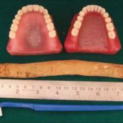
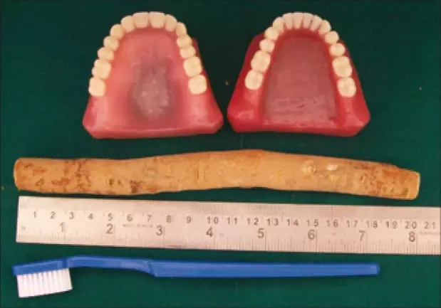
The values of plant-based products have taken on an expanding relevance in dentistry. Salvadora persica chewing stick (miswak) has been practiced for centuries and is recommended by the World Health Organization as a customary oral hygiene tool. The therapeutic effects of S. persica chewing stick are contributed by its mechanical cleansing action, active chemicals released, or the combination of these two actions. However, the S. persica chewing stick in its natural form can be difficult to maneuver in certain parts of the mouth. This concern has inspired the innovation of the S. persica toothbrush that is designed to merge the ease of use of a toothbrush with the beneficial natural properties of S. persica preserved in its bristle. The present study aimed to compare the clinical effectiveness between S. persica toothbrush, S. persica chewing stick and the standard toothbrush in plaque and gingivitis control. In this single-blinded and parallel randomized controlled trial, 78 participants were randomly divided into three groups to either use (i) S. persica toothbrush (MTB); (ii) S. persica chewing stick (MCS); or (iii) standard toothbrush (STB) in a standardized manner for three weeks. Plaque Index (PI) and Periodontal Inflamed Surface Area (PISA) values, measuring plaque levels and severity of gingivitis, respectively, were evaluated at baseline, one- and three-week post-interventions. Results: The MCS group showed a significant improvement in the mean PISA values of the anterior teeth compared to the MTB and STB groups (MCS: from 16.35 ± 10.03 to 3.41 ± 1.14; MTB: from 25.20 ± 14.01 to 3.57 ± 1.19; STB: from 26.54 ± 8.64 to 6.17 ± 0.86; p < .050). All three groups reported significant improvements (p < .001) in the plaque levels and the severity of gingivitis from baseline to three weeks after the intervention. Following correct techniques, S. persica toothbrush and chewing sticks are as effective as the standard toothbrush in plaque control and gingival health, which represent the reputed anti-plaque and anti-gingivitis properties of S. persica.

Reference:

Azizan, N.F., Mohd, N., Nik Azis, N.M. et al. Effectiveness of Salvadora persica toothbrush and Salvadora persica chewing stick in plaque and gingivitis control: a randomized control trial. BMC Complement Med Ther 23, 456 (2023). https://doi.org/10.1186/s12906-023-04295-z

Keywords:

S. persica, chewing sticks, toothbrush, standard toothbrush, equally reliable, controlling, plaque, gingivitis, study, BMC Complementary Medicine and Therapies, Azizan, N.F., Mohd, N., Nik Azis, N.M, Biofilm, Inflammation, Natural product, Oral hygiene, Salvadora persica

Powered by WPeMatico

Anemia may contribute to higher female mortality during heart surgery: JACC

USA: Women are at higher risk of death when undergoing heart bypass surgery than men. Researchers at Weill Cornell Medicine have determined that this disparity is mediated, to a large extent, by intraoperative anemia-the loss of red blood cells during surgery.

The study, published on March 5, in the Journal of the American College of Cardiology, suggests that strategies for minimizing anemia that occurs during this procedure could lead to better outcomes for women with cardiovascular disease.

This study set out to discover why women are less likely to survive coronary artery bypass grafting, a surgical procedure for restoring blood flow to the heart. The team, led by senior author Dr. Mario Gaudino, the Stephen and Suzanne Weiss Professor in Cardiothoracic Surgery at Weill Cornell Medicine, analyzed information obtained from the Society of Thoracic Surgeons Adult Cardiac Surgery Database on more than one million patients. Dr. Lamia Harik, fellow in Cardiothoracic Surgery Research at Weill Cornell Medicine, was first author on the paper.

They examined patient demographics (such as age and ethnicity), risk factors (including disease severity, previous heart attacks and the co-occurrence of other health conditions) and surgical data (including the time spent on the bypass machine and the volume of the components of blood, such as red blood cells).

Crunching the numbers, Dr. Gaudino and his team previously confirmed that women had a higher mortality associated with the procedure than men: 2.8 percent versus 1.7 percent, a nearly 50 percent difference. Now, using sophisticated statistical analyses to assess all the possible variables, the researchers found that a substantial portion of this enhanced risk—38 percent—could be attributed to severe intraoperative anemia.

This depletion of red blood cells is an inevitable side effect of using blood-diluting fluids to prime the heart-lung bypass machine that takes over the job of pumping blood throughout the body during surgery. Women may be even more susceptible to the effects of intraoperative anemia because they tend to arrive in surgery with lower red blood cell counts and have smaller body size compared to their male counterparts.

The study does not establish that intraoperative anemia is causing greater female mortality, but the two factors are associated. It suggests that clinicians and researchers should consider interventions to prevent or minimize severe intraoperative anemia, which can lead to dangerously reduced oxygen delivery to the body’s tissues, including the heart.

Using heart-lung bypass machines with shorter circuits, for example, would limit the volume of blood-diluting solution needed to run the pump. Randomized trials to assess whether methods for curtailing anemia could improve outcomes for women undergoing heart bypass surgery are “urgently needed,” wrote Dr. Gaudino, who is also a cardiovascular surgeon at NewYork-Presbyterian/Weill Cornell Medical Center.

References: Lamia Harik et al, Intraoperative Anemia Mediates Sex Disparity in Operative Mortality After Coronary Artery Bypass Grafting, Journal of the American College of Cardiology (2024). DOI: 10.1016/j.jacc.2023.12.032 10.1001/jamasurg.2022.8156 Journal information: Journal of the American College of Cardiology

Powered by WPeMatico