Echo-intensity characterization at implant sites: Unveiling novel diagnostic ultrasonographic markers for peri-implantitis

USA: A recent study published in the Journal of Clinical Periodontology revealed a significant difference in high-frequency ultrasound (HFUS) echo intensity (EI) characterization of peri-implant tissues between healthy and disease sites; healthy tissues exhibited higher EI values.

“HFUS EI and the absence/presence of a hypoechoic supracrestal area (HSA) may be valid diagnostic ultrasonographic markers to discriminate peri-implant health status,” the researchers wrote.

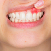
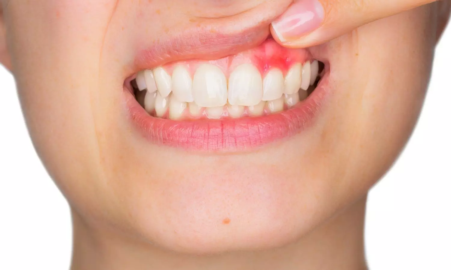
In the realm of dental implantology, peri-implantitis stands as a significant challenge, threatening the longevity and success of dental implants. With conventional diagnostic methods often lacking in sensitivity and specificity, researchers have turned to ultrasonography as a promising avenue for early detection and characterization of peri-implant tissue changes. Recent advancements in this field have unveiled novel diagnostic ultrasonographic markers, shedding light on echo-intensity characterization at implant sites and offering hope for improved management of peri-implantitis.

Peri-implantitis, characterized by inflammation and bone loss around dental implants, poses a considerable risk to implant stability and patient oral health. Timely detection and accurate assessment of peri-implant tissue changes are crucial for effective intervention and prevention of further complications. Traditional diagnostic methods, such as probing and radiography, have limitations in detecting early stages of peri-implantitis and assessing soft tissue alterations.

Ultrasonography emerges as a non-invasive, cost-effective imaging modality with the potential to overcome these limitations. By utilizing high-frequency sound waves, ultrasonography enables real-time visualization of peri-implant tissues, including soft tissue thickness, bone morphology, and vascularity. Considering this, Maria Elisa Galarraga-Vinueza, Tufts University School of Dental Medicine, Boston, Massachusetts, USA, and colleagues aimed to apply HFUS echo intensity for characterizing peri-implant tissues at healthy and diseased sites and to investigate the possible ultrasonographic markers of health versus disease.

For this purpose, the researcher assessed sixty patients presenting 60 implants diagnosed as healthy (N = 30) and peri-implantitis (N = 30) with HFUS. HFUS scans were imported into software where first-order greyscale outcomes [i.e., mean echo intensity] and second-order greyscale outcomes were assessed.

Other ultrasonographic outcomes of interest involved the vertical extension of the hypoechoic supracrestal area (HSA), buccal bone dehiscence (BBD), and soft-tissue area (STA) among others.

The study led to the following findings:

  • HFUS EI mean values obtained from peri-implant soft tissue at healthy and diseased sites were 122.9 ± 19.7 and 107.9 ± 24.7 grey levels (GL), respectively.
  • All the diseased sites showed the appearance of an HSA that was not present in healthy implants (area under the curve = 1).
  • The proportion of HSA/STA was 37.9% ± 14.8%.
  • Regression analysis showed that the EI of the peri-implant soft tissue was significantly different between healthy and peri-implantitis sites (odds ratio 0.97).

“Our study observed significant differences in mean EI between healthy and peri-implantitis sites, with healthy tissues exhibiting higher EI values. The presence of a hypoechoic supracrestal area was exclusively found in diseased sites, serving as a reliable marker for peri-implantitis,” the researchers wrote.

In conclusion, HFUS is a real-time and non-invasive diagnostic tool that provides a comprehensive assessment of peri-implant tissue structures and might be used for diagnosing peri-implant health status.

Reference:

Galarraga-Vinueza, M. E., Barootchi, S., Mancini, L., Sabri, H., Schwarz, F., Gallucci, G. O., & Tavelli, L. Echo-intensity characterization at implant sites and novel diagnostic ultrasonographic markers for peri-implantitis. Journal of Clinical Periodontology. https://doi.org/10.1111/jcpe.13976

Powered by WPeMatico

Individuals with dry eye more likely to experience worse sleep quality, study shows distinct patterns

China: A recent meta-analysis published in BMC Ophthalmology has revealed worse sleep quality in individuals with dry eye than in the healthy population. They also reported a higher risk of unhealthy sleep duration such as insufficient sleep or excessive sleepiness, longer sleep latency, and poorer subjective sleep quality.

Dry eye disease (DED) is a common ocular condition affecting millions worldwide, characterized by discomfort, visual disturbances, and potential damage to the ocular surface. Recent research has begun to uncover a potential link between DED and sleep quality, suggesting that the health of our eyes may be intertwined with the quality of our sleep.

Against the above background, Lei Tian, Capital Medical University, Beijing, China, and colleagues aimed to review the association between dry eye and sleep quality systematically.

For this purpose, they searched the online databases for observational studies published before April 2023. Meta-analysis was performed using STAT15 software. The meta-analysis included a total of 21 studies with 419,218 participants.

Based on the review, the researchers reported the following findings:

  • Dry eye subjects had a worse sleep quality than the healthy population, with poorer subjective sleep quality, a higher risk of unhealthy sleep duration such as insufficient sleep or excessive sleep, and longer sleep latency.
  • The Pittsburgh Sleep Quality Index (PSQI) scores of subjects with dry eye were significantly higher than those of the control subjects (WMD = 1.78).
  • The dry eye subjects scored higher than the control subjects in sleep quality, sleep latency, and sleep disturbance in PSQI; there was no difference between the dry eye individuals and control subjects in sleep duration, sleep efficiency, daytime dysfunction, and sleep medication scores.
  • The risk of sleep disorders in the dry eye subjects was significantly higher than that in the non-dry eye subjects (RR = 2.20); the risk of insufficient sleep in the dry eye subjects was higher than that in the control subjects (RR = 3.76), and the prevalence of excessive sleepiness in dry eye subjects was higher than that in the control subjects (RR = 5.53).
  • The ESS scores of the dry eye subjects were significantly higher than those of the control subjects (WMD = 3.02).

The findings indicate that dry eye patients have a lower sleep quality than the healthy population, with longer sleep latency, poorer subjective sleep quality, and a higher risk of unhealthy sleep duration such as insufficient sleep or excessive sleep.

“However, so far, there is insufficient evidence to establish a causal relationship and related mechanisms between dry eye and sleep disorder,” the researchers wrote. “In the future, there will be a need for more large-scale prospective studies to provide more assistance in patient management and treatment.”

Reference:

Gu, Y., Cao, K., Li, A. et al. Association between sleep quality and dry eye disease: a literature review and meta-analysis. BMC Ophthalmol 24, 152 (2024). https://doi.org/10.1186/s12886-024-03416-7

Powered by WPeMatico

Progressive resistance training as good as neuromuscular exercise for relieving pain in hip OA patients: Study

Denmark: In patients with hip osteoarthritis (OA), progressive resistance training (PRT) is not superior to neuromuscular exercise (NEMEX) for improving hip pain, functional performance, or hip-related quality of life (QoL), a recent study published in Annals of Internal Medicine has revealed.

NEMEX emphasized sensorimotor control & stability, while PRT focused on high-intensity resistance training. The primary outcome, change in the 30-second chair stand test (30s-CST), showed no significant difference between the groups.

Hip osteoarthritis is a prevalent and debilitating condition characterized by stiffness, joint pain, and reduced mobility. Traditional management approaches often include medication and lifestyle modifications. However, emerging research suggests that targeted exercise interventions, such as progressive resistance training or neuromuscular exercise hold significant promise in managing hip OA symptoms and improving overall function. However, randomized controlled trials providing evidence for the optimal exercise type are lacking.

To fill this knowledge gap, Troels Kjeldsen, Næstved-Slagelse-Ringsted Hospitals, Slagelse, Denmark (T.K.), and colleagues aimed to investigate whether PRT is superior to NEMEX for improving functional performance in patients with hip OA.

For this purpose, the researchers conducted a multicenter, cluster-randomized, controlled, parallel-group, assessor-blinded, superiority trial enrolling 160 participants with clinically diagnosed hip OA from 2021 to 2023. They were randomly assigned to PRT (n = 82) or NEMEX (n = 78).

They received twelve weeks of PRT or NEMEX with 2 supervised 60-minute group sessions each week. The PRT intervention comprised of high-intensity resistance training exercises targeting muscles at the hip and knee joints. The NEMEX intervention comprised 10 exercises and emphasized functional stability and sensorimotor control.

The primary outcome was a change in the 30-second chair stand test. Key secondary outcomes included changes in scores on the pain and hip-related QoL subscales of the Hip Disability and Osteoarthritis Outcome Score (HOOS).

Following were the study’s key findings:

  • The mean changes from baseline to 12-week follow-up in the 30s-CST were 1.5 chair stands with PRT and 1.5 chair stands with NEMEX (difference, 0.0 chair stands).
  • For the HOOS pain subscale, mean changes were 8.6 points with PRT and 9.3 points with NEMEX (difference, −0.7 points).
  • For the HOOS QoL subscale, mean changes were 8.0 points with PRT and 5.7 points with NEMEX (difference, 2.3 points).

The main limitation of the study was that the participants and physiotherapists were not blinded.

In conclusion, both progressive resistance training and neuromuscular exercise showed comparable efficacy in improving functional performance, hip pain, and hip-related quality of life over 12 weeks in hip osteoarthritis patients.

Reference:

Kjeldsen T, Skou ST, Dalgas U, Tønning LU, Ingwersen KG, Birch S, Holm PM, Frydendal T, Garval M, Varnum C, Bibby BM, Mechlenburg I. Progressive Resistance Training or Neuromuscular Exercise for Hip Osteoarthritis : A Multicenter Cluster Randomized Controlled Trial. Ann Intern Med. 2024 Apr 9. doi: 10.7326/M23-3225. Epub ahead of print. PMID: 38588540.

Powered by WPeMatico

Physical activity reduces stress-related brain activity and lower CVD risk, suggests study

New research indicates that physical activity lowers cardiovascular disease risk in part by reducing stress-related signaling in the brain.

In the study, which was led by investigators at Massachusetts General Hospital (MGH), a founding member of the Mass General Brigham healthcare system and published in the Journal of the American College of Cardiology, people with stress-related conditions such as depression experienced the most cardiovascular benefits from physical activity.

To assess the mechanisms underlying the psychological and cardiovascular disease benefits of physical activity, Ahmed Tawakol, MD, an investigator and cardiologist in the Cardiovascular Imaging Research Center at Massachusetts General Hospital, and his colleagues analyzed medical records and other information of 50,359 participants from the Mass General Brigham Biobank who completed a physical activity survey.

A subset of 774 participants also underwent brain imaging tests and measurements of stress-related brain activity.

Over a median follow-up of 10 years, 12.9% of participants developed cardiovascular disease. Participants who met physical activity recommendations had a 23% lower risk of developing cardiovascular disease compared with those not meeting these recommendations.

Individuals with higher levels of physical activity also tended to have lower stress-related brain activity. Notably, reductions in stress-associated brain activity were driven by gains in function in the prefrontal cortex, a part of the brain involved in executive function (i.e., decision making, impulse control) and is known to restrain stress centers of the brain. Analyses accounted for other lifestyle variables and risk factors for coronary disease.

Moreover, reductions in stress-related brain signaling partially accounted for physical activity’s cardiovascular benefit.

As an extension of this finding, the researchers found in a cohort of 50,359 participants that the cardiovascular benefit of exercise was substantially greater among participants who would be expected to have higher stress-related brain activity, such as those with pre-existing depression.

“Physical activity was roughly twice as effective in lowering cardiovascular disease risk among those with depression. Effects on the brain’s stress-related activity may explain this novel observation,” says Tawakol, who is the senior author of the study.

“Prospective studies are needed to identify potential mediators and to prove causality. In the meantime, clinicians could convey to patients that physical activity may have important brain effects, which may impart greater cardiovascular benefits among individuals with stress-related syndromes such as depression.”

Effect of Stress-Related Neural Pathways on the Cardiovascular Benefit of Physical Activity, Journal of the American College of Cardiology (2024). DOI: 10.1016/j.jacc.2024.02.02

Powered by WPeMatico

New evidence links passive smoking with dangerous heart rhythm disorder

Exposure to secondhand smoke-even at small amounts-is linked with greater risk of a serious heart rhythm disorder, according to research presented at EHRA 2024, a scientific congress of the European Society of Cardiology (ESC).  The likelihood of atrial fibrillation increased as the duration of passive smoking lengthened.

“The dangers of secondhand smoke were significant regardless of whether individuals were at home, outdoors, or at work, indicating that exposure universally elevates the risk of atrial fibrillation,” said study author Dr. Kyung-Yeon Lee of Seoul National University Hospital, Seoul, Republic of Korea. “We should all make every effort to avoid spending time in smoky environments. The findings should also drive policymakers to further curb smoking in public areas and support smoking cessation programmes to improve public health.”

Atrial fibrillation is the most common heart rhythm disorder worldwide. Symptoms include palpitations, shortness of breath, fatigue, and difficulty sleeping. It is estimated that one in three Europeans will develop the condition during their lifetime. People with atrial fibrillation are five times more likely to have a stroke than their healthy peers. 

It is well established that passive smoking is linked to coronary artery disease and premature death.3-8 However, the links between secondhand smoke and atrial fibrillation have been unclear. This study examined the association between secondhand smoke exposure and the long-term risk of incident atrial fibrillation. The study included adults aged 40 to 69 years who had used the UK National Health Service (NHS) for any reason and were enrolled in the UK Biobank. Current smokers and those with atrial fibrillation at baseline were excluded from the study.

A total of 400,493 adults were included in the analysis. The average age of participants was 56.5 years and 55.2% were women. A touchscreen questionnaire was used to ask participants the number of hours they had been exposed to other people’s smoke in a typical week over the past year at home and in other environments. Participants were categorised into the ‘exposed group’ if they had any contact with secondhand smoke and the ‘non-exposed group’ if they had no contact with secondhand smoke. Some 85,984 (21%) participants had been exposed to secondhand smoke in the previous year, with an average exposure of 2.2 hours per week. During a median follow-up of 12.5 years, atrial fibrillation developed in 23,471 (6%) participants.

The researchers analysed the association between exposure to secondhand smoke and incident atrial fibrillation after adjusting for factors that could potentially affect the relationship, including age, sex, ethnicity, body mass index, daily alcohol consumption, moderate-to-vigorous physical activity, diabetes, high blood pressure, high blood lipids, socioeconomic status, and enrolment centre. The group exposed to secondhand smoke had a 6% higher risk of incident atrial fibrillation during follow-up compared with the non-exposed group after adjusting for the previously mentioned factors (hazard ratio 1.06, 95% confidence interval 1.03–1.10, p <0.001).

A dose-dependent relationship was observed, whereby each increase in the duration of weekly passive smoking was linked with an even greater risk of atrial fibrillation. For example, 7.8 hours of passive smoking per week was associated with an 11% higher likelihood of the heart rhythm disorder compared with no passive smoking. The risk of atrial fibrillation for passive smokers was raised in homes, workplaces, and outside spaces.

Dr. Lee said: “According to our study, once exposed to secondhand smoke, the likelihood of developing atrial fibrillation begins to increase, with the risk escalating significantly as the exposure time lengthens. The finding that passive smoking is harmful not only in enclosed indoor spaces but also outdoor environments underscores the importance of smoking bans to protect public health.” 

Powered by WPeMatico

PET/MRI accurately classifies prostate cancer patients, offers potential to avoid unnecessary biopsies: Study

China: Reston, VA-PET/MRI can improve diagnostic accuracy for prostate cancer patients and help avoid unnecessary biopsies, according to new research published in the April issue of The Journal of Nuclear Medicine.

By applying the PRIMARY scoring system to PET/MRI results, researchers found that more than 80 percent of unnecessary biopsies could be avoided at the expense of missing one in eight clinically significant prostate cancer cases.

The Prostate Imaging Reporting and Data System (PI-RADS) is a five-point scale used to evaluate suspected prostate cancer on MR images. PI-RADS category 3, which presents an unclear suggestion of clinically significant prostate cancer, remains a diagnostic challenge. Although biopsy is recommended under the current guidelines, less than 20 percent of PI-RADS 3 lesions contain clinically significant prostate cancer.

“PI-RADS 3 lesions present a dilemma to both urologists and patients because immediate biopsy could be unnecessary; however, a monitoring strategy could lead to some missed diagnoses of clinically significant prostate cancer,” stated Hongqian Guo, MD, a urologist at Nanjing Drum Tower Hospital at the Affiliated Hospital of Nanjing University Medical School in Nanjing, China. “Hence, specifically ruling out clinically significant prostate cancer among PI-RADS 3 lesions has significant clinical implications.”

In this study, 56 men with PI-RADS 3 lesions underwent 68Ga-PSMA PET/MRI. The five-level PRIMARY system, which is based on a combination of 68Ga-PSMA pattern, localization, and intensity information, was used to report prostate 68Ga-PSMA PET/MRI findings. After imaging, all patients underwent prostate systematic biopsy in combination with targeted biopsy to determine clinically significant prostate cancer.

Among the 56 patients, clinically significant prostate cancer was detected in eight patients (14.3 percent) by biopsy. When a PRIMARY score of at least four was used to make a biopsy decision in men with PI-RADS 3 lesions, 40 of 48 (83.3 percent) participants could have avoided unnecessary biopsies, at the expense of missing 1 in 8 (12.5 percent) of clinically significant prostate cancer cases.

“By demonstrating the additive value of 68Ga-PSMA PET/MRI in classifying PI-RADS 3 lesions, this study provides new insight into the clinical indication for 68Ga-PSMA PET/MRI,” noted Guo. “In the future, PI-RADS 3 patients could be referred for 68Ga-PSMA PET/MRI before prostate biopsy.”

References: Jingyan Shi, Danyan Li, Mengxia Chen, Yao Fu, Shan Peng, Qing Zhang, Jing Liang, Qun Lu, Jiaming Lu, Shuyue Ai, Feng Wang, Xuefeng Qiu and Hongqian Guo Journal of Nuclear Medicine April 2024, 65 (4) 555-559; DOI: https://doi.org/10.2967/jnumed.123.266742

Powered by WPeMatico

Can Probiotics Keep My Gut Healthy?

There’s convincing evidence that probiotics can be helpful in maintaining gastrointestinal health in certain select situations.

But like many natural approaches to maintaining health, there’s been a tendency toward generalizing the benefits, so probiotics are frequently touted as a panacea for all types of health problems. Many advertisements and Internet postings say probiotics are effective for the treatment of asthma, dermatitis, and a wide diversity of other conditions. At best, there is marginal evidence that probiotics are helpful for many of these conditions.

There’s been a lot of work that’s been done on probiotics in the management of irritable bowel, which is important since it’s such a common condition: it’s estimated that upwards of 15% of the U.S. adult population has irritable bowel. I would say that all the studies summarize that certain probiotics seem to help a little bit with irritable bowel, but quite frankly, the magnitude of the benefit tends to be very small. and it’s probably not adequate as the management of irritable bowel by itself.

Where they have been convincingly shown to be beneficial is in the treatment or prevention of certain kinds of diarrhea. Rotavirus is a common cause of infectious diarrhea in infants and children. Once a child has the ‘gastrointestinal flu’, there’s strong evidence to show that probiotics reduce the duration and severity of the illness. There’s less convincing evidence that suggests probiotics can be used to prevent it.

Let’s say your child’s school sends home a note that says 30% of the class is out with gastrointestinal flu. You could give your child the appropriate probiotic with the hope that it might prevent them from developing it. But if they get sick and you administer it to them, it’ll probably shorten the duration of the disease.

The probiotic most consistently shown to be effective in this situation is Lactobacillus GG, which was developed at Tufts University by Sherwood Gorbach and Barry Goldin of the medical school. You can buy it at drugstores under the name Culturelle. A yeast called Saccharomyces boulardii, sold in stores under the name Florastor, also seems to be effective in managing childhood infectious diarrhea.

A second instance where probiotics can be useful is with antibiotic-associated diarrhea. When people go on antibiotics—whatever kind that may be—they frequently develop diarrhea. There have been a number of trials in which people were administered certain probiotics along with the antibiotics used to treat an infection, and that helped prevent diarrhea. Two probiotics have been shown to be effective for this problem: Lactobacillus GG and Saccharomyces boulardii.

For people who often go on antibiotics—say, a woman who gets a lot of bladder infections and then gets diarrhea from the antibiotic—it would make sense to take one of these products along with the antibiotic and extending for a few days after the antibiotic stops. The data suggests that to be effective in the prevention of antibiotic-associated diarrhea, the probiotic should be taken for upwards of a week after you stop taking the antibiotic.

In some cases of antibiotic-associated diarrhea, a very nasty bug called Clostridium difficile takes over because the antibiotics have knocked out the good bacteria in your gut and this bug fills the void. C. diff, as it is known, can cause dysentery, bloody diarrhea and fever, and can even be life-threatening. The only organism that has been shown to convincingly prevent C. difficile is Saccharomyces boulardii.

A major addition to the list of things that probiotics seem to be effective for is traveler’s diarrhea. There are now pretty convincing data that these same two probiotics, Lactobacillus GG and Saccharomyces boulardii, are each effective in reducing the risk of traveler’s diarrhea.

For people who want to avoid developing this kind of traveler’s diarrhea, taking one of these two probiotics seems to sizably reduce the risk. You also can take Pepto Bismol tablets, two tablets four times a day. This has been shown to also be effective. So, there are a couple ways that people can try to protect themselves from developing traveler’s diarrhea aside from being careful about what they eat and drink.

Many people eat yogurt or kefir because it contains probiotic organisms, but studies looking at the potential usefulness of probiotics in a rigorous scientific manner have generally used pure preparations in tablets or capsules, not yogurt. And in many brands, the organisms used to make the yogurt or kefir aren’t necessarily the ones are effective in clinical trials. I can’t say that fermented dairy products aren’t effective. But whether the helpful bacteria you get by eating yogurt are really as effective as pure probiotics is up for debate right now.

People taking probiotics should also be aware that they are not totally innocuous for all individuals. A lot of people say, “This is natural; it might help, and it won’t hurt, so I may as well take it.” Remember that when you’re taking a probiotic, you’re swallowing billions of bacterial spores, and there are rare situations in which those spores can set up a serious infection in your body. Lest I create too much anxiety: this complication seems to occur only in people whose immune systems are suppressed by drugs or other diseases, or who are critically ill.

The most important message is to pay attention to which probiotics you take. There’s a myth that all probiotics are the same, but because they’re not viewed as drugs, they are not regulated by the Food and Drug Administration, so there can be problems with quality control. When you take a probiotic, you assume you’re ingesting billions of bacterial spores that will hatch and result in live bacteria in your gut.

But there are probiotics you can buy in which 99 percent of the spores are dead—you may as well throw your money away. Moreover, if you’re going to approach this scientifically, you have to pay attention to the particular species of bacteria or yeast that you’re ingesting. When I put patients on probiotics, I’m very specific about what products to take. 

Powered by WPeMatico

High glucose and TG levels and low HDL-C levels tied to risk of depression, anxiety, and stress-related disorders: JAMA

Sweden: A recent study published in JAMA Network Open has suggested the involvement of carbohydrate and lipid metabolism in the development of common psychiatric disorders.

The population-based cohort study of more than 200,000 individuals revealed that high levels of triglycerides and glucose and a low level of high-density lipoprotein (HDL) were associated with a higher risk of anxiety, depression, and stress-related disorders. The findings support closer follow-up of people with metabolic dysregulations for the diagnosis and prevention of psychiatric disorders.

Anxiety, depression, and stress-related disorders are common psychiatric disorders that affect approximately one-third of individuals during their life course. Increasing evidence suggests that biomarkers of lipid, apolipoprotein, and carbohydrate metabolism may be associated with the risk for anxiety, depression, and stress-related disorders, but results are inconsistent.

Against the above background, Charilaos Chourpiliadis, Karolinska Institutet, Stockholm, Sweden, and colleagues aimed to investigate whether the biomarkers of lipid, carbohydrate, and apolipoprotein metabolism are associated with the risk of anxiety, depression, and stress-related disorders.

For this purpose, the researchers conducted a population-based cohort study with longitudinal data collection that assessed 211 200 participants from the Apolipoprotein-Related Mortality Risk (AMORIS) cohort who underwent occupational health screening between 1985 and 1996, mainly in the Stockholm region in Sweden.

The main outcomes were the associations between biomarker levels and the risk of developing anxiety, depression, and stress-related disorders through the end of 2020. Nested case-control analyses were conducted within the cohort, including all cases of anxiety, depression, and stress-related disorders, and up to 10 control individuals per case who were individually matched to the case by sex, year of birth, and year of enrollment to the AMORIS cohort, using incidence density sampling.

Population trajectories were used to illustrate the temporal trends in biomarker levels for controls and cases.

The study led to the following findings:

  • 211 200 individuals (mean age at first biomarker measurement, 12.6 years; 58.0% male; 89.4% born in Sweden) participated in the study. During a mean follow-up of 21.0 years, a total of 16,256 individuals were diagnosed with depression, anxiety, or stress-related disorders.
  • High levels of triglycerides (HR, 1.15) and glucose (hazard ratio [HR], 1.30) were associated with an increased subsequent risk of all tested psychiatric disorders, whereas high levels of high-density lipoprotein (HR, 0.88) were associated with a reduced risk. These results were similar for female and male participants and all tested disorders.
  • The nested case-control analyses demonstrated that patients with depression, anxiety, or stress-related disorders had higher levels of glucose, triglycerides, and total cholesterol during the 20 years preceding diagnosis, as well as higher levels of apolipoprotein A-I and apolipoprotein B during the ten years preceding diagnosis, compared with control participants.

In conclusion, the researchers found elevated levels of triglycerides and glucose and reduced levels of HDL-C are linked to a higher risk of subsequent diagnosis of anxiety, depression, and stress-related disorders. The study provides further longitudinal evidence that metabolic syndrome or metabolic dysregulation raises the risk of developing common psychiatric disorders.

“These results add further evidence of the association between cardiometabolic health and psychiatric disorders and potentially advocate for a closer follow-up of individuals with metabolic dysregulations for prevention and early diagnosis of psychiatric disorders,” the researchers wrote.

“There is a need for additional studies to explore whether rigorous or earlier interventions for cardiometabolic diseases could counteract such an association,” they concluded.

Reference:

Chourpiliadis C, Zeng Y, Lovik A, et al. Metabolic Profile and Long-Term Risk of Depression, Anxiety, and Stress-Related Disorders. JAMA Netw Open. 2024;7(4):e244525. doi:10.1001/jamanetworkopen.2024.4525

Powered by WPeMatico

Elevated calcitonin gene-related peptide levels linked to rosacea: A Groundbreaking Study

Denmark: A recent study published in the Journal of the European Academy of Dermatology and Venereology has revealed significantly higher calcitonin gene-related peptide (CGRP) levels in rosacea patients even after adjusting for sex and age.

CGRP levels were unaffected by several factors, including sex, age, BMI, rosacea subtypes, migraine, comorbidities, or ongoing treatments. The findings suggest CGRP’s role in the pathogenesis of rosacea. Targeting CGRP signaling might hold therapeutic promise in individuals affected by this disease.

Rosacea, a chronic inflammatory skin condition affecting millions worldwide, has long confounded sufferers and clinicians with its elusive causes and variable symptomatology. Capsaicin, a well-known trigger of rosacea flushing, has been demonstrated to increase dermal skin blood flow in the forearm by activating transient receptor potential vanilloid (TRPV) channels and promoting the release of CGRP.

Traditional treatments for rosacea, such as oral antibiotics and topical agents, primarily address symptoms rather than underlying mechanisms. However, modulating CGRP signaling could represent a more targeted approach, potentially offering improved efficacy and tolerability for patients with refractory or severe disease.

Against the above background, Messoud Ashina, Copenhagen University Hospital – Rigshospitalet, Copenhagen, Denmark, and colleagues aimed to compare plasma levels of CGRP between individuals with rosacea and healthy controls.

For this purpose, the researchers conducted a cross-sectional case-control study in Copenhagen, Denmark. They collected blood samples from the antecubital vein from adults with rosacea and healthy controls. 123 individuals with rosacea and 68 healthy controls were enrolled.

The study revealed the following findings:

  • After adjusting for age and sex, plasma levels of CGRP were significantly higher in individuals with rosacea (mean, 95% confidence interval: 140.21 pmol/L, 128.50–151.92 pmol/L), compared with controls (110.77 pmol/L, 99.91–120.14 pmol/L).
  • Plasma levels of CGRP were not affected by age, sex, BMI, rosacea sub- or phenotype, concomitant migraine, concomitant disease, or current treatment.

The main limitation was that the participants were not age-, sex- and BMI-matched.

In conclusion, the study provides evidence that plasma CGRP levels are elevated in individuals with rosacea versus healthy controls. However, the researchers did not find a correlation between CGRP levels and rosacea severity or subtype, indicating that CGRP might not be a reliable biomarker for diagnosing or assessing rosacea severity.

“Nevertheless, our findings suggest that CGRP might be involved in the pathophysiology of rosacea,” the researchers wrote. “Thus, there is a need for further research to investigate the potential of medications targeting CGRP signaling in managing rosacea.”

Reference:

F. Wienholtz, N. K., Christensen, C. E., Ashina, H., Jørgensen, N. R., Egeberg, A., Thyssen, J. P., & Ashina, M. Elevated plasma levels of calcitonin gene-related peptide in individuals with rosacea: A cross-sectional case–control study. Journal of the European Academy of Dermatology and Venereology. https://doi.org/10.1111/jdv.19954

Powered by WPeMatico

Diabetes, anemia and old age may increase Dacron-cuffed catheter related infection risk among hemodialysis patients: Study

Diabetes, anemia and old age may increase risk Dacron-cuffed catheter related infection among hemodialysis patients suggests a study published in the BMC Nephrology.

A study was done to provide theoretical basis for prevention of a Dacron-cuffed catheter related infection (CRI), the risk factors of CRI in hemodialysis patients were systematically evaluated. Eight databases, including PubMed, Cochrane library, EMBASE, Web of Science, China National Knowledge Infrastructure (CNKI), Chinese Biomedical Database (CBM), Wanfang Database and Chinese Scientific Journal Database (VIP), were searched to screen out literatures related to the risk factors of long-term indwelling a Dacron-cuffed CRI in hemodialysis. Meta-analysis of risk factors for a Dacron-cuffed CRI in hemodialysis and publication bias test were performed using RevMan 5.4 software. Results: After screening, 13 literatures involving a Dacron-cuffed CRI were included, with a total of 625 patients, and the infection rate was 11.7%. The combined OR value and 95% confidence interval (CI) of all factors were: Combined with Diabetes (1.94, 1.51 ~ 2.50), Hb (1.82, 1.35 ~ 2.44), age (2.38, 1.06 ~ 5.34), catheter indwelling time (1.79, 1.21 ~ 2.66), serum albumin (2.26, 1.25 ~ 4.08), catheter indwelling site (3.29, 1.74 ~ 6.23) and the number of tube placement (5.40, 2.65 ~ 11.02).The main risk factors for a Dacron-cuffed CRI in hemodialysis were combined with diabetes, hemoglobin level, age, catheter indwelling time, serum albumin level, femoral vein catheter indwelling and catheterization times. In other words, hemodialysis patients are at higher risk of CRI if they have diabetes, or if they have a lower hemoglobin level, or if they are older, or if they have a longer duration of catheterization, or if they have a lower serum albumin level, or if they have a femoral vein catheter, or if they have more catheters.

Reference:

Chen, W., Wang, Z., Wang, G. et al. A meta-analysis of risk factors for a Dacron-cuffed catheter related infection in hemodialysis. BMC Nephrol 25, 126 (2024). https://doi.org/10.1186/s12882-024-03568-0

Keywords:

Diabetes, anemia and old age, increase risk, Dacron-cuffed catheter, infection, hemodialysis patients, study, BMC Nephrology, Chen, W., Wang, Z., Wang, G.

Powered by WPeMatico