Splanchnic nerve neurolysis: promising tool for treatment of cancer pain of the upper abdomen

The research paper presents a systematic review and meta-analysis evaluating the analgesic efficacy of neurolytic splanchnic nerve block (NSNB) in treating chronic upper abdominal pain caused by malignancies involving the liver, gall bladder, stomach, and pancreas. The study also assesses the impact of NSNB on quality of life, opioid consumption, procedural safety, and patient survival. The review includes the use of structured Population, Intervention, Control, Outcome, Study (PICOS) criteria to select relevant studies and incorporates a comprehensive literature search of various databases.

The primary objective of the review was to evaluate the analgesic efficacy of NSNB, while the secondary objectives included the impact on quality of life, opioid consumption, procedural safety, and patient survival. The review included studies investigating NSNB for chronic abdominal pain unresponsive to conservative treatment, including randomised controlled trials (RCTs) and non-randomised before-and-after studies from 2001 to 2022. The studies were evaluated for methodological quality and underwent a comprehensive data analysis using a random-effect model with an inverse variance method. The review found that NSNB provided significant pain relief, reduced opioid consumption, and improved quality of life with minimal procedure-related complications.

Study Findings and Heterogeneity

Additionally, the review revealed substantial heterogeneity in the included studies due to differences in participant characteristics, disease severity, comorbidities, treatment responses, and methodological variations. The review highlights the need for high-quality multicentric clinical trials with larger sample sizes and substantial follow-up evaluations to strengthen the evidence and address the identified limitations in the current literature.

Conclusion and Further Research

In conclusion, the review suggests that NSNB is a promising intervention for chronic upper abdominal pain, but further high-quality research is needed to enhance the predictive strength of the evidence and validate the efficacy of NSNB in improving patient outcomes. No conflicts of interest were reported by the authors.

Reference-

Goyal, Sonal1; Kumar, Ajit2; Goyal, Divakar3; Attar, Pradeep2; Bhandari, Baibhav2; Purohit, Gaurav4; Mahiswar, Aditya Pal5; Gupta, Shiwam2. Efficacy of splanchnic nerve neurolysis in the management of upper abdominal cancer pain: A systematic review and meta-analysis. Indian Journal of Anaesthesia 67(12):p 1036-1050, December 2023. | DOI: 10.4103/ija.ija_439_23

Powered by WPeMatico

AI-assisted focused cardiac ultrasound useful modality for assessment of left ventricular ejection fraction

Cardiovascular disease is the global leading cause of death worldwide, with disease prevalence increasing day by day. The rising prevalence of the disease has dramatically increased the financial burden on healthcare systems and has further constrained access to limited resources.

Focused cardiac ultrasound (FoCUS) is recently becoming standard practice in a wide spectrum of clinical settings. However there is a limited data evaluating the real-world use of FoCUS with artificial intelligence (AI) and a recent study aimed to determine the accuracy of FoCUS AI-assisted left ventricular ejection fraction (LVEF) assessment and compare its accuracy between novice and experienced users.

The study published in NPJ Digital Medicine reveals that FoCUS AI-assisted LVEF assessments provide highly reproducible LVEF estimations in comparison to formal transthoracic echocardiogram (TTE).

In this prospective, multicentre study, participants requiring a TTE were recruited to have a FoCUS done by a novice or experienced user. The AI-assisted device calculated LVEF at the bedside, which was subsequently compared to TTE.

The key findings of the study are

• A total of 449 participants were enrolled with 424 studies included in the final analysis. The overall intraclass coefficient was 0.904, and 0.921 in the novice (n = 208) and 0.845 in the experienced (n = 216) cohorts.

• There was a significant bias of 0.73% towards TTE (p = 0.005) with a level of agreement of 11.2%. Categorical grading of LVEF severity had excellent agreement to TTE (weighted kappa = 0.83).

• The area under the curve (AUC) was 0.98 for identifying an abnormal LVEF (<50%) with a sensitivity of 92.8%, specificity of 92.3%, negative predictive value (NPV) of 0.97 and a positive predictive value (PPV) of 0.83.

• In identifying severe dysfunction (<30%) the AUC was 0.99 with a sensitivity of 78.1%, specificity of 98.0%, NPV of 0.98 and PPV of 0.76.

Researchers concluded that FoCUS AI-assisted LVEF assessments provide highly reproducible LVEF estimations in comparison to formal TTE. This finding was consistent among senior and novice echocardiographers suggesting applicability in a variety of clinical settings.

They added even though TTE is the standard for determining LVEF in clinical practice, it is noted that it is “often not readily available” for immediate bedside evaluation and in low-resource settings. Assessing LVEF is fundamental in FoCUS exams.

Reference: Motazedian, P., Marbach, J.A., Prosperi-Porta, G. et al. Diagnostic accuracy of point-of-care ultrasound with artificial intelligence-assisted assessment of left ventricular ejection fraction. npj Digit. Med. 6, 201 (2023). https://doi.org/10.1038/s41746-023-00945-1.

Powered by WPeMatico

Natural daylight photodynamic therapy with keratolytics improve actinic keratoses outcomes

A recent study published in the Journal of the European Academy of Dermatology and Venereology challenges the conventional method of treating actinic keratoses (AKs) through Methyl Aminolevulinate (MAL) photodynamic therapy (PDT). Traditionally, this process involves the removal of crusts and scales through curettage followed by the application of MAL cream which often causes intense pain for the patients.

This research was conducted on a total of 40 patients with multiple AKs on the face and/or scalp to compare the efficacy, safety, and patient satisfaction of standard natural daylight PDT (n-DL-PDT) with curettage versus a new approach. In this new app8roach, curettage was omitted and patients were prepared at home with keratolytics with a 30% urea cream applied twice daily for a week.

After the completion of this study, the findings indicated similarity in efficacy between the two methods analyzed. The mean reduction in the number of AK lesions three months post-treatment was 54.7% in the group without curettage (-Cur) and 58.7% in the group with curettage (+Cur). Also, the differences in efficacy and patient satisfaction were statistically insignificant. The pain scores reported during and after daylight exposure were low and comparable in both the groups, and no unexpected adverse events were observed.

The key highlights of this research suggest that curettage might not be essential for the full treatment effect of n-DL-PDT. This study was conducted in a real-life setting and underscores that the n-DL-PDT protocol could be revolutionized by replacing curettage with keratolytics. This offers a more patient-friendly approach and also presents a major advancement in the field of dermatological treatments.

Source:

Caccavale, S., Boccellino, M. P., Brancaccio, G., Alfano, R., & Argenziano, G. (2023). Keratolytics can replace curettage in daylight photodynamic therapy for actinic keratosis on the face/scalp: A randomized clinical trial. In Journal of the European Academy of Dermatology and Venereology. Wiley. https://doi.org/10.1111/jdv.19687

Powered by WPeMatico

Non-invasive ultrasound therapy safe and feasible for treating calcified aortic stenosis: Lancet

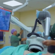
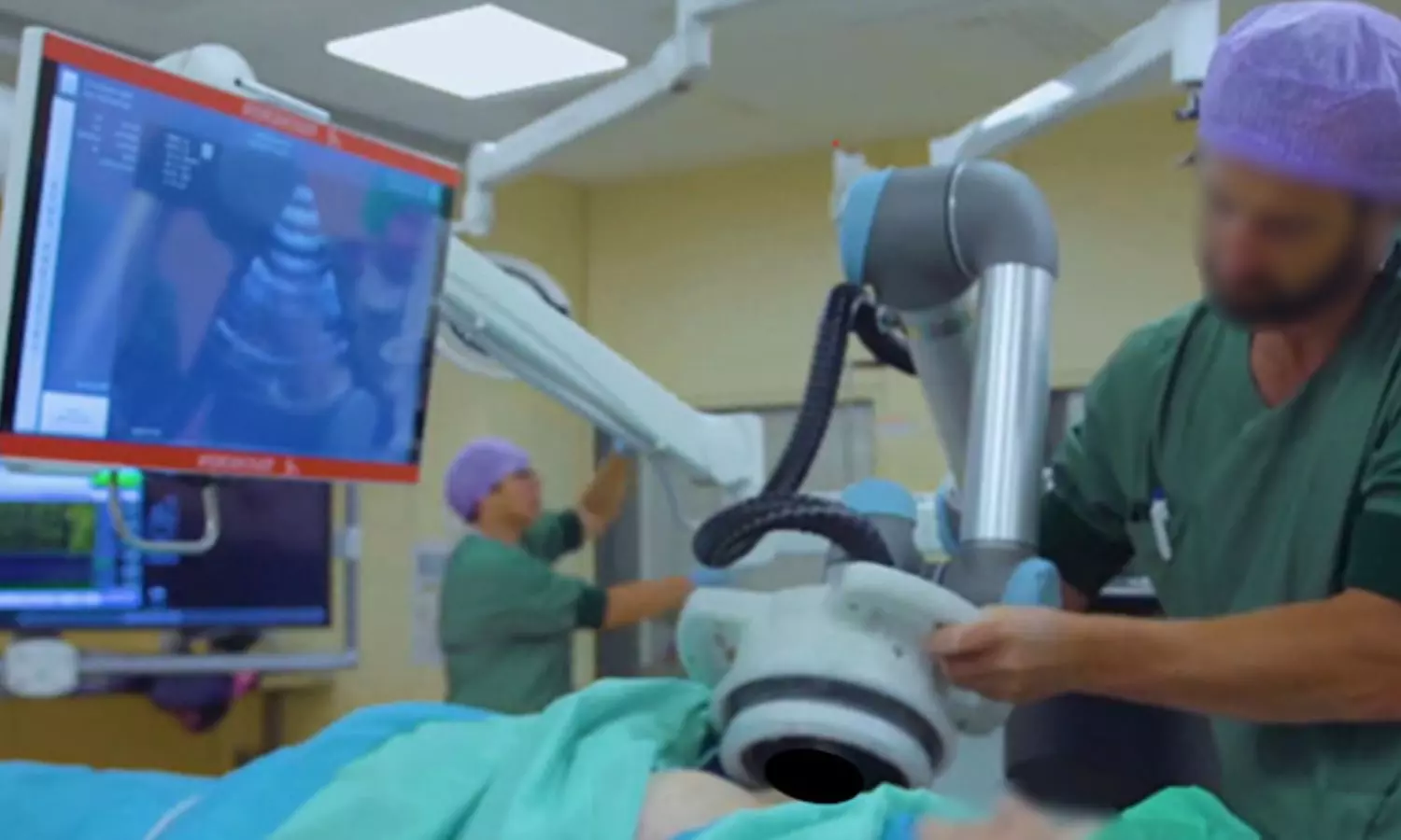
France: In a recent study published in The Lancet, French researchers have shown the safety and feasibility of a non-invasive ultrasound therapy for treating calcified aortic stenosis.

Surgical or transcatheter aortic valve replacement (TAVR) is commonly used for the treatment of calcified aortic valve stenosis, which affects approximately 5 million patients in Europe and 5 million in the United States. However, due to severe comorbidities and limited life expectancy, many patients are not considered suitable candidates for these interventions. As such, non-invasive therapies might offer alternative therapeutic possibilities in these patients.

Emmanuel Messas, a cardiologist and co-founder of the startup Cardiawave, which developed the non-invasive device, and colleagues aimed to evaluate the safety of non-invasive ultrasound therapy and its ability to improve valvular function by softening calcified valve tissue.

The multicenter, prospective, single-arm series enrolled 40 adult patients with severe symptomatic aortic valve stenosis at three hospitals in the Netherlands, France, and Serbia between 2019 and 2022. Patients were treated with transthoracically delivered non-invasive ultrasound therapy. Follow-ups were done at 1, 3, 6, 12, and 24 months.

The primary endpoints of the study were improved valve function and procedure-related deaths within 30 days. In the study, the researchers reported the 6-month data.

Foruty high-risk patients with a mean Society of Thoracic Surgeons score of 5·6% and multiple severe comorbidities were included in the study.

The study revealed the following findings:

  • The primary endpoint, procedure-related mortality, did not occur; also, no life-threatening or cerebrovascular events were reported.
  • Improved valve function was confirmed up to 6 months, reflected by a 10% increase in mean aortic valve area from 0·58 cm2 at baseline to 0·64 cm2 at follow-up, and a 7% decrease in mean pressure gradient from 41·9 mm Hg to 38·8 mm Hg.
  • At six months, the New York Heart Association score had improved or stabilised in 96% of the patients, and the mean Kansas City Cardiomyopathy Questionnaire score had improved by 33%, from 48·5 to 64·5.
  • One serious procedure-related adverse event occurred in a patient who presented with a transient decrease in peripheral oxygen saturation.
  • Non-serious adverse events included pain, discomfort during treatment, and transient arrhythmias.

“This novel, non-invasive ultrasound therapy for calcified aortic stenosis proved to be feasible and safe,” the researchers concluded.

Reference:

Messas E, Ijsselmuiden A, Trifunović-Zamaklar D, Cholley B, Puymirat E, Halim J, Karan R, van Gameren M, Terzić D, Milićević V, Tanter M, Pernot M, Goudot G. Treatment of severe symptomatic aortic valve stenosis using non-invasive ultrasound therapy: a cohort study. Lancet. 2023 Dec 16;402(10419):2317-2325. doi: 10.1016/S0140-6736(23)01518-0. Epub 2023 Nov 14. PMID: 37972628.

Powered by WPeMatico
Benralizumab reduces inhaled corticosteroid usage and help in asthma control

Asthma is a prevalent respiratory disease, affecting over 300 million people worldwide. Approximately 3-5% of asthma patients have severe disease, which involves poor symptom control, frequent exacerbations, and high exposure to oral corticosteroids. These patients often have eosinophilic asthma with uncontrolled inflammation.

According to The Lancet, Patients controlled on benralizumab can significantly reduce ICS therapy while maintaining asthma control.

Stepwise intensification of inhaled corticosteroids is routine for severe eosinophilic asthma, although some patients may not respond to high-dose ICS. Dose reductions are advised for patients responding to biologics, but limited safety evidence exists.

The SHAMAL study was conducted at 22 sites across four countries, involving adults with severe eosinophilic asthma and low Asthma Control Questionnaire scores. Participants received three doses of benralizumab before screening. The study randomly assigned participants to either reduce their high-dose inhaled corticosteroid (ICS) dose to a medium, low, or as-needed dose (reduction group) or continue (reference group) their ICS-formoterol therapy for 32 weeks, followed by a 16-week maintenance period. The primary endpoint was the proportion of participants who successfully reduced their ICS-formoterol dose by week 32, assessed in the reduction group.

Key results are:

· 208 patients were screened and enrolled in the run-in period.

· 168 patients were randomly assigned to either the reduction (n=125) or reference (n=43) arms.

· 110 patients reduced their ICS-formoterol dose: 18 to medium-dose, 20 to low-dose, and 72 to as-needed only.

· By week 48, in 113 patients, reductions were maintained. One hundred fourteen patients in the reduction group had zero exacerbations during tapering.

· The adverse events rate was similar between groups: 91 in the reduction and 35 in the reference group.

· 17 serious adverse events were reported: 12 in the reduction and five in the reference group.

· No deaths were reported.

The results suggest that individuals treated with benralizumab can significantly reduce their use of inhaled corticosteroids (ICS) while maintaining effective asthma control. They added that our findings underscore the opportunity to shift away from high-dose ICS towards a precision-medicine approach with improved patient outcomes.

AstraZeneca funded the study.

Reference:

Jackson, D. J et al. Reduction of daily maintenance inhaled corticosteroids in patients with severe eosinophilic asthma treated with benralizumab (SHAMAL): a randomised, multicentre, open-label, phase 4 study. The Lancet. https://doi.org/10.1016/s0140-6736(23)02284-5

Powered by WPeMatico

Serotonin loss linked to early cognitive impairment in Alzheimer’s disease patients

Comparing PET scans of more than 90 adults with and without mild cognitive impairment (MCI), Johns Hopkins Medicine researchers say relatively lower levels of the so-called “happiness” chemical, serotonin, in parts of the brain of those with MCI may play a role in memory problems including Alzheimer’s disease.

The findings, first published online Sept. 13 in the Journal of Alzheimer’s Disease, lend support to growing evidence that measurable changes in the brain happen in people with mild memory problems long before an Alzheimer’s diagnosis, and may offer novel targets for treatments to slow or stop disease progression.

“The study shows that people with mild cognitive impairment already display loss of the serotonin transporter. This measure that reflects serotonin degeneration is associated with problems with memory, even when we take into account in our statistical model MRI measures of neurodegeneration and PET measures of the amyloid protein that are associated with Alzheimer’s Disease,” says Gwenn Smith, Ph.D., professor of psychiatry and behavioral sciences at the Johns Hopkins University School of Medicine.

MCI describes the diagnostic stage between normal brain function in aging and Alzheimer’s Disease (AD). Symptoms of MCI include frequent forgetfulness of recent events, word finding difficulty, and loss of the sense of smell. Those with MCI may stay in this stage indefinitely, or progress to more severe forms of cognitive deficits, giving urgency to the search for predictive markers, and possible early prevention interventions, investigators say.

The investigators cautioned that their study showed a correlation between lower serotonin transporter levels and memory problems in MCI, and was not designed to show causation or the role of serotonin in the progression from MCI to AD. To answer these questions, further research is needed to study over time healthy controls and individuals with MCI to demonstrate the role of serotonin in disease progression.

For the study, the Hopkins scientists recruited 49 volunteers with MCI, and 45 healthy adults ages 55 and older who underwent an MRI to measure changes in brain structure and two positron emission tomography (PET) scans of their brains at Johns Hopkins between 2009 and 2022. The research team used PET scans to look specifically at the serotonin transporter – a neurotransmitter, or brain chemical long associated with positive mood, appetite and sleep – and to look at the amyloid-beta protein (Aβ) distribution in the brain. Aβ is thought to play a central role in the pathology of AD. Studies in mice done at Johns Hopkins have shown that serotonin degeneration occurs before the development of widespread beta-amyloid deposits in the brain. Loss of serotonin is often associated with depression, anxiety, and psychological disorders.

Researchers found that MCI patients had lower levels of the serotonin transporter, and higher levels of Aβ than healthy controls. The MCI patients had up to 25% lower serotonin transporter levels in cortical and limbic regions than healthy controls. In particular, they report, lower serotonin transporter levels were found in cortical, limbic, and subcortical regions of the brains in those with MCI, areas specifically responsible for executive function, emotion, and memory.

“The correlation we observed between lower serotonin transporters and memory problems in MCI is important because we may have identified a brain chemical that we can safely target that may improve cognitive deficits and, potentially, depressive symptoms,” says Smith. “If we can show that serotonin loss over time is directly involved in the transition from MCI to AD, recently developed antidepressant medications may be an effective way to improve memory deficits and depressive symptoms and thus, may be a powerful way forward to slow disease progression.”

Researchers say future studies include longitudinal follow up of individuals with MCI to compare serotonin degeneration to the increase in and Aβ levels, as well as the increase in levels of the Tau protein that is also associated with AD compared to healthy adults. They are also studying multi-modal antidepressant drugs to treat depression and memory deficits in hopes of mitigating and halting symptoms.

Reference:

Smith, Gwenn S. et al. ‘Serotonin Degeneration and Amyloid-β Deposition in Mild Cognitive Impairment: Relationship to Cognitive Deficits’. 1 Jan. 2023 : 215 – 227

Powered by WPeMatico

Prednisone combined with mycophenolate mofetil effective in Immunoglobulin A nephropathy with Renal Dysfunction

A recent study identified a promising treatment for Immunoglobulin A Nephropathy (IgAN), a prevalent form of chronic glomerulonephritis which is a leading cause of chronic renal failure in China. The findings were published in the World Journal of Clinical Cases.

Mei-Juan Meng and team from the Wuxi People’s Hospital and Nanjing Medical University conducted this study from January 2011 to December 2020. This study included 200 patients with moderate-to-severe IgAN. The renal function at the time of diagnosis is crucial as the prognosis of IgAN varies widely. The research evaluated the effectiveness and safety of prednisone alone and in combination with mycophenolate mofetil (MMF) for treating IgAN with moderate-to-severe renal impairment.

The patients were divided into a glucocorticoid therapy group (GTG) and an immunosuppressive therapy group (ITG). Both the groups received general treatments, and the GTG was administered prednisone while the ITG received MMF. The study found that both therapy significantly reduced 24-hour urinary protein levels and stabilized renal function. Also, at the nine-month mark, the ITG exhibited a more significant decrease in protein levels and serum creatinine compared to the GTG. By the  end of 12th month, both groups continued to show improvements but the ITG demonstrated a higher overall response rate.

The outcome of the study suggest that the combination of prednisone and MMF proved highly effective for treating IgAN with moderate-to-severe renal dysfunction. This discovery holds significant promise for improving the prognosis and outcomes for patients grappling with this challenging condition. Also, further research and clinical trials will be imperative to validate and build upon these promising results from this study.

Reference:

Meng, M.-J., Hu, L., Fan, Y., Gao, H., Chen, H.-Z., Chen, C.-M., Qi, Z., & Liu, B. (2023). Efficacy of prednisone combined with mycophenolate mofetil for immunoglobulin A nephropathy with moderate-to-severe renal dysfunction. World Journal of Clinical Cases, 11(35), 8300–8309. https://doi.org/10.12998/wjcc.v11.i35.8300

Powered by WPeMatico

Delaying frozen embryo transfer for six months after preceding pregnancy loss improves pregnancy outcomes

When it comes to the interpregnancy interval (IPI) following a clinical pregnancy loss (CPL) during in vitro fertilization (IVF) treatment, finding the right timing has remained a subject of debate. A recent retrospective cohort study conducted in China aimed to shed light on this matter by evaluating the association between various IPI lengths and the outcomes of the next frozen embryo transfer (FET). This study suggests that the timing of the interpregnancy interval is a crucial factor in achieving successful pregnancy outcomes, particularly for women undergoing IVF treatment after experiencing a clinical pregnancy loss.

This study was published in JAMA Network Open by Ze Wang and colleagues. This was a retrospective cohort study conducted at the Center for Reproductive Medicine of Shandong University in China. The study aimed to evaluate the association between different interpregnancy interval (IPI) lengths following a preceding clinical pregnancy loss (CPL) and the subsequent pregnancy outcomes of the next frozen embryo transfer (FET).

The study included women who had undergone frozen-thawed blastocyst transfer within 1 year after experiencing a CPL during in vitro fertilization (IVF) treatment. The data were collected from women who received IVF treatment between July 1, 2017, and June 30, 2022. Follow-up for pregnancy outcomes was completed for all participants on March 31, 2023.

The study included a total of 2433 women with a mean age of 31.8 years. These women were divided into three groups based on the length of their interpregnancy interval following a CPL: less than 3 months, 3 to less than 6 months, and 6 to 12 months. The study assessed several key pregnancy outcomes related to the next FET after a preceding CPL. These outcomes included:

Clinical Pregnancy: The achievement of a clinical pregnancy following the FET.

Live Birth: The successful delivery of a live infant following the FET.

Healthy Live Birth: The delivery of a live infant without adverse health conditions.

Total Pregnancy Loss: The occurrence of pregnancy loss, including both clinical and subclinical losses.

The outcomes were evaluated by calculating adjusted odds ratios (AORs) to determine the association between the length of interpregnancy intervals and these pregnancy outcomes. The study focused on comparing the outcomes between women with shorter IPIs (<3 and 3 to <6 months) and those with an IPI of 6 to 12 months.

Compared with women with an IPI of 6 to 12 months, those with shorter IPIs (<3 and 3 to <6 months) had the following results:

Clinical Pregnancy:

An IPI of less than 3 months: Adjusted Odds Ratio (AOR) of 0.70 (95% CI: 0.53-0.92)

An IPI of 3 to less than 6 months: AOR of 0.79 (95% CI: 0.65-0.95)

Live Birth:

An IPI of less than 3 months: AOR of 0.64 (95% CI: 0.48-0.85)

An IPI of 3 to less than 6 months: AOR of 0.74 (95% CI: 0.61-0.90)

Healthy Live Birth:

An IPI of less than 3 months: AOR of 0.63 (95% CI: 0.46-0.87)

An IPI of 3 to less than 6 months: AOR of 0.79 (95% CI: 0.64-0.98)

Total Pregnancy Loss:

An IPI of less than 3 months: AOR of 1.87 (95% CI: 1.31-2.67)

An IPI of 3 to less than 6 months: AOR of 1.29 (95% CI: 1.00-1.66)

These results indicate that shorter interpregnancy intervals (<3 and 3 to <6 months) were associated with a decreased likelihood of achieving clinical pregnancy, live birth, and healthy live birth. Additionally, women with shorter IPIs had a higher risk of total pregnancy loss compared to those with an IPI of 6 to 12 months.

Delaying the next frozen embryo transfer for at least 6 months after a preceding clinical pregnancy loss was found to be associated with more favorable pregnancy outcomes, emphasizing the significance of interpregnancy interval length in the context of IVF treatment following a CPL. Further prospective studies are warranted to validate and expand upon these findings.

Reference:Wang, Z., Meng, Y., Shang, X., Suo, L., Zhao, D., Han, X., Yang, M., Yin, M., Miao, H., Wang, Y., Yang, H., Yu, Y., Wei, D., & Chen, Z.-J. Interpregnancy interval after clinical pregnancy loss and outcomes of the next frozen embryo transfer. JAMA Network Open,2023;6(10):e2340709. https://doi.org/10.1001/jamanetworkopen.2023.40709 

Powered by WPeMatico

Cerasorb reliable bone graft substitute with low complication rates for curettage of bone tumors

Artificial bone graft substitutes (ABGS) for curettage of bone tumors are becoming increasingly popular. Ulrike Susanne Wittig et al conducted a retrospective analysis to determine the efficacy of the ABGS Cerasorb (Curasan-AG, Kleinostheim, Germany), a beta-tricalcium phosphate (beta-TCP), concerning resorption profile, bone healing, and remodeling after surgery and to evaluate potential complications. The study has been published in “Indian Journal of Orthopaedics.”

Cerasorb is a pure-phase beta-tricalcium phosphate (β-TCP) with precisely defined physical and chemo-crystalline properties and a high level of uniformity of chemical composition and purity. In contrast to alpha-tricalcium phosphates (α-TCP), β-TCP is thermodynamically stable in a biological environment and within a normal temperature range. Biodegradation is faster than that of α-TCP.

Forty-three patients suffering from benign and low-grade malignant bone tumors were treated with curettage and refilling of the bony cavity using the ABGS Cerasorb were included in the final analysis. Clinical follow-up exams with X-rays in two planes were performed 6 weeks, 3 months, 6 months, and 1 year after surgery.

Key findings of the study were:

• The study cohort consisted of 21 male and 22 female patients.

• Patients’ mean age at surgery was 42 years (range 15–70 years).

• Seventeen lesions were located in the distal femur, one was in the femoral diaphysis, one was in the proximal fibula, three lesions were in the proximal tibia, one in the distal tibia, one was in the acetabulum, two in the metacarpal bones, and one in the medial phalanx of the fifth finger, three lesions were in the proximal femur, four were located in the humeral diaphysis, and nine lesions were located in the proximal humerus.

• After a mean follow-up period of 14.6 months, radiological consolidation following curettage was observed in all patients.

• Total resorption was observed in 16.3% of patients; in the other 83.7%, resorption was partial.

• In four patients, of whom two had a tumor in the distal femur and two in the humeral diaphysis, fractures occurred within 6 weeks after primary surgery.

The authors commented – “In conclusion, the beta-TCP Cerasorb seems to be a reliable bone graft substitute with low complication rates and is a suitable alternative to autologous bone grafts or allografts. Nonetheless, it shows a tendency of delayed resorption.”

Level of Evidence III – retrospective cohort study.

Further reading:

Artificial Bone Graft Substitutes for Curettage of Benign and Low Grade Malignant Bone Tumors: Clinical and Radiological Experience with Cerasorb

Ulrike Susanne Wittig, Jorg Friesenbichler et al

Indian Journal of Orthopaedics (2023) 57:1409–1414

https://doi.org/10.1007/s43465-023-00919-1

Powered by WPeMatico

Urinary Iodine Concentration inversely and independently associated with testosterone levels

According to recent reports from the World Health Organization, iodine intake, crucial for thyroid function, is sufficient or even excessive in numerous countries due to iodization policies. While this may seem beneficial for addressing iodine deficiency, concerns have emerged regarding the potential adverse effects of excess iodine on communities. A groundbreaking study has delved into the intricate relationship between iodine exposure and testosterone levels in men, shedding light on a previously unexplored aspect of reproductive health. The study revealed that low urinary iodine concentration is an independent factor causing increased testosterone levels and hence men should be cautioned while taking iodine supplementation. 

The study results were published in the journal JAMA Network Open. 

The study, drawing on data from the National Health and Nutrition Examination Survey (NHANES), a comprehensive survey of the US population, aimed to investigate the association between urinary iodine concentration (UIC) and testosterone levels. The rationale behind this inquiry lies in the identification of iodine transporters in extrathyroidal tissues, including the testis, raising questions about the impact of iodine on male reproductive hormones.

In a cross-sectional analysis encompassing five NHANES cycles spanning from 1999 to 2002 and 2011 to 2016, the study focused on 11,433 male participants with available data on total testosterone (TT). After excluding individuals with missing UIC data and those below 18 years old, 2,934 men formed the basis for the investigation. Adhering to the Strengthening the Reporting of Observational Studies in Epidemiology (STROBE) reporting guidelines, the levels of total testosterone (TT) and urinary iodine concentration (UIC) were examined through inductively coupled plasma dynamic reaction cell mass spectroscopy and isotope dilution high-performance liquid chromatography–tandem mass spectrometry, respectively. Free testosterone (cFT) was calculated using the Vermeulen formula.

To assess distinctions among UIC groups, the Wilcoxon rank-sum test, or χ2 test, was employed. Covariates selected for univariate regressions were based on pre-existing pathophysiological knowledge. Subsequently, multivariable linear regression analysis, applied to log-transformed values, gauged the independent association between continuous UIC and TT levels.

Results:

  • The findings revealed a noteworthy correlation between UIC and testosterone levels.
  • Men with low UIC exhibited higher levels of both total testosterone and calculated free testosterone (cFT) compared to those with normal and high UIC.
  • Additionally, the low-UIC group displayed a more favorable metabolic and glycolipid profile, including a lower body mass index.
  • Through multivariable linear regression analysis, adjusting for various factors such as age and features of metabolic syndrome, the negative association between UIC and TT persisted.
  • This intriguing evidence challenges previous assumptions about the effects of iodine on male reproductive hormones.

Thus, the research shows a correlation between lower urinary iodine concentration (UIC) and higher testosterone levels, irrespective of factors affecting androgen status. The study suggests caution in excessive iodine supplementation due to potential effects on testicular steroidogenesis.

While the study acknowledges its limitations, including the cross-sectional design that does not establish cause-effect relationships, the implications are significant. The observed link between lower UIC and higher testosterone levels prompts further investigation into the causal directionality of this association. The study also underscores the need for a nuanced understanding of iodine’s impact on reproductive health, moving beyond its well-established role in thyroid function.

Further reading: Barbonetti A, Castellini C, Di Giulio F, et al. Iodine Intake and Testosterone. JAMA Netw Open. 2023;6(12):e2348573. doi:10.1001/jamanetworkopen.2023.48573

Powered by WPeMatico